• 검색 결과가 없습니다.

CT-Guided Percutaneous Transthoracic Needle Biopsy Using the Additional Laser Guidance System by a Pulmonologist with 2 Years of Experience in CT-Guided Percutaneous Transthoracic Needle Biopsy

N/A
N/A
Protected

Academic year: 2021

Share "CT-Guided Percutaneous Transthoracic Needle Biopsy Using the Additional Laser Guidance System by a Pulmonologist with 2 Years of Experience in CT-Guided Percutaneous Transthoracic Needle Biopsy"

Copied!
9
0
0

로드 중.... (전체 텍스트 보기)

전체 글

(1)

Introduction

Conventional computed tomography (CT)–guided per- cutaneous transthoracic needle biopsy (PTNB) is a well- established method and is the most common imaging mo- dality used to obtain tissue from a lung lesion

1

. Cone-beam CT (CBCT)–guided PTNB has recently been in the spotlight because of its real-time fluoroscopic capability, greater flex- ibility in orientating the detector around the patient, and rela- tively low radiation exposure compared with CT fluoroscopy- guided PTNB

2,3

. The advantage of CBCT-guided PTNB over conventional CT-guided PTNB is its real-time properties that enable operators to avoid dangerous structures and access the target lesion in patients unable to cooperate with breath hold-

CT-Guided Percutaneous Transthoracic Needle Biopsy Using the Additional Laser Guidance System by a Pulmonologist

with 2 Years of Experience in CT-Guided Percutaneous Transthoracic Needle Biopsy

Min-Cheol Jeon, Ph.D.

1

, Ju Ock Kim, M.D., Ph.D.

2

, Sung Soo Jung, M.D., Ph.D.

2

, Hee Sun Park, M.D., Ph.D.

2

, Jeong Eun Lee, M.D., Ph.D.

2

, Jae Young Moon, M.D., Ph.D.

2

, Chae Uk Chung, M.D., Ph.D.

2

, Da Hyun Kang, M.D.

2

and Dong Il Park, M.D.

2

1

Department of Radiology, Daejeon Health Institute of Technology, Daejeon,

2

Division of Pulmonary and Critical Care Medicine, Department of Internal Medicine, Chungnam National University Hospital, Daejeon, Korea

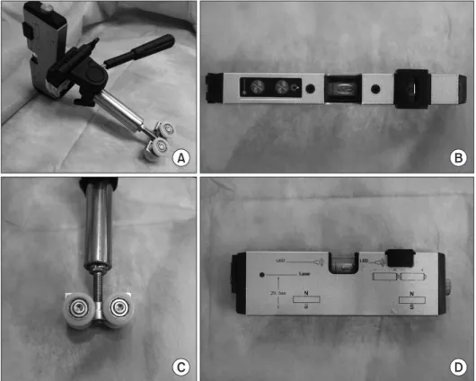
Background: We developed an additional laser guidance system to improve the efficacy and safety of conventional computed tomography (CT)–guided per cutaneous transthoracic needle biopsy (PTNB), and we conducted this study to evaluate the efficacy and safety of our system.

Methods: We retrospectively analyzed the medical records of 244 patients who underwent CT-guided PTNB using our additional laser guidance system from July 1, 2015, to January 20, 2016.

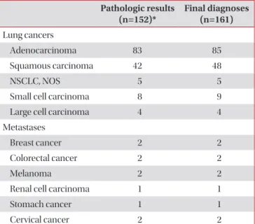
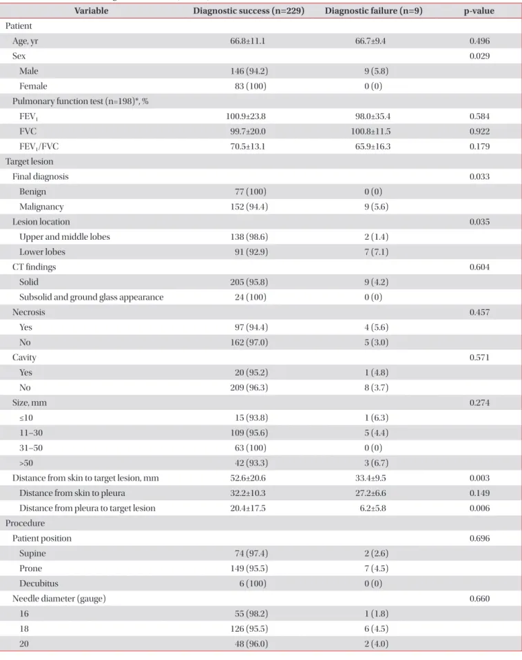
Results: There were nine false-negative results among the 238 total cases. The sensitivity, specificity, positive predictive value, negative predictive value, and diagnostic accuracy of our system for diagnosing malignancy were 94.4% (152/161), 100% (77/77), 100% (152/152), 89.5% (77/86), and 96.2% (229/238), respectively. The results of univariate analysis showed that the risk factors for a false-negative result were male sex (p=0.029), a final diagnosis of malignancy (p=0.033), a lesion in the lower lobe (p=0.035), shorter distance from the skin to the target lesion (p=0.003), and shorter distance from the pleura to the target lesion (p=0.006). The overall complication rate was 30.5% (74/243). Pneumothorax, hemoptysis, and hemothorax occurred in 21.8% (53/243), 9.1% (22/243), and 1.6% (4/243) of cases, respectively.

Conclusion: The additional laser guidance system might be a highly economical and efficient method to improve the diagnostic efficacy and safety of conventional CT-guided PTNB even if performed by inexperienced pulmonologists.

Keywords: Lasers; Solitary Pulmonary Nodule; Lung Neoplasms; Biopsy, Needle

Address for correspondence: Dong Il Park, M.D.

Division of Pulmonary and Critical Care Medicine, Department of Internal Medicine, Chungnam National University Hospital, 282 Munhwa-ro, Jung-gu, Daejeon 35015, Korea

Phone: 82-42-280-7147, Fax: 82-42-257-5753 E-mail: [email protected]

Received: Dec. 3, 2017 Revised: Jan. 16, 2018 Accepted: Feb. 20, 2018 Published online: Jun. 19, 2018

cc It is identical to the Creative Commons Attribution Non-Commercial License (http://creativecommons.org/licenses/by-nc/4.0/).

Copyright © 2018

The Korean Academy of Tuberculosis and Respiratory Diseases.

(2)

ing

3

. Lee et al.

3

reported the outcomes of CBCT-guided PTNB in 1,108 patients. The results showed that the sensitivity, specificity, diagnostic accuracy, and incidence of complica- tions (pneumothorax and hemoptysis) were 95.7% (733/766), 100% (323/323), 97.0% (1,056/1,089), and 23.9% (276/1,153), respectively. However, several studies on conventional CT- guided PTNB involving more than 100 patients have been re- ported since 2010, all of which showed similar results to those of CBCT-guided PTNB (sensitivities, 90%–96%; specificities, 92%–100%; diagnostic accuracies, 93%–97%)

5–11

. Choi et al.

11

performed conventional CT-guided PTNB on patients with pulmonary nodules <1 cm and reported a sensitivity, specific- ity, and diagnostic accuracy of 93.1% (148/159), 98.8% (81/82), and 95.0% (229/241), respectively. These results are compara- ble with those of a study that performed CBCT on pulmonary nodules ≤1 cm, in which the sensitivity, specificity, and diag- nostic accuracy were 96.7% (58/60), 100% (38/38), and 98.0%

(96/98), respectively

12

. CBCT is very expensive and requires a large physical space for the equipment. Therefore, consider- ing the diagnostic yield and patient safety of conventional CT reported in recent studies, which are comparable with those of CBCT, it is preferable to improve existing conventional CT-guided PTNB than to purchase a CBCT system

1-4,11,12

. We developed the additional laser guidance system to improve the efficacy and safety of conventional CT-guided PTNB. This study was conducted to evaluate the efficacy and safety of our laser guidance system.

Materials and Methods

1. Patients

We performed a retrospective analysis of the medical re- cords of 244 patients who underwent PTNB from July 1, 2015, to January 20, 2016. Approval for this study was given by the Chungnam National University Hospital Institutional Review Board (CNUH 2016-02-004), and requirement for informed consent was waived.

2. Procedures

1) Installation and alignment of the additional laser guidance system

The installation and alignment of the additional laser guid- ance system were as follows. (1) We mounted the laser level (HG-909A; X-CLOVE, China), which adhered to a wheel bracket, on a stainless steel frame and positioned it on the opposite side of the CT gantry (Figures 1, 2A). (2) By adjust- ing the wheel bracket (to the left or right), we matched both vertical laser beam lines, which came from the laser level (HG- 909A; X-CLOVE) and the CT gantry (Figure 2B). (3) The sec- ond laser level, also mounted on an iron frame, was placed on the opposite side of the patient to the operator, perpendicular to the other laser (Figure 2C).

A B

C D

Figure 1. Laser level (HG-909A; X-

CLOVE) and a wheel bracket. (A) Laser

level, adhered to a wheel bracket. (B, D)

Overhead and side view of laser level. (C)

Wheel part of a bracket.

(3)

2) CT-guided PTNB procedure

All PTNBs were performed under CT (Siemens SOMATOM Sensation 64 CT scanner; Siemens Medical Solutions, Forch- heim, Germany) guidance by one pulmonologist (D.I. Park with 2 years of experience in CT-guided PTNB). Before CT

was initiated, the importance of breathing was explained to each patient, who was asked to practice breathing in at tidal volume and holding it three times. After obtaining a CT scout image, low-dose (100 kVp, 20 mA) CT was performed at the area of interest using a 2-mm section thickness and with the Figure 2. (A) The laser level, adhered to a wheel bracket, mounted on a stainless steel frame and positioned on the op- posite side of the computed tomography (CT) gantry. Horizontal movement is possible (blue arrows). (B) Both verti- cal laser beam lines, which came from the laser level and CT gantry, had been aligned, such that they looked like a line (red arrows). (C) The second laser level was placed on the side of the patient opposite to the operator, perpendicular to the other laser. (D) In the case of an oblique approach, a digital level (DWL- 80E, Sincon) was used to tilt the laser level. The digital level was 29.9° (red round arrows).

Figure 3. Adjustment of the laser lines.

(A, B) The laser line originating from the

head end was located at the entry point

on the skin and ran along the coaxial

needle. (C, D) The intersection of the

two laser lines originating from the head

end and the side opposite to the opera-

tor were matched at the entry point on

the skin and ran long the coaxial needle.

(4)

patient practicing breath-holding after inspiration. The subse- quent procedures were as follows. (1) The skin entry site was determined by the CT gantry laser lights and radiopaque grid displayed on the skin and was sterilized using Betadine. (2) Local anesthesia from the skin to the pleural surfaces was ad- ministered using a 1% lidocaine solution. (3) By adjusting the laser position when the patient inhaled and held their breath, the intersection of the two laser lines originating from the head end and the side opposite to the operator was located at the entry point on the skin (Figure 3). In the case of an oblique approach, we used a digital level (DWL-80E; Sincon, China) to tilt the laser level (HG-909A; X-CLOVE) before perform- ing procedure (Figure 2D). (4) A coaxial needle was inserted along the two laser lines into the target lesion via the skin entry site (Figure 3D). (5) A CT scan was conducted to ensure that the coaxial needle tip was in the correct position (Figure 4). (6) Then, a semi-automatic cutting needle ranging in size from 20 to 16 gauge (Stericut; TSK Laboratory, Tochigi, Japan) was inserted via the coaxial introducer, and the biopsy was performed. Specimen acquisition was repeated until a visually adequate amount of tissue was obtained. To check for biopsy- related complications, CT was performed after obtaining the tissue.

3. Statistical analysis

To determine the risk factors for diagnostic failure, the study population was divided into a diagnostic success group (i.e.,

true-positive and true-negative results) and a diagnostic fail- ure group (i.e., false-positive and false-negative results). The factors related to diagnostic failure were evaluated by univari- ate analyses using the Mann-Whitney U test for numerical values and the chi-square test for categorical values. Statistical analysis was performed using PASW Statistics version 18.0 (IBM Corp., Armonk, NY, USA). A p-value of < 0.05 was con- sidered to indicate statistical significance.

Results

1. Patient and target lesion characteristics

The baseline patient characteristics and procedures per- formed are summarized in Table 1 (n=244). The mean age of the patients was 66.5±11.3 years, and 64.8% (n=158) of all cases were male. Among all target lesions, 41.4% (n=101) were located in the lower lobes. The mean longest diameter was 33.8±20.1 mm. The numbers of solid, sub-solid, and ground- glass lesions were 218 (89.3%), 14 (5.7%), and 12 (4.9%), re- spectively.

2. Procedure and radiation dose

During the procedure, 79 (32.4%) patients were placed in the supine position, 159 (65.2%) in the prone position, and six (2.4%) in the decubitus position; 58 (23.8%) 16-gauge bi- opsy needles, 136 (55.7%) 18-gauge needles, and 50 (20.5%) 20-gauge needles were used. The mean distance from the skin to the target lesion was 51.8±21.5 mm. The total proce- dure time was 8.8±2.9 minutes, and the needle dwelling time was 2.2±1.3 minutes. The number of biopsies performed was 2.2±1.2. The calculated mean effective radiation dose was 1.0±0.4 mSv.

3. Pathological results and final diagnoses

A lung lesion was recorded as ‘benign’ if the nodule met the following criteria. (1) The pathologic results showed defini- tive benign features (e.g., aspergilloma, tuberculosis). (2) The pathologic results did not show definitive benign features (e.g., inflammation, fibrosis); for example, (a) the CT findings favored a benign lesion, (b) the size of the lesion decreased or showed no interval change on follow-up CT after more than 18 months, or (c) the physician decided to discontinue follow- up because the lesion was regarded as benign. (3) The patho- logic results confirmed the nodule as “benign” on more than two occasions. All lesions not determined to be benign or ma- lignant were classified as “indeterminate” and were excluded from the statistical analysis.

Among the technically successful cases (n=243), 161 (66.3%), 77 (31.7%), and five (2.1%) were diagnosed as malig- Figure 4. A 76-year-old woman who was diagnosed with non-small

cell lung cancer (adenocarcinoma) by percutaneous transthoracic

needle biopsy. (A) Axial view. (B) Sagittal view.

(5)

nant, benign, and indeterminate lesions, respectively. Of the malignant lesions (n=161), 152 (94.4%) were diagnosed by PTNB. False-negative cases (n=9) were finally diagnosed as malignancy by re-PTNB (n=4), bronchoscopy (n=2), surgical biopsy (n=1), endobronchial ultrasound (n=1), and pleural effusion cytology (n=1). Of the 77 (31.7%) lesions diagnosed as benign, 32 (13.2%) had specific benign pathologic features, with the most common benign lesion being tuberculosis (n=16, 6.6%). There were five indeterminate cases. In the re- maining technically successful cases, excluding the indetermi- nate cases, the sensitivity, specificity, positive predictive value (PPV), negative predictive value (NPV), and diagnostic ac- curacy for the diagnosis of malignancy were 94.4% (152/161), 100% (77/77), 100% (152/152), 89.5% (77/86), and 96.2%

(229/238), respectively. The pathologic results and final diag- noses of the malignant lesions are summarized in Table 2.

4. Risk factors for diagnostic failure

There were nine false-negative results among the 238 total cases. The results of the univariate analyses showed that the risk factors for a false-negative result were male sex (p=0.029), a final diagnosis of malignancy (p=0.033), a lesion in the lower lobe (p=0.035), and a shorter distance from the pleura to the target lesion (p=0.006). The longest diameter of the target le- sion was not related to diagnostic failure (p=0.274) (Table 3).

Table 1. Baseline characteristics and procedure records (n=244)

Variable Value

Patient

Age, yr 66.5±11.3

Sex

Male 158 (64.8)

Female 86 (35.2)

Pulmonary function test (n=198)*, %

FEV

1

100.8±24.6

FVC 99.8±19.6

FEV

1

/FVC 70.3±13.4

Target lesion Lesion location

Upper and middle lobes 143 (58.6)

Lower lobes 101 (41.4)

CT findings

Solid 218 (89.3)

Subsolid 14 (5.7)

Ground glass appearance 12 (4.9) Longest diameter of the target lesion, mm 33.8±20.1 Target lesion size, mm

≤10 17 (7.0)

11–30 118 (48.4)

31–50 63 (25.8)

51–70 35 (14.3)

>70 11 (4.5)

Procedure Patient position

Supine 79 (32.4)

Prone 159 (65.2)

Decubitus 6 (2.4)

Needle diameter (gauge)

16 58 (23.8)

18 136 (55.7)

20 50 (20.5)

Distance from skin to target lesion, mm 51.8±21.5 Distance from skin to pleura, mm 31.9±10.1 Distance from pleura to target lesion, mm 19.8±17.4

Total procedure time, min 8.8±2.9

Needle dwelling time, min 2.2±1.3

No. of tissue samplings 2.2±1.2

Radiation dose, mGy 60.3±24.1

Effective radiation dose, mSv 1.0±0.4 Values are presented as means±SD or numbers (%) unless other- wise indicated.

*Missing data were excluded from the analysis.

FEV

1

: forced expiratory volume in 1 second; FVC: forced vital ca- pacity; CT: computed tomography.

Table 2. Pathological results and final diagnoses of malignancies (n=161)

Pathologic results

(n=152)* Final diagnoses (n=161) Lung cancers

Adenocarcinoma 83 85

Squamous carcinoma 42 48

NSCLC, NOS 5 5

Small cell carcinoma 8 9

Large cell carcinoma 4 4

Metastases

Breast cancer 2 2

Colorectal cancer 2 2

Melanoma 2 2

Renal cell carcinoma 1 1

Stomach cancer 1 1

Cervical cancer 2 2

*Nine malignant lesions had been diagnosed as benign lesions by PTNB.

NSCLC: non-small cell lung cancer; NOS: not otherwise specified.

(6)

Table 3. Risk factors for diagnostic failure (n=238)

Variable Diagnostic success (n=229) Diagnostic failure (n=9) p-value Patient

Age, yr 66.8±11.1 66.7±9.4 0.496

Sex 0.029

Male 146 (94.2) 9 (5.8)

Female 83 (100) 0 (0)

Pulmonary function test (n=198)*, %

FEV

1

100.9±23.8 98.0±35.4 0.584

FVC 99.7±20.0 100.8±11.5 0.922

FEV

1

/FVC 70.5±13.1 65.9±16.3 0.179

Target lesion

Final diagnosis 0.033

Benign 77 (100) 0 (0)

Malignancy 152 (94.4) 9 (5.6)

Lesion location 0.035

Upper and middle lobes 138 (98.6) 2 (1.4)

Lower lobes 91 (92.9) 7 (7.1)

CT findings 0.604

Solid 205 (95.8) 9 (4.2)

Subsolid and ground glass appearance 24 (100) 0 (0)

Necrosis 0.457

Yes 97 (94.4) 4 (5.6)

No 162 (97.0) 5 (3.0)

Cavity 0.571

Yes 20 (95.2) 1 (4.8)

No 209 (96.3) 8 (3.7)

Size, mm 0.274

≤10 15 (93.8) 1 (6.3)

11–30 109 (95.6) 5 (4.4)

31–50 63 (100) 0 (0)

>50 42 (93.3) 3 (6.7)

Distance from skin to target lesion, mm 52.6±20.6 33.4±9.5 0.003

Distance from skin to pleura 32.2±10.3 27.2±6.6 0.149

Distance from pleura to target lesion 20.4±17.5 6.2±5.8 0.006

Procedure

Patient position 0.696

Supine 74 (97.4) 2 (2.6)

Prone 149 (95.5) 7 (4.5)

Decubitus 6 (100) 0 (0)

Needle diameter (gauge) 0.660

16 55 (98.2) 1 (1.8)

18 126 (95.5) 6 (4.5)

20 48 (96.0) 2 (4.0)

(7)

5. Complications and management

The overall complication rate was 30.5% (74/243). Pneu- mothorax, hemoptysis, and hemothorax occurred in 21.8%

(53/243), 9.1% (22/243), and 1.6% (4/243) of cases, respec- tively. In terms of pneumothorax management, aspiration by syringe (3.7%, 9/243) and chest tube drainage (5.3%, 13/243) were performed. All hemoptysis (n=22) and hemothorax (n=4) cases recovered after oxygen therapy and close observation.

Discussion

In this study, we performed CT-guided PTNB using the additional laser guidance system. Our additional laser guid- ance system facilitated the ideal trajectory angle of the coaxial needle though the skin and pleura. The results showed that the sensitivity, specificity, PPV, NPV, and diagnostic accuracy for the diagnosis of malignancy were 94.4% (152/161), 100%

(77/77), 100% (152/152), 89.5% (77/86), and 96.2% (229/238), respectively.

These results are comparable with the outcomes

2

of the initial experience with CBCT-guided PTNB, performed in 71 patients with lung nodules ≤30 mm. The sensitivity, specificity, diagnostic accuracy, and incidence of complications in that study were 97% (35/36), 100% (25/25), 98.4% (60/61), and

38.0%, respectively.

In terms radiation exposure, the mean effective radiation dose in our study was 1.0±0.4 mSv. This is less than the 7.3 mSv and 4.6 mSv doses of CBCT reported by Lee et al.

3

and Hwang et al.

13

, respectively, and is similar to the mean effective dose of the National Lung Screening Trial (1.4±0.5 mSv)

14

. The additional laser guidance system provides operators guidance and confidence regarding the correct insertion angle of the coaxial needle, thereby decreasing the time and number of adjustments to the needle location required to approach the target lesion. We believe that these factors can reduce radia- tion exposure.

Regarding diagnostic failure, there was one technical failure case and nine false-negative cases. The one technical failure was caused by a marking error (marking the left lung lesion as the right lung lesion) made by the radiologic technologist when determining the skin entry site, and this case was not included in the analysis of the diagnostic yield. In this study, male sex (p=0.029), a final diagnosis of malignancy (p=0.033), a lesion in the lower lobe (p=0.035), a shorter distance from the skin to the target lesion (p=0.003), and a shorter distance from the pleura to the target lesion (p=0.006) were signifi- cantly related to false-negative results. Our finding that a ma- lignant final diagnosis was a risk factor for diagnostic failure corresponds to the results of previous studies

8,15

. A lower lobe lesion is a well-known risk factor for diagnostic failure and is Table 3. Continued

Variable Diagnostic success (n=229) Diagnostic failure (n=9) p-value Procedure time, min

Total procedure time 8.7±2.6 7.2±2.4 0.152

Needle dwelling time 2.2±0.9 2.7±2.3 0.803

Tissue acquisition

No. of tissue samplings 2.2±1.2 2.2±1.1 0.823

Length of obtained tissue, mm 32.5±15.4 31.7±13.7 0.990

Complications

Overall 0.465

Yes 70 (94.6) 4 (5.4)

No 159 (97.0) 5 (3.0)

Pneumothorax 0.114

Yes 49 (92.5) 4 (7.5)

No 180 (97.3) 5 (2.7)

Hemoptysis 1.000

Yes 22 (100) 0 (0)

No 207 (95.8) 9 (4.2)

Values are presented as means±SD or numbers (%) unless otherwise indicated.

*Missing data were excluded from the analysis.

FEV

1

: forced expiratory volume in 1 second; FVC: forced vital capacity; CT: computed tomography.

(8)

explained by the greater respiratory motion of the lower lobe during the procedure

3,8,15

. In several studies, male sex has been reported as a risk factor for pneumothorax and for diagnostic failure

3,8,16

. Known patient-related risk factors for pneumotho- rax, such as chronic obstructive pulmonary disease, emphyse- ma, and a history of smoking, are more common in males

17,18

.

Interestingly, our results showed that shorter distances from the pleura to the target lesion (p=0.006) were significantly related to diagnostic failure. In studies analyzing the relation- ship between the distance from the pleura to the target lesion and diagnostic failure, there was a tendency toward increased diagnostic failure if the lesion was too close or too distant from the pleura

3,15,19

. Ko et al.

20

reported that adjacent pleural disease is a risk factor for pneumothorax. Other studies found that the incidence of pneumothorax increases with decreasing distance between the lesion and pleura

3,21

. Biopsy of lesions adjacent to the pleura is technically challenging, because it is very difficult to reposition the needle tip. We believe that pneumothorax and technical difficulty may have influenced our results.

In terms of complications, the rates of overall complications (30.5%, 74/243), pneumothorax (21.8%, 53/243), and hemop- tysis (9.1%, 22/243) in the current study were comparable with those of other studies

1-3

. The incidence of chest tube placement for pneumothorax was 5.3% (13/243).

There are several disadvantages of CBCT-guided PTNB compared with conventional CT-guided PTNB. First, the op- erator is exposed to radiation in CBCT-guided PTNB but not in conventional CT-guided PTNB. The radiation exposure to patients is much higher in CBCT-guided PTNB than in con- ventional CT-guided PTNB, especially when using the low- dose CT protocol

22,23

. Second, the image quality of CBCT is inferior to that of conventional CT

24,25

. In general, reduced image quality does not negatively affect patient safety or the accuracy of PTNB

23,26

. However, if the target lesion is adjacent to the mediastinum or main vessels or is surrounded by pleu- ral effusion, an inferior resolution could affect patient safety and the diagnostic yield. Third, CBCT is very expensive and requires a large physical space for the equipment

27

. Our laser guidance system, however, is extremely economical and re- quires approximately 200 USD to build.

One of the limitations of this study was its retrospective de- sign, which is associated with unknown bias. The efficacy of our laser guidance system could have been underestimated as a result of patient selection bias, which is a crucial factor for PTNB results, especially considering that the operator was a pulmonologist with only 2 years of experience. Recently, we conducted a review of medical records to identify rejection cases from October 2016 to May 2017. Surprisingly, there were only seven out of 298 PTNB cases that were rejected for any reason. Another limitation was that we did not compare our method with that of conventional CT-guided PTNB or CBCT- guided PTNB directly. Prospective randomized controlled tri-

als with specific enrollment criteria are needed to determine the efficacy and safety of our method.

In conclusion, the additional laser guidance system might be a highly economical and efficient method to improve the diagnostic efficacy and safety of conventional CT-guided PTNB even performed by a pulmonologist (2 years of experi- ence) who is unexperienced.

Authors’ Contributions

Conceptualization: Park DI, Jeon MC. Methodology: Park DI, Jeon MC. Formal analysis: Park DI, Jeon MC. Data cura- tion:  Park DI, Jeon MC. Software: Park DI, Jeon MC. Validation:

Kim JO, Jung SS, Park HS, Lee JE, Moon JY, Chung CU, Kang DH. Writing - original draft preparation:  Park DI, Jeon MC.

Writing - review and editing:  Kim JO, Jung SS, Park HS, Lee JE, Moon JY, Chung CU, Kang DH. Approval of final manuscript:

all authors.

Conflicts of Interest

No potential conflict of interest relevant to this article was reported.

References

1. DiBardino DM, Yarmus LB, Semaan RW. Transthoracic nee- dle biopsy of the lung. J Thorac Dis 2015;7(Suppl 4):S304-16.

2. Jin KN, Park CM, Goo JM, Lee HJ, Lee Y, Kim JI, et al. Initial experience of percutaneous transthoracic needle biopsy of lung nodules using C-arm cone-beam CT systems. Eur Radiol 2010;20:2108-15.

3. Lee SM, Park CM, Lee KH, Bahn YE, Kim JI, Goo JM. C-arm cone-beam CT-guided percutaneous transthoracic needle biopsy of lung nodules: clinical experience in 1108 patients.

Radiology 2014;271:291-300.

4. Yan GW, Bhetuwal A, Yan GW, Sun QQ, Niu XK, Zhou Y, et al.

A systematic review and meta-analysis of C-arm cone-beam CT-guided percutaneous transthoracic needle biopsy of lung nodules. Pol J Radiol 2017;82:152-60.

5. Zhuang YP, Wang HY, Zhang J, Feng Y, Zhang L. Diagnostic accuracy and safety of CT-guided fine needle aspiration bi- opsy in cavitary pulmonary lesions. Eur J Radiol 2013;82:182- 6.

6. Li Y, Du Y, Yang HF, Yu JH, Xu XX. CT-guided percutaneous core needle biopsy for small (≤20 mm) pulmonary lesions.

Clin Radiol 2013;68:e43-8.

7. Loh SE, Wu DD, Venkatesh SK, Ong CK, Liu E, Seto KY, et al.

CT-guided thoracic biopsy: evaluating diagnostic yield and

complications. Ann Acad Med Singapore 2013;42:285-90.

(9)

8. Takeshita J, Masago K, Kato R, Hata A, Kaji R, Fujita S, et al.

CT-guided fine-needle aspiration and core needle biopsies of pulmonary lesions: a single-center experience with 750 biop- sies in Japan. AJR Am J Roentgenol 2015;204:29-34.

9. Yang W, Sun W, Li Q, Yao Y, Lv T, Zeng J, et al. Diagnostic ac- curacy of CT-guided transthoracic needle biopsy for solitary pulmonary nodules. PLoS One 2015;10:e0131373.

10. Yaffe D, Koslow M, Haskiya H, Shitrit D. A novel technique for CT-guided transthoracic biopsy of lung lesions: improved biopsy accuracy and safety. Eur Radiol 2015;25:3354-60.

11. Choi SH, Chae EJ, Kim JE, Kim EY, Oh SY, Hwang HJ, et al.

Percutaneous CT-guided aspiration and core biopsy of pul- monary nodules smaller than 1 cm: analysis of outcomes of 305 procedures from a tertiary referral center. AJR Am J Roentgenol 2013;201:964-70.

12. Choo JY, Park CM, Lee NK, Lee SM, Lee HJ, Goo JM. Percuta- neous transthoracic needle biopsy of small (≤1 cm) lung nod- ules under C-arm cone-beam CT virtual navigation guidance.

Eur Radiol 2013;23:712-9.

13. Hwang HS, Chung MJ, Lee JW, Shin SW, Lee KS. C-arm cone- beam CT-guided percutaneous transthoracic lung biopsy:

usefulness in evaluation of small pulmonary nodules. AJR Am J Roentgenol 2010;195:W400-7.

14. Larke FJ, Kruger RL, Cagnon CH, Flynn MJ, McNitt-Gray MM, Wu X, et al. Estimated radiation dose associated with low- dose chest CT of average-size participants in the National Lung Screening Trial. AJR Am J Roentgenol 2011;197:1165-9.

15. Hiraki T, Mimura H, Gobara H, Iguchi T, Fujiwara H, Sakurai J, et al. CT fluoroscopy-guided biopsy of 1,000 pulmonary lesions performed with 20-gauge coaxial cutting needles:

diagnostic yield and risk factors for diagnostic failure. Chest 2009;136:1612-7.

16. Chami HA, Faraj W, Yehia ZA, Badour SA, Sawan P, Rebeiz K, et al. Predictors of pneumothorax after CT-guided trans- thoracic needle lung biopsy: the role of quantitative CT. Clin Radiol 2015;70:1382-7.

17. Boskovic T, Stanic J, Pena-Karan S, Zarogoulidis P, Drevelegas K, Katsikogiannis N, et al. Pneumothorax after transthoracic needle biopsy of lung lesions under CT guidance. J Thorac Dis 2014;6 Suppl 1:S99-107.

18. van Haren-Willems J, Heijdra Y. Increasing evidence for gen- der differences in chronic obstructive pulmonary disease.

Womens Health (Lond) 2010;6:595-600.

19. Yeow KM, Tsay PK, Cheung YC, Lui KW, Pan KT, Chou AS.

Factors affecting diagnostic accuracy of CT-guided coaxial cutting needle lung biopsy: retrospective analysis of 631 pro- cedures. J Vasc Interv Radiol 2003;14:581-8.

20. Ko JP, Shepard JO, Drucker EA, Aquino SL, Sharma A, Sabloff B, et al. Factors influencing pneumothorax rate at lung biopsy:

are dwell time and angle of pleural puncture contributing fac- tors? Radiology 2001;218:491-6.

21. Khan MF, Straub R, Moghaddam SR, Maataoui A, Gurung J, Wagner TO, et al. Variables affecting the risk of pneumothorax and intrapulmonal hemorrhage in CT-guided transthoracic biopsy. Eur Radiol 2008;18:1356-63.

22. Rotolo N, Floridi C, Imperatori A, Fontana F, Ierardi AM, Mangini M, et al. Comparison of cone-beam CT-guided and CT fluoroscopy-guided transthoracic needle biopsy of lung nodules. Eur Radiol 2016;26:381-9.

23. Adiga S, Athreya S. Safety, efficacy, and feasibility of an ultra- low dose radiation protocol for CT-guided percutaneous needle biopsy of pulmonary lesions: initial experience. Clin Radiol 2014;69:709-14.

24. Lechuga L, Weidlich GA. Cone beam CT vs. fan beam CT: a comparison of image quality and dose delivered between two differing CT imaging modalities. Cureus 2016;8:e778.

25. Schegerer AA, Lechel U, Ritter M, Weisser G, Fink C, Brix G.

Dose and image quality of cone-beam computed tomogra- phy as compared with conventional multislice computed to- mography in abdominal imaging. Invest Radiol 2014;49:675- 84.

26. Ravenel JG, Scalzetti EM, Huda W, Garrisi W. Radiation expo- sure and image quality in chest CT examinations. AJR Am J Roentgenol 2001;177:279-84.

27. Hirota S, Nakao N, Yamamoto S, Kobayashi K, Maeda H, Ishi-

kura R, et al. Cone-beam CT with flat-panel-detector digital

angiography system: early experience in abdominal interven-

tional procedures. Cardiovasc Intervent Radiol 2006;29:1034-

8.

수치

Figure 1. Laser level (HG-909A; X- X-CLOVE) and a wheel bracket. (A) Laser  level, adhered to a wheel bracket
Figure 3. Adjustment of the laser lines.
Table 2. Pathological results and final diagnoses of malignancies  (n=161) Pathologic results  (n=152)* Final diagnoses (n=161) Lung cancers     Adenocarcinoma 83 85     Squamous carcinoma 42 48     NSCLC, NOS 5 5
Table 3. Risk factors for diagnostic failure (n=238)

참조

관련 문서

The free semester system, which was implemented with the introduction of the Irish transition school system, is a system that provides diverse

First, one of the coaching factors perceived by superior hurdlers is guidance authority.. The subcategories of guidance authority consisted of

Comparison of pain scale between the conventional and 2-step needle insertion technique according to the injection area ··· 11... The combination of pain scale and

 In the present study, as a preliminary study, a LACC-based Compton CT was developed to estimate the activity of the spot inside the radioactive waste drum. To

- Mechanical-collimation-based Gamma CT system uses a mechanical collimator to record the unscattered gamma rays events, and to block the scattered events and other

US-guided FNAB diagnosis and corresponding histologic findings after thyroid surgery of infracentimetric nodule --- 16 Table 3..

Grunder U, Hürzeler MB, Schüpbach P, Strub JR: Treatment of ligature-induced peri-implantitis using guided tissue regeneration: A clinical and histologic study

CT (Computed Tomography) – TDLAS (Tunable Diode Laser Absorption Spectroscopy) is a non-intrusive diagnostic technique that allows for spatially resolved measurements