Introduction
Recently, several reports have argued that coronal alignment after total knee arthroplasty (TKA) does not have a significant impact on function and longevity13). Nevertheless, restoration of neutral alignment is still regarded as the most important goal of TKA46). Accurate bone resection of the femur and tibia is critical
for restoring the neutral alignment. Therefore, in conventional TKA, the femur and tibia are resected using an intramedullary or extramedullary alignment guide such that they can be vertical to each mechanical axis. In case of resection of the proximal tibia, an intramedullary or extramedullary alignment guide is used depending on the preference of the surgeon. However, the intra
medullary alignment guide is used in most cases of distal femur resection. According to the analysis of the alignment of lower extremity, the angle between the anatomical and mechanical axes of the femur ranges from 5° to 6°7,8). Therefore, a lot of surgeons perform the resection of the distal femur based on the valgus cor
rection angle of about 5° to 6°9).
However, recent studies on the alignment of lower extrem
ity report that bowing of the femoral shaft in the coronal plane is prevalent in Asians and that such femoral bowing affects the postoperative coronal alignment1014). Several authors have argued that a navigation system is useful for achieving better postopera
Effects of Femoral Lateral Bowing on Coronal
Alignment and Component Position after Total Knee Arthroplasty: A Comparison of Conventional and NavigationAssisted Surgery
ChangWan Kim, MD and ChangRack Lee, MD
Department of Orthopedic Surgery, Inje University Busan Paik Hospital, Busan, Korea
Purpose: To evaluate the effects of femoral lateral bowing on coronal alignment after total knee arthroplasty (TKA) and examine whether the use of navigation helps obtain better postoperative coronal alignment and component position.
Materials and Methods: Radiological results and outlier rates were compared between the conventional TKA group and the navigationassisted TKA group, and factors associated with postoperative alignment were evaluated. For clinical assessment, patientreported outcomes were used.
Results: A total of 297 knees were retrospectively reviewed. Among the patients with femoral lateral bowing, a significant difference was observed between the conventional TKA group (n=72) and the navigationassisted TKA group (n=96) in postoperative mechanical femorotibial angle (mFTA, 1.6° vs. 0.8°; p=0.005) and femoral component alignment angle (89.0° vs 90.0°; p=0.017). Preoperative mFTA (p<0.001), femoral bowing angle (p<0.001), and mechanical lateral distal femoral angle (p=0.032) had effects on postoperative mFTA in the conventional TKA group. In the navigationassisted TKA group, only preoperative mFTA (p<0.001) had effects on postoperative mFTA.
Conclusions: Despite the individualized determination of the valgus correction angle through preoperative planning, in the cases with severe lateral bowing, the outlier rate was higher in the conventional TKA group than in the navigationassisted TKA group. However, there was no significant difference in the clinical results between the two groups in the shortterm followup.
Keywords: Knee, Arthroplasty, Varus deformity, Bowing, Navigation pISSN 2234-0726 · eISSN 2234-2451
Knee Surgery & Related Research
Received July 27, 2017; Revised (1st) August 29, 2017;
(2nd) September 16, 2017; Accepted September 18, 2017 Correspondence to: ChangRack Lee, MD
Department of Orthopedic Surgery, Inje University Busan Paik Hospital, 75 Bokjiro, Busanjingu, Busan 47392, Korea
Tel: +82518906257, Fax: +82518926619 Email: [email protected]
64
This is an Open Access article distributed under the terms of the Creative Commons Attribution NonCommercial License (http://creativecommons.org/licenses/bync/4.0/) which permits unrestricted noncommercial use, distribution, and reproduction in any medium, provided the original work is properly cited.
Copyright © 2018 KOREAN KNEE SOCIETY www.jksrr.org
tive alignment after TKA in patients with femoral bowing11,12). In navigationassisted TKA, bone resection of the femur and tibia is implemented based on the anatomical landmarks that are regis
tered in the navigation system without using an intramedullary or extramedullary alignment guide. Whether the use of naviga
tion actually improves postoperative alignment and component position is still controversial1518). However, considering the recent studies on the effect of anatomical variances on the postoperative alignment in TKA, we believe that an anatomical feature such as femoral bowing should be accounted for when evaluating the accuracy of a navigation system in TKA. Huang et al.11) reported that the use of a navigation system can be effective for compo
nent positioning and neutral alignment restoration in patients with severe femoral lateral bowing although the component position was not evaluated on weight bearing whole leg antero
posterior (AP) radiographs in their study. Lasam et al.12) reported that navigationassisted TKA can reduce outlier rates more than conventional TKA in patients with femoral lateral bowing. How
ever, there is insufficient research that evaluates the accuracy of navigation systems in patients with femoral bowing and the fac
tors associated with postoperative alignment.
The purpose of this study was to evaluate the effects of femoral lateral bowing on coronal alignment after TKA and examine whether the use of navigation helps obtain better postoperative coronal alignment and component position. We assumed that the navigationassisted TKA group would have better postoperative coronal alignment and component positioning than the conven
tional TKA group among patients with femoral lateral bowing.
Materials and Methods
1. Patients
This study was approved by the Institutional Review Board of our institution. A total of 335 knees (292 patients) that received TKA from March 2012 to September 2013 were retrospectively reviewed. All TKAs were done by one experienced knee surgeon who had performed over 100 cases of navigationassisted TKA.
There was no specific indication for navigationassisted TKA.
Among the subjects, 38 knees (11.2%) were excluded due to the following reasons: 1) valgus alignment (10 knees), 2) inflammato
ry arthritis (7 knees), 3) medical history of fracture in the affected extremity (2 knees), 4) medical history of prior total hip arthro
plasty (2 knees), 5) followup period shorter than six months (5 cases) and 6) followup radiographs that are inappropriate for evaluating the radiological results (12 knees). A total of 297 knees (277 patients) were enrolled in this study and conventional TKA
was implemented on 128 knees and navigationassisted TKA was implemented on 169 knees (Table 1).
2. Surgical Technique
In all knees, the surgery was performed using an anterior midline longitudinal skin incision and a medial parapatellar arthrotomy regardless of the technique. In conventional TKA, bone resection progressed from distal femur and proximal tibia to anterior and posterior femur. Resection of the distal femur was implemented by using an intramedullary alignment guide. Distal femoral cutting block was placed in valgus condition of an angle formed by anatomical and mechanical femoral axes that were measured on the preoperative weight bearing whole leg AP ra
diograph. The intramedullary alignment guide was inserted into the intramedullary canal through the center of the intercondylar notch of the femur from 1 cm above the femoral attachment of the posterior cruciate ligament. However, there were some cases that had severe femoral lateral bowing where the required valgus correction angle exceeded the permissible angle in the intra
medullary alignment guide. In these cases, the intramedullary alignment guide was inserted into the canal by placing it slightly lateral to the center of the intercondylar notch of the femur to re
sect the distal femur at 90° to the mechanical axis of the femur on the coronal plane19) (Fig. 1). In proximal tibial resection, a poste
rior tibial slope of 3°–5° was targeted by using an extramedullary alignment guide. The femoral component was placed in a condi
tion of 3°–5° external rotation to the posterior condylar axis.
The OrthoPilot (Aesculap AG, Tuttlingen, Germany) image
free navigation was used for navigationassisted TKA. This navigation system has a minimum measurable angle change of 1°. Anatomical landmarks were registered after the navigation tracker was fixed on the distal femur and proximal tibia. After palpating medial and lateral epicondyles with fingers, the most Table 1. Demographic Data
Variable Conventional
TKA (n=128) Navigationassisted TKA (n=169) pvalue Age (yr) 67.7±6.4 (53–84) 67.6±7.2 (54–83) 0.853
Sex (M:F) 12:118 14:156 0.742
BMI (kg/m2) 25.2±3.3 (15.8–36.1) 24.9±3.0 (16.0–32.4) 0.911 Implant (case)
Vega system 90 169
Nexgen LPS 22
Triathlon 16
Values are presented as mean±standard deviation (range).
TKA: total knee arthroplasty, BMI: body mass index.
prominent part was registered as medial and lateral epicondyles in the navigation system. Bone resection was implemented from proximal tibia and distal femur to anterior and posterior femur.
The proximal tibia was resected to be 90° to the mechanical axis of tibia on the coronal plane and it was resected such that the tibia had a 3° posterior slope on the sagittal plane. In distal femo
ral resection, the distal transverse plane cut was targeted to be 90°
to the mechanical axis of the femur. The rotation of the femoral component was determined by a measured resection technique in conventional TKA and by a gap technique in navigationassist
ed TKA. In the measured resection technique, the AP axis (whi
teside line) and transepicondylar line were used as a reference for determining the rotation of the femoral component, and the femoral component was 3° to 5° externally rotated relative to the posterior condylar line. VEGA system (Aesculap AG), Triathlon (Stryker, Mahwah, NJ, USA), Nexgen LPS (Zimmer, Warsaw, IN, USA) were used in conventional TKA. VEGA system was used in navigationassisted TKA.
3. Evaluation
Preoperative mechanical femorotibial angle (mFTA), femoral bowing angle, tibial bowing angle, mechanical lateral distal femo
ral angle (mLDFA), and mechanical medial proximal tibial angle (mMPTA) were measured on preoperative weight bearing whole leg AP radiographs. The mFTA was defined as an angle formed by the mechanical axes of the femur and tibia (Fig. 2A). The mechanical axis of the femur was defined as a line that connects the center of the femoral head to the center of the intercondylar notch. The mechanical axis of the tibia was defined as a line that
connects the center of the tibial spine to the center of the tibial plafond. A valgus alignment was given a negative value and a varus alignment was given a positive value. The femoral bowing angle and tibial bowing angle were measured using a method suggested by Yau et al.14). Femoral bowing was evaluated by di
viding the diaphysis of the femur into four sections (from the lowest level of the lesser trochanter to 5 cm above the lowest level of the lateral femoral condyle). The femoral bowing angle was defined as an angle formed by the proximal and distal quarter axes extending from the proximal quarter and the distal quarter, respectively, and crossing at the midpoint of the endosteal canal (Fig. 2B). Each case was categorized into straight or medial bow
ing (≤0°), mild (0°–5°), and severe (>5°) according to the value of the femoral bowing angle. As the number of cases with medial bowing was small in the pilot study and most had mild bowing smaller than 3°, a medial bowing group was not categorized sepa
rately. The tibial bowing angle was defined as an angle formed by the proximal and distal onethird axes by dividing the diaphysis of the tibia in three sections (from 5 cm below the articular sur
face of the proximal tibia to 5 cm above the tibial plafond) (Fig.
2C). The proximal and distal onethird axes were defined as lines that extend from the proximal and distal end, respectively, and cross at the midpoint of the endosteal canal. For femoral and tibial bowing angles, lateral bowing was given a positive value and medial bowing was given a negative value. The mLDFA was defined as an angle formed by the mechanical axis of the femur and the line connecting the distal ends of the medial and lateral femoral condyles of the femur. The mMPTA was defined as an Fig. 1. Insertion of an intramedullary alignment guide in a case with se
vere femoral lateral bowing. Fig. 2. Measurement of preoperative radiological parameters. A: me
chanical femorotibial angle, B: femoral bowing angle, C: tibial bowing angle.
A
B
C
angle formed by the mechanical axis of the tibia and the articular surface of the proximal tibia (Fig. 3)20).
On weight bearing whole leg AP radiographs that were taken six months to one year after the surgery, postoperative mFTA, femoral component alignment angle, and tibial component align
ment angle were measured. Postoperative mFTA was measured by a method identical to preoperative mFTA. Femoral compo
nent alignment angle was defined as a medial angle formed by a line that connects the distal ends of the femoral component and the mechanical axis of the femur. Tibial component alignment angle was defined as a medial angle formed by a line that is paral
lel to the tibial component baseplate and the mechanical axis of the tibia (Fig. 4)21,22).
At the last followup, weight bearing whole leg AP radiographs were taken and compared to previous radiographs to examine the change of component position and occurrence of a radio
lucent line using the Knee Society roentgenographic evaluation system23). In the clinical evaluation, Knee Society knee and func
tion scores24), Western Ontario and McMaster Universities Os
teoarthritis index25), and complications were assessed. This study aimed to evaluate the effect of femoral lateral bowing on the coronal alignment after TKA. Hence, postoperative mFTA and femoral component alignment angle were used as main parame
ters. However, we used additional parameters including mLDFA, mMPTA, and tibial component alignment angle to evaluate di
verse factors that can affect the postoperative alignment.
All radiological parameters were measured using a picture archiving and communication system (PACS, Infinitt, Seoul, Ko
rea) on a highresolution LCD monitor. Minimum measurable
angle of this software is 0.1°.
Age and body mass index were measured before surgery and used as demographic data.
4. Statistical Analysis
We used IBM SPSS ver. 22.0 (IBM Corp., Armonk, NY, USA) to conduct statistical analysis with a significance level set at p<0.05.
Priori power analysis was conducted by setting the alpha level at 0.5 and power at 0.8 to identify the sample size required for detecting difference in postoperative mFTA. A pilot study was implemented on 34 cases that had femoral bowing. The mean postoperative mFTA was 2.1 (standard deviation [SD], 2.2) in the conventional TKA group (n=17) and 1.1 (SD, 2.0) in the navigationassisted TKA group (n=17). The sample size required for detecting the difference in postoperative mFTA between the two groups was 71 cases in each group. In this study, the number of cases with femoral bowing was 72 in the conventional TKA group and 96 in the navigationassisted TKA group.
Independent ttest and chisquare test were used to compare the demographic factors and radiological parameters between the conventional TKA group and the navigationassisted TKA group.
Comparison analysis was conducted by using all the patients in each group and then subgroup analysis was conducted by cat
egorizing the patients according to the degree of femoral bowing (mild or severe femoral bowing).
Pearson correlation analysis was conducted between femoral bowing angle, mLDFA and postoperative mFTA, femoral com
ponent alignment angle as well as between tibial bowing angle, mMPTA and postoperative mFTA, tibial component alignment
Fig. 3. Measurement of mechanical lateral distal femoral angle (mLDFA) (A) and mechanical medial proximal tibial angle (mMPTA) (B).
A B
mMPTA
mLDFA mLDFA
Fig. 4. Measurement of postoperative radiological parameters. A: femo
ral component alignment angle, B: tibial component alignment angle.
B A
angle.
Multiple linear regression analysis (stepwise method) was con
ducted to evaluate the factors that can affect postoperative mFTA.
Postoperative mFTA was used as a dependent variable and femoral bowing angle, preoperative mFTA, tibial bowing angle, mLDFA, and mMPTA were used as independent variables.
Fisher exact test was used to evaluate the incidence of outliers in cases that had femoral lateral bowing. An outlier was defined as
>3° or <–3° for postoperative mFTA and >93° or <87° for femoral and tibial component alignment angles.
Radiological parameters were measured by two orthopedic surgeons twice with a twoweek interval. Reliability of the mea
surements was assessed by using intraclass correlation coefficient (ICC). An ICC>0.75 was regarded as good agreement. In this study, the interobserver reliability was between 0.77 and 0.85 and intraobserver reliability was between 0.80 and 0.91.
Results
Femoral lateral bowing was observed in 168 knees (56.6%) and severe lateral bowing was observed in 67 knees (22.6%). Among all the patients, no significant difference in preoperative and postoperative radiographic parameters was observed between the conventional TKA group and the navigationassisted TKA group (p>0.05, all parameters). However, in the analysis involving only the patients with femoral lateral bowing, a significant difference
was observed between the two groups in postoperative mFTA (1.6° in the conventional TKA group vs. 0.8° in the navigation
assisted TKA group; p=0.005) and femoral component align
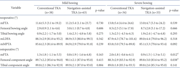
ment angle (89.0° in the conventional TKA group vs 90.0° in the navigationassisted TKA group; p=0.018) (Table 2). On the subgroup analysis that was conducted according to the degree of femoral lateral bowing, no parameter showed a significant dif
ference between the two groups in cases of mild femoral lateral bowing (femoral bowing angle ≤5°); however, in cases of severe femoral lateral bowing (femoral bowing angle >5°), there was a significant difference in postoperative mFTA (2.0° in the conven
tional TKA group vs. 0.9° in the navigationassisted TKA group;
p=0.012) and femoral component alignment angle (88.3° in the conventional TKA group vs. 89.8° in the navigationassisted TKA group; p=0.038) (Table 3).
Table 4 describes the results of the correlation analysis. In the conventional TKA group, femoral bowing angle showed a corre
lation with femoral component alignment angle and postopera
tive mFTA (Pearson correlation coefficient [PCC]=–0.199 and 0.410, respectively) and mLDFA showed a correlation with femo
ral component alignment angle (PCC=–0.360). Tibial bowing angle did not show a correlation with either postoperative mFTA or tibial component alignment, and mMPTA showed a nega
tive correlation with postoperative mFTA (PCC=–0.193). In the navigationassisted TKA group, mLDFA showed a negative cor
relation with femoral component alignment angle and mMPTA
Table 2. Comparison of Radiological Results between Conventional TKA Group and NavigationAssisted TKA Group in Total Cases and Cases with Femoral Lateral Bowing
Variable
Total Femoral lateral bowing
Conventional
TKA (n=128) Navigationassisted
TKA (n=169) pvalue Conventional
TKA (n=72) Navigationassisted
TKA (n=96) pvalue Preoperative (°)
mFTA 11.4±6.1 (0 to 37.4) 11.5±5.7 (0.6 to 39.5) 0.874 12.6±5.4 (3.1 to 24.6) 11.9±5.2 (1.1 to 24.2) 0.341 Femoral bowing angle 3.1±3.7 (–1.5 to 17.8) 2.8±3.5 (–1.8 to 17.2) 0.502 5.5±3.4 (1.1 to 17.8) 5.0±3.3 (0.7 to 17.2) 0.289 Tibial bowing angle 1.1±2.3 (–4.5 to 7.6) 1.4±2.4 (–6.5 to 7.3) 0.300 1.1±2.3 (–4.5 to 6.3) 1.6±2.4 (–4.8 to 6.8) 0.170 mLDFA 89.2±3.8 (78.7 to 101.6) 89.0±3.6 (79.8 to 99.5) 0.722 88.1±3.6 (78.7 to 101.6) 88.9±3.8 (79.8 to 99.5) 0.151 mMPTA 85.1±2.8 (79.5 to 91.3) 85.0±3.0 (79.0 to 93.4) 0.823 84.8±2.8 (79.5 to 89.9) 85.0±3.0 (79.0 to 93.4) 0.581 Postoperative (°)
mFTA 1.1±1.8 (–1.1 to 7.0) 0.6±1.9 (–1.6 to 6.8) 0.056 1.6±1.8 (–1.1 to 6.1) 0.8±1.8 (–1.6 to 6.8) 0.005a) Femoral component angle 89.4±2.9 (83.5 to 97.9) 90.0±2.5 (83.6 to 95.7) 0.055 89.0±2.7 (83.5 to 94.0) 90.0±2.5 (83.6 to 95.2) 0.018a) Tibial component angle 89.7±2.0 (85.3 to 94.4) 89.7±2.1 (84.9 to 95.0) 0.856 89.4±2.0 (85.5 to 93.5) 89.9±2.3 (85.3 to 95.0) 0.211 Values are presented as mean±standard deviation (range).
TKA: total knee arthroplasty, mFTA: mechanical femorotibial angle, mLDFA: mechanical lateral distal femoral angle, mMPTA: mechanical medial proximal tibial angle.
a)Statistically significant.
showed a positive correlation with tibial component alignment angle (PCC=–0.189 and 0.237, respectively). However, neither variable showed a correlation with postoperative mFTA. Neither the femoral bowing angle nor the tibial bowing angle showed a correlation with postoperative mFTA and femoral component alignment angle.
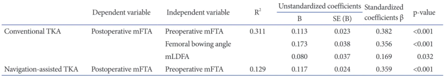
According to the results of the multiple linear regression analy
sis that was conducted in each group, it was proven that preop
erative mFTA (p<0.001), femoral bowing angle (p<0.001), and
mLDFA (p=0.032) had effects on postoperative mFTA in the conventional TKA group (R2=0.311). However, only preoperative mFTA (p<0.001) showed an impact on postoperative mFTA in the navigationassisted TKA group (R2=0.129). Multicollinearity in each regression analysis was evaluated by the variance inflation factor and none of them exceeded 10 (Table 5).
In the outlier analysis, the navigationassisted TKA group showed a lower rate of outliers in mFTA and femoral component alignment angle than the conventional TKA group (p=0.006 and Table 3. Comparison of Radiological Results between Conventional TKA Group and NavigationAssisted TKA Group according to the Degree of Femoral Lateral Bowing
Variable
Mild bowing Severe bowing
Conventional TKA
(n=38) Navigationassisted
TKA (n=63) pvalue Conventional TKA
(n=34) Navigationassisted
TKA (n=33) pvalue Preoperative (°)
mFTA 11.6±5.3 (3.1 to 19.2) 11.2±5.4 (1.1 to 23.7) 0.730 13.8±5.4 (4.4 to 24.6) 13.0±4.7 (5.3 to 24.2) 0.539 Femoral bowing angle 2.9±0.9 (1.1 to 4.6) 3.0±1.1 (0.7 to 4.8) 0.606 8.5±2.5 (5.1 to 17.8) 8.7±2.8 (5.1 to 17.2) 0.666 Tibial bowing angle 0.9±2.2 (–1.7 to 5.0) 1.4±2.3 (–4.8 to 5.8) 0.275 1.3±2.5 (–4.5 to 6.3) 1.9±2.4 (–4.7 to 6.8) 0.293 mLDFA 88.3±2.9 (81.8 to 93.2) 88.9±3.5 (80.8 to 99.5) 0.342 87.9±4.3 (78.7 to 101.6) 89.0±4.4 (79.8 to 96.2) 0.318 mMPTA 85.6±2.3 (81.6 to 89.9) 84.9±2.9 (79.0 to 91.8) 0.239 83.8±3.0 (79.5 to 89.8) 85.1±3.3 (79.6 to 93.4) 0.092 Postoperative (°)
mFTA 1.3±1.8 (–1.1 to 5.5) 0.8±1.9 (–1.6 to 6.8) 0.163 2.0±1.8 (–0.6 to 6.1) 0.9±1.5 (–1.3 to 5.1) 0.012a) Femoral component angle 89.7±2.2 (85.6 to 94.0) 90.1±2.1 (87.0 to 95.0) 0.415 88.3±2.9 (83.5 to 92.9) 89.8±3.0 (83.6 to 95.2) 0.038a) Tibial component angle 89.8±2.1 (86.3 to 92.9) 89.9±2.1 (87.0 to 93.0) 0.884 89.0±1.8 (85.5 to 93.5) 89.8±2.6 (85.3 to 95.0) 0.141 Values are presented as mean±standard deviation (range).
TKA: total knee arthroplasty, mFTA: mechanical femorotibial angle, mLDFA: mechanical lateral distal femoral angle, mMPTA: mechanical medial proximal tibial angle.
a)Statistically significant.
Table 4. Correlation Analysis on the Anatomical Features and Postoperative Radiological Results
Variable Postoperative mFTA Femoral component alignment angle Tibial component alignment angle Conventional TKA
Femoral bowing angle 0.410 (<0.001)a) –0.199 (0.025)a) N/A
mLDFA 0.001 (0.993) –0.360 (<0.001)a) N/A
Tibial bowing angle 0.098 (0.273) N/A –0.045 (0.557)a)
mMPTA –0.193 (0.029)a) N/A 0.144 (0.104)
Navigationassisted TKA
Femoral bowing angle 0.100 (0.197) –0.027 (0.730) N/A
mLDFA 0.149 (0.053) –0.189 (0.014)a) N/A
Tibial bowing angle –0.017 (0.830) N/A –0.036 (0.640)
mMPTA –0.021 (0.782) N/A 0.237 (0.002)
Values are presented as Pearson correlation coefficient (pvalue).
mFTA: mechanical femorotibial angle, TKA: total knee arthroplasty, N/A: not applicable, mLDFA: mechanical lateral distal femoral angle, mMPTA:
mechanical medial proximal tibial angle.
a)Statistically significant.
p=0.012, respectively) (Table 6).
Fig. 5 presents the patientreported outcomes. No significant difference between the conventional TKA group and navigation
assisted TKA group was observed (p>0.05, all parameters). As for the complications, there was one case of infection in the naviga
tionassisted TKA group. However, other complications, includ
ing osteolysis and loosening, were not observed in either group.
Discussion
The principle finding of this study is that navigationassisted TKA resulted in a better postoperative coronal alignment than conventional TKA in patients with femoral lateral bowing al
though no significant difference was observed when patients without femoral lateral bowing was included in the analysis. Re
gression analysis results indicated that preoperative mFTA, femo
ral bowing angle and mLDFA affected postoperative mFTA in the conventional TKA group, whereas only preoperative mFTA had an impact on postoperative mFTA in the navigationassisted TKA group.
A number of authors reported that the use of a navigation sys
tem improved postoperative alignment and component position and reduced the incidence of outliers2629). However, other authors have reported that there is no significant difference in postopera
tive alignment or outlier incidence between navigationassisted TKA and conventional TKA1517). Although whether TKA using navigation improves postoperative alignment and component position is still controversial, we believe that anatomical features such as femoral bowing should be accounted for when evaluating the accuracy of using a navigation system in TKA. Yau et al.14) re
ported that 41/93 knees (44%) had femoral lateral bowing of >2°
in research conducted in China. Since then, several more authors have reported that anatomical variations such as femoral lateral Table 5. Evaluation of Factors that Affect Postoperative mFTA in Conventional TKA and NavigationAssisted TKAa)
Dependent variable Independent variable R2 Unstandardized coefficients Standardized
coefficients β pvalue
B SE (B)
Conventional TKA Postoperative mFTA Preoperative mFTA 0.311 0.113 0.023 0.382 <0.001
Femoral bowing angle 0.173 0.038 0.356 <0.001
mLDFA 0.080 0.037 0.169 0.032
Navigationassisted TKA Postoperative mFTA Preoperative mFTA 0.129 0.117 0.024 0.359 <0.001 mFTA: mechanical femorotibial angle, TKA: total knee arthroplasty, mLDFA: mechanical lateral distal femoral angle.
a)Multiple linear regression analysis using stepwise method.
Table 6. Comparison of Outlier Rate between Conventional TKA Group and NavigationAssisted TKA Group
Variable Conventional TKA (%) Navigationassisted TKA (%)
pvaluea) Mild (n=38) Severe (n=34) Total (n=72) Mild (n=63) Severe (n=33) Total (n=96)
Postoperative mFTA (°) 6 (15.8) 8 (23.5) 14 (19.4) 3 (4.8) 2 (6.1) 5 (5.2) 0.006b)
Femoral component alignment angle (°) 4 (10.5) 11 (32.4) 15 (20.8) 1 (1.6) 6 (18.2) 7 (7.3) 0.012b) Tibial component alignment angle (°) 2 (0.8) 7 (20.6) 9 (12.5) 0 (0.0) 4 (12.1) 4 (4.2) 0.077 TKA: total knee arthroplasty, mFTA: mechanical femorotibial angle.
a)Comparison between conventional TKA group and navigationassisted TKA group in total patients with femoral lateral bowing.
b)Statistically significant.
Fig. 5. Comparison of clinical results between the conventional and navi
ga tionassisted total knee arthroplasty (TKA) groups. Preop: preopera
tive, KS: Knee Society, WOMAC: Western Ontario and McMaster Uni
versities Osteoarthritis index, Postop: postoperative.
Preop KS knee score 0
100 90 80 70 60 50 40 30 20 10
Conventional TKA Navigation-assisted TKA
Preop KS function score
Preop WOMAC
Postop KS knee score
Postop KS function score
Postop WOMAC
bowing are prevalent in Asians and that such features affect post
operative alignment and component position after TKA. Lasam et al.12) reported that femoral lateral bowing and varus condylar orientation of the femur are related to postoperative alignment in conventional TKA, but navigationassisted TKA did not show such correlation. Moreover, they reported that the outlier rate was higher in cases of conventional TKA compared to navigation
assisted TKA.
In this study, radiological results showed no significant differ
ence between the conventional TKA group and the navigation
assisted TKA group when all the patients were included in the analysis as subjects. Of note, the navigationassisted TKA group showed a better postoperative alignment and femoral component alignment angle when we limited the subjects to only patients with femoral lateral bowing. In this study, the resection of the distal femur was implemented to match the angle formed by the anatomical axis and the mechanical axis of the femur on preop
erative weight bearing whole leg AP radiographs in conventional TKA. The relatively lower accuracy compared to navigation
assisted TKA is conjectured to be caused by the inaccuracy of the intramedullary alignment guide. Despite the efforts to insert an intramedullary alignment guide into the femoral canal follow
ing the preoperative plan, it is difficult to insert the guide in the desired direction from the designated point in cases of femoral bowing referring to the twodimensional simple radiographs, which we believe eventually lowers the accuracy. The problem of accuracy becomes more prominent in cases that have severe fem
oral lateral bowing than in cases that have mild femoral lateral bowing. Additionally, if a valgus correction angle for the resec
tion of the femur is excessive (>9°), the surgeon can feel pressure when performing resection of the distal femur as planned due to the possibility that the preoperative simple radiograph was not accurately taken.
In this study, preoperative mFTA and mLDFA were proven to have an influence on postoperative alignment in the conventional TKA group, in addition to femoral lateral bowing. However, only preoperative mFTA showed a relevance to postoperative alignment in the navigationassisted TKA group. There was no correlation between anatomical features such as femoral bowing, mLDFA, and postoperative alignment. This can be attributed to the fact that bone resection was executed vertical to the mechani
cal axis when using navigation, regardless of femur anatomy.
Tibial bowing and mMPTA did not affect postoperative mFTA in either the navigationassisted TKA group or the conventional TKA group. This can be attributed to the use of an extramedul
lary alignment guide in cases of proximal tibia resection. Ko et
al.30) reported that cutting error is more likely to occur in bone resection when using an intramedullary guide system in cases with tibial bowing. In this study, different results were likely if an intramedullary alignment guide system was used in proximal tibia resection when conventional TKA was performed.
In the outlier analysis, the navigationassisted TKA group showed a lower outlier rate in mFTA and femoral component alignment angle than the conventional TKA group. However, the outlier rate of postoperative mFTA was as high as 5.2% and the outlier rate of femoral component alignment angle was 7.3% in the navigationassisted TKA group. This can be attributed to an error during the bone resection or implantation process. It can also be attributed to the patient’s posture during weight bearing whole leg AP radiography or an error in the measurement of ra
diological parameters. Hence, we could learn that outlier occurs regardless of the surgical technique. Postoperative malalignment can occur in diverse phases, including preoperative planning, bone resection, and implantation. It might be difficult to avoid the occurrence of malalignment in all TKAs. However, we believe that careful preoperative physical examination and planning as well as accurate surgical procedure can reduce the outlier rate.
Recently, there was some controversy over whether limb alignment after TKA is correlated with midterm or longterm results13). However, an absence of difference in clinical results such as function and longevity between outlier and neutral align
ment does not mean that the restoration of neutral alignment is not important in TKA. We believe that every surgery requires a certain standard. Efforts to reduce outliers will ultimately give benefits to patients. Hence, more studies that investigate the cor
relation between postoperative alignment and clinical results will be required.
In terms of the clinical results, there was no significant differ
ence between the two groups. Although the navigationassisted TKA group had higher accuracy, the difference with the conven
tional TKA group was only about 1°. Hence, we believe that it is difficult to obtain meaningful clinical results in a shortterm followup. To address this point, a longterm followup in sur
vival analysis will be required in the future.
This study had several limitations. First, we included only a small number of cases. Second, this study used only one naviga
tion system whose minimal measurable angular change was 1°.
Hence, the results of this study cannot represent all navigation systems, and different results will likely be obtained according to the performance of each navigation system. Third, despite the high testretest reliability, errors in the measurement of radiologi
cal parameters were possible due to the position and flexion con
tracture of patients when simple radiographs were taken. Finally, rotation of the femoral component was not assessed identically between the two groups in this study, which could affect the postoperative alignment. However, the rotation of the femoral component was not assessed in this study.
Conclusions
Despite the individualized determination of the valgus correc
tion angle through preoperative planning, conventional TKA resulted in a higher outlier rate than navigationassisted TKA in patients with severe lateral bowing. However, there was no sig
nificant difference in the clinical results in the shortterm follow
up.
Conflict of Interest
No potential conflict of interest relevant to this article was re
ported.
References
1. Gothesen O, Espehaug B, Havelin LI, Petursson G, Hallan G, Strom E, Dyrhovden G, Furnes O. Functional outcome and alignment in computerassisted and conventionally operated total knee replacements: a multicentre parallelgroup ran
domised controlled trial. Bone Joint J. 2014;96:60918.
2. Magnussen RA, Weppe F, Demey G, Servien E, Lustig S.
Residual varus alignment does not compromise results of TKAs in patients with preoperative varus. Clin Orthop Relat Res. 2011;469:344350.
3. Morgan SS, Bonshahi A, Pradhan N, Gregory A, Gambhir A, Porter ML. The influence of postoperative coronal align
ment on revision surgery in total knee arthroplasty. Int Or
thop. 2008;32:63942.
4. Abdel MP, Oussedik S, Parratte S, Lustig S, Haddad FS. Cor
onal alignment in total knee replacement: historical review, contemporary analysis, and future direction. Bone Joint J.
2014;96:85762.
5. Liow MH, Goh GS, Pang HN, Tay DK, Lo NN, Yeo SJ.
Computerassisted stereotaxic navigation improves the ac
curacy of mechanical alignment and component positioning in total knee arthroplasty. Arch Orthop Trauma Surg. 2016;
136:117380.
6. Ritter MA, Faris PM, Keating EM, Meding JB. Postoperative alignment of total knee replacement. Its effect on survival.
Clin Orthop Relat Res. 1994;(299):1536.
7. Kharwadkar N, Kent RE, Sharara KH, Naique S. 5 degrees to 6 degrees of distal femoral cut for uncomplicated primary total knee arthroplasty: is it safe? Knee. 2006;13:5760.
8. McGrory JE, Trousdale RT, Pagnano MW, Nigbur M. Pre
operative hip to ankle radiographs in total knee arthroplasty.
Clin Orthop Relat Res. 2002;(404):196202.
9. Shi X, Li H, Zhou Z, Shen B, Yang J, Pei F. Comparison of postoperative alignment using fixed vs individual valgus correction angle in primary total knee arthroplasty with lat
eral bowing femur. J Arthroplasty. 2016;31:97683.
10. Bardakos N, Cil A, Thompson B, Stocks G. Mechanical axis cannot be restored in total knee arthroplasty with a fixed valgus resection angle: a radiographic study. J Arthroplasty.
2007;22(6 Suppl 2):859.
11. Huang TW, Hsu WH, Peng KT, Hsu RW. Total knee replace
ment in patients with significant femoral bowing in the coronal plane: a comparison of conventional and computer
assisted surgery in an Asian population. J Bone Joint Surg Br.
2011;93:34550.
12. Lasam MP, Lee KJ, Chang CB, Kang YG, Kim TK. Femoral lateral bowing and varus condylar orientation are prevalent and affect axial alignment of TKA in Koreans. Clin Orthop Relat Res. 2013;471:147283.
13. Mullaji AB, Shetty GM, Kanna R, Vadapalli RC. The influ
ence of preoperative deformity on valgus correction angle:
an analysis of 503 total knee arthroplasties. J Arthroplasty.
2013;28:207.
14. Yau WP, Chiu KY, Tang WM, Ng TP. Coronal bowing of the femur and tibia in Chinese: its incidence and effects on total knee arthroplasty planning. J Orthop Surg (Hong Kong).
2007;15:326.
15. Kim YH, Kim JS, Choi Y, Kwon OR. Computerassisted sur
gical navigation does not improve the alignment and orien
tation of the components in total knee arthroplasty. J Bone Joint Surg Am. 2009;91:149.
16. Kim YH, Kim JS, Yoon SH. Alignment and orientation of the components in total knee replacement with and without navigation support: a prospective, randomised study. J Bone Joint Surg Br. 2007;89:4716.
17. Matziolis G, Krocker D, Weiss U, Tohtz S, Perka C. A pro
spective, randomized study of computerassisted and con
ventional total knee arthroplasty: threedimensional evalu
ation of implant alignment and rotation. J Bone Joint Surg Am. 2007;89:23643.
18. Roberts TD, Clatworthy MG, Frampton CM, Young SW.
Does computer assisted navigation improve functional outcomes and implant survivability after total knee arthro
plasty? J Arthroplasty. 2015;30(9 Suppl):5963.
19. Thippanna RK, Kumar MN. Lateralization of femoral entry point to improve the coronal alignment during total knee arthroplasty in patients with bowed femur. J Arthroplasty.
2016;31:19438.
20. Song MH, Yoo SH, Kang SW, Kim YJ, Park GT, Pyeun YS.
Coronal alignment of the lower limb and the incidence of constitutional varus knee in korean females. Knee Surg Relat Res. 2015;27:4955.
21. Abane L, Anract P, Boisgard S, Descamps S, Courpied JP, Hamadouche M. A comparison of patientspecific and conventional instrumentation for total knee arthroplasty: a multicentre randomised controlled trial. Bone Joint J. 2015;
97:5663.
22. Huang TW, Lee CY, Lin SJ, Lee MS, Hsu RW, Shen WJ. The influence of alignment on midterm outcome after total knee arthroplasty in patients with marked coronal femoral bow
ing. J Arthroplasty. 2015;30:15316.
23. Ewald FC. The Knee Society total knee arthroplasty roent
genographic evaluation and scoring system. Clin Orthop Relat Res. 1989;(248):912.
24. Insall JN, Dorr LD, Scott RD, Scott WN. Rationale of the Knee Society clinical rating system. Clin Orthop Relat Res.
1989;(248):134.
25. Bellamy N, Buchanan WW, Goldsmith CH, Campbell J, Stitt
LW. Validation study of WOMAC: a health status instrument for measuring clinically important patient relevant outcomes to antirheumatic drug therapy in patients with osteoarthritis of the hip or knee. J Rheumatol. 1988;15:183340.
26. Cheng T, Zhao S, Peng X, Zhang X. Does computerassisted surgery improve postoperative leg alignment and implant positioning following total knee arthroplasty? A meta
analysis of randomized controlled trials? Knee Surg Sports Traumatol Arthrosc. 2012;20:130722.
27. HernandezVaquero D, SuarezVazquez A, SandovalGarcia MA, NoriegaFernandez A. Computer assistance increases precision of component placement in total knee arthroplasty with articular deformity. Clin Orthop Relat Res. 2010;468:
123741.
28. Lee DH, Park JH, Song DI, Padhy D, Jeong WK, Han SB.
Accuracy of soft tissue balancing in TKA: comparison be
tween navigationassisted gap balancing and conventional measured resection. Knee Surg Sports Traumatol Arthrosc.
2010;18:3817.
29. Weng YJ, Hsu RW, Hsu WH. Comparison of computer
assisted navigation and conventional instrumentation for bilateral total knee arthroplasty. J Arthroplasty. 2009;24:668
73.
30. Ko PS, Tio MK, Ban CM, Mak YK, Ip FK, Lam JJ. Radiolog
ic analysis of the tibial intramedullary canal in Chinese varus knees: implications in total knee arthroplasty. J Arthroplasty.
2001;16:2125.