• 검색 결과가 없습니다.

Confirmation of Supraorbital Nerve and Its Branch in the Supraorbital Notch with Ultrasound Guidance

N/A
N/A
Protected

Academic year: 2021

Share "Confirmation of Supraorbital Nerve and Its Branch in the Supraorbital Notch with Ultrasound Guidance"

Copied!
5
0
0

로드 중.... (전체 텍스트 보기)

전체 글

(1)

© Copyright

Keimyung University School of Medicine 2016 Received: September 01, 2016

Revised: November 10, 2016 Accepted: November 21, 2016 Corresponding Author: Ji Hee Hong, M.D., Department of Anesthesiology, Keimyung University School of Medicine, 56 Dalseong-ro, Jung-gu, Daegu 41931, Korea Tel: +82-53-250-7288

E-mail: [email protected]

• The authors report no conflict of interest in this work.

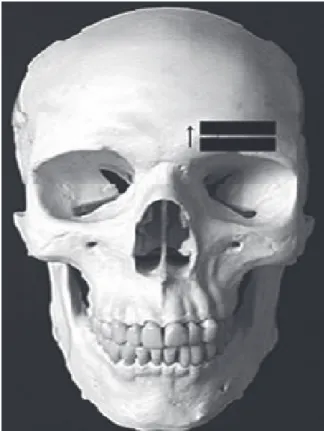
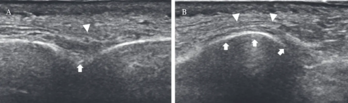
A 55-year-old female with severe herpes zoster related forehead pain radiating to anterior and posterior scalp visited our pain clinic. The right side forehead pain with numerical rating score of 7 had persisted in spite of antiviral and anticonvulsant medication. We blocked the right supraorbital nerve under ultrasound guidance, and obtained the proper pain relief. In this case, we would like to present the method to identify the supraorbital notch or foramen and possible visualization of the supraorbital nerve.

Keywords: Forehead pain, Herpes zoster, Supraorbital notch, Ultrasound guidance

Introduction

Herpes zoster occurs due to the reactivation of varicella zoster virus.

Among adult population, the ophthalmic division of the trigeminal nerve (CN V) is one of the most common site of involvement. The incidence of herpes zoster ophthalmicus is known to be 20 times more common when compared with either maxillary or mandibular division, being exceeded only by zoster of thoracic dermatome [1].

Following herpes zoster ophthalmicus, various ocular complications associated with poor visual sight include optic neuritis, uveitis, retinitis and acute corneal lesions. Ultimately, these process may cause permanent visual loss with substantial health care utilization [1,2].

Superficial trigeminal nerve block of ophthalmic division using local anesthetics is one of the modality to relieve the zoster related pain and minimize the ocular complication [3].

We report a case of severe zoster related forehead pain and its successful treatment using superficial trigeminal nerve block under Department of Anesthesiology and Pain Medicine, Radiology

1

,

Keimyung University School of Medicine, Daegu, Korea

Ji Hee Hong, M.D., Sung Mun Lee

1

, M.D., Kyeong Hwan Seo, M.D.

Confirmation of Supraorbital Nerve and Its Branch in the

Supraorbital Notch with Ultrasound Guidance

(2)

ultrasound guidance. In addition, we would like to present the method to identify the supraorbital notch or foramen and possible visualization of the supraorbital nerve.

Case Report

A 55-year-old female with severe zoster related forehead pain radiating to anterior and posterior scalp visited our pain clinic. This patient has been diagnosed with herpes zoster affecting the right ophthalmic nerve of the trigeminal nerve (V1) area 2 weeks previously. The right side forehead pain with numerical rating score of 7 had persisted in spite of antiviral and anticonvulsant medication. During this period, the medications included 250 mg famciclovior, 300 mg gabapentin, and 37.5 mg tramadol/375 mg acetaminophen combination tablet three times daily, however, they showed little effect. She demonstrated shooting, paroxysmal and sharp pain on her forehead. When she touched her hair on the scalp, it aggravated the original scalp pain.

Therefore, we planned a superficial supraorbital nerve block in the V1 area. We obtained a consent from the patient related to the nerve block and explained all possible risks and benefits of the procedure.

The patient was placed in a supine position and the skin around orbital rim and forehead was draped aseptically with betadine. The transducer (linear transducer, 5-12 MHz, EPIQ 5G, Philips, Bothell, Washington, USA) was prepared with a sterile transparent sheath and aseptic ultrasound gel was applied to the draped skin. The transducer was applied just below the eyebrow. At first, we tried to identify the supraorbital notch or foramen according to the method suggested by Tsui [4]. The transducer was applied transversely to the upper side of the orbital rim and then rotated slightly to obtain an ideal

image of longitudinal view of supraorbital notch or foramen. We scanned her forehead from lower to upper direction in the coronal plane along the right upper orbital margin (Fig. 1). After identifying a supraorbital notch, subtle cranial or caudal tilting of transducer position could capture the location of supraorbital nerve and its branch (Fig. 2A&B). After confirming the nerve and its branch, ultrasound guided injection was done with the mixture of 1 mL of 0.2% ropivacaine and 10 mg triamcinolone using transverse and longitudinal view (Fig. 3A&B). During ultrasound guided injection, the patient did not complain any paresthesia except some painful sensation during skin puncture with the needle.

When injection was performed successfully, we checked the sensory distribution of V1 dermatome area, and confirmed the decreased or numbing

Fig. 1. The direction of transducer position (arrow)

scanning the forehead to identify the supraorbital

notch or foramen.

(3)

sensation on the forehead and anterior scalp area.

One day after the supraorbital nerve block, the numerical rating score decreased to 2 or 3 and this pain relief maintained during her admission period.

One month after a discharge from our hospital, the patient visited our pain clinic again, and her pain was managed successfully with intermittent aid of acetaminophen and tramadol combination tablet.

Discussion

The anatomy of the supraorbital nerve and notch

is especially important to perform supraorbital nerve block. The supraorbital nerve is the terminal branch of the ophthalmic nerve (CN V1). The ophthalmic nerve begins at the superior part of the trigeminal ganglion as a flattened plexiform band and leaves the skull through the superior orbital fissure. The V1 carries sensory information from the scalp and forehead, the upper eyelid, the conjunctiva, the cornea, and the nose tip and nasal mucosa [5,6]. As the supraorbital nerve comes from the orbit, it passes through a foramen or notch and sends deep and superficial branches [7]. Cadaver work has demonstrated that the supraorbital nerve passes Fig. 2. Transverse (A) and longitudinal (B) view of supraorbital nerve (arrows) and its branch (arrow heads) in the supraorbital notch.

Fig. 3. An ultrasound image of the block needle with longitudinal (A) and transverse (B) view. The needle through the supraorbital notch is indicated with arrow heads. The supraorbital nerve is shown just below the needle with an arrow.

A

A

B

B

(4)

through a notch (83%) more frequently than a bony foramen (27%) [8].

Ultrasound imaging is a non-invasive and safe method through which nerve, soft tissue and bony structure can be easily identified when combined with thorough understanding of regional anatomy.

However, due to its small size of superficial supraorbital nerve, definite identification of terminal branch of V1 was challenging. Previously, there have been efforts to localize a supraorbital notch or foramen for superficial trigeminal nerve block [4,7].

Bony structure appears as a hyperechoic linear edge with an underlying anechoic shadow under ultrasound guidance. Any disruption or discontinuity within the hyperechoic line may indicate the presence of foramen. Tsui suggested a simple ultrasound guided approach to easily locate the important landmark of foramen for superficial trigeminal nerve block [4].

Garg et al. [7] reported that sonography could distinguish a supraorbital notch from a foramen with 100% sensitivity through the studies of cadaver work.

According to their report, a foramen was suspected when transverse views revealed a bony acoustic landmark corresponding to the rim of the foramen, whereas this bony landmark was absent with a notch.

Therefore, we think that the supraorbital nerve and its branch shown in figure 2 are coming from the supraorbital notch, because we could not find any bony acoustic landmark constituting the rim of foramen.

Obviously, if the supraorbital nerve passing through a foramen or notch could be directly identified under ultrasound guidance, the nerve block would be performed more easily and safely. At first, we thought that the identification of superficial supraorbital nerve would be impossible due to its small size and superficial distribution, although its notch or foramen might be visible. However, the technical improvement providing high quality image

and proper handling of ultrasound probe enabled the identification of superficial supraorbital nerve and even its branch.

In this case, we used the linear transducer to perform the supraorbital nerve block, however, we think that hockey stick probe would be more valuable if the ultrasound guided injection should be combined. The image obtained from the hockey stick probe would be more limited compared to linear probe, however, it is enough to include the whole image of supraorbital notch or foramen and its nerve.

Although we could not use this hockey stick probe due to a transient technical error, we would like to recommend the use of the hockey stick probe.

Lim et al . [6] presented a case of pulsed radio- frequency treatment of infraorbital nerve for intractable postherpetic neuralgia. According to their report, they confirmed the infraorbital foramen through the protuberance within the hyperechoic line under ultrasound guidance, but no efforts were described to identify the infraorbital nerve.

In this case, we tilted the linear transducer into cranial or caudal direction to optimize the visualization of the supraorbital nerve. It is well known that the ultrasound beam should penetrate the target tissue with 90 degrees to obtain the best image [9]. Therefore, subsequent tilting or movement of transducer into cranial or caudal direction which adjusts the ultrasound beam to the target neve would provide the best image showing the supraorbital nerve.

If a supraorbital foramen or notch without the visualization of nerve is identified under ultrasound imaging, the block might be performed successfully.

However, in our clinical practice, we can encounter

the painful sensation of paresthesia during peripheral

nerve block even guided with the ultrasound. In such

cases, we can suspect very close approximation of

nerve and needle or direct puncture of a needle to the

adjacent nerve. Therefore, if the location of nerve is

(5)

confirmed under ultrasound guidance, we can pay attention more easily to ovoid direct needle puncture of nerve and maintain a proper space between the nerve and needle.

We suggest that the effort to localize the superficial nerve should be combined to improve the block quality and safety of patient.

Acknowledgement

We would like to give special thanks to Eun Suk Kim, Jin A Lee and Eun Bi Kim who did a contribution making high quality figures.

References

1. Bandeira F, Roizenblatt M, Levi GC, Freitas D, Belfort R, Jr. Herpes zoster ophthalmicus and varicella zoster virus vasculopathy. Arq Bras Oftalmol 2016;79:126-9.

2. Kaufman SC. Anterior segment complications of herpes zoster ophthalmicus. Ophthalmology 2008;115:S24-32.

3. Kuo YC, Hsieh LF, Chiou HJ. Ultrasound-guided musculocutaneous nerve block in postherpetic neuralgia.

Am J Phys Med Rehabil 2016;95:e1-6.

4. Tsui BC. Ultrasound imaging to localize foramina for superficial trigeminal nerve block. Can J Anaesth 2009;56:704-6.

5. Henssen DJ, Kurt E, Kozicz T, van Dongen R, Bartels RH, van Cappellen van Walsum AM. New insights in trigeminal anatomy: a double orofacial tract for nociceptive input. Front Neuroanat 2016;10:53.

6. Lim SM, Park HL, Moon HY, Kang KH, Kang H, Baek CH, et al. Ultrasound-guided infraorbital nerve pulsed radiofrequency treatment for intractable postherpetic neuralgia - a case report. Korean J Pain 2013;26:84-8.

7. Garg RK, Lee KS, Kohn SC, Baskaya MK, Afifi AM.

Can sonography distinguish a supraorbital notch from a foramen? J Ultrasound Med 2015;34:2089-91.

8. Fallucco M, Janis JE, Hagan RR. The anatomical morphology of the supraorbital notch: clinical relevance to the surgical treatment of migraine headaches. Plast Reconstr Surg 2012;130:1227-33.

9. Cook CR. Ultrasound imaging of the musculoskeletal

system. Vet Clin North Am Small Anim Pract 2016;46:355-71.

수치

Fig. 1. The direction of transducer position (arrow)  scanning the forehead to identify the supraorbital  notch or foramen.
Fig. 3. An ultrasound image of the block needle with longitudinal (A) and transverse (B) view

참조

관련 문서