․투고일: 2015.01.12, 심사일: 2015.03.11, 게재확정일: 2015.03.17
․교신저자: 김영철 서울시 동대문구 회기동 1번지 경희대학교 대학원 한의학과 간계내과학교실 TEL: 02-958-9118 FAX: 02-958-9120 E-mail: [email protected]
漆皮 추출물의 담도암 세포주 SNU-1079와 SNU-1196에 대한 항암효과
정보배, 김영철
경희대학교 한의과대학 간계내과학교실
Study on Anti-Cancer Effects of Rhus Verniciflua Stokes Extracted with Sterile Distilled Water on Two Cholangiocarcinoma Cell Lines, SNU-1079 and SNU-1196
Bo-bae Joung, Young-chul Kim
Dept. of Internal Medicine, College of Korean Medicine, Kyung-Hee University
ABSTRACT
Objectives:
This study was performed to investigate the anti-cancer effects of Rhus verniciflua Stokes (RVS) extracted with sterile distilled water on cholangiocarcinoma cell lines.
Materials and Methods:
Two cholangiocarcinoma cell lines, SNU-1079 and SNU-1196, were used in this study. Cells were treated with different concentrations of RVS for 24, 48, and 72 hours. Cell count, viability, apoptosis, and mRNA expression of Bax, Bcl-2, Mcl-1, survivin, caspase-3, and cyclin D1 and P21 were determined with an automatic cell counter (ADAM-MC), MTT assay, apoptosis assay (Annexin-V/PI staining), and RT-PCR.
Results:
All cells treated with RVS showed decreased cell counts in a dose-dependent manner. RVS inhibited proliferation of SNU-1196 in a dose-dependent manner, but SNU-1079 proliferation was inhibited in the long-time culture group in a dose-dependent manner. The proportion of early and late-stage apoptotic cells was increased by RVS in a dose-dependent manner in SNU-1196. In contrast, it was increased significantly in SNU-1079 treated with high-dose RVS. After treatment with RVS, the mRNA expression of Bcl-2 was decreased while Bax was increased in SNU-1079. Cyclin D1 mRNA levels were decreased in SNU-1196 in a dose-dependent manner. P21 expression was increased in all cells after the treatment with RVS.
Conclusions:
RVS appears to have potential as a therapeutic agent for cholangiocarcinoma.
Key words:
Rhus verniciflua Stokes, Cholangiocarcinoma, Apoptosis
Ⅰ. 서 론
담관의 상피세포에서 발생되는 담도암은 나이가 들수록 발생빈도가 급격하게 증가하고, 특히 간질 환과 비만 등에서 더욱 발생빈도가 증가한다. 담도
암 발생 부위의 해부학적 위치에 따라, 간내담도암, 간문부담도암, 간외담도암의 3종류로 구분하며, 그 중 간문부담도암의 발생률이 가장 높은 것으로 알 려져 있다
1.
우리나라 통계청에서 발표한 2010년 기준, 담도
암의 5년(2006년-2010년)상대 생존율은 26.7%로,
기타 다른 암에 비해 기대되는 생존율이 낮게 보고
되고 있다
2. 담도암은 발병초기 임상적인 특이 증상
이 없기 때문에 조기 발견과 진단이 어려워, 진단
후 의학적으로 기대되는 생존기간이 2년 정도로 예후가 매우 불량한 편이다
3.
현재 시행되고 있는 담도암의 주된 치료 방법은 선행 화학요법 이후의 절제술이나 간이식 등의 수 술요법이다. 하지만 진단 당시, 이미 암세포가 주변 조직을 광범위하게 침범되어 있거나, 환자의 연령 이나 건강상태 등의 요인으로 제한된 수술만이 가능 한 경우가 대부분이다
4. 따라서 담도암 환자의 생존 율을 높이기 위한 치료방법에 대하여 더 많은 연구 가 필요한 실정이다.
漆皮는 옻나무의 껍질로, 이미 15세기경부터 동 아시아 지역에서 암을 포함하여, 다양한 질환의 치 료를 목적으로 사용되어온 약재이다
5,6. 최근에는 약 리학적으로 漆皮에 함유되어있는 flavonoid 성분이 암세포에 대한 항증식효과와 세포자멸 활성효과가 보고되면서
7,8임상에서 주목을 받고 있다. 그러나, 담도암과 관련된 漆皮의 항암 효과에 대한 실험이 나 임상응용 등은 아직까지 보고된 바 없다.
이에 저자는 기원이 다른 2종류의 담도암 세포주 SNU-1079와 SNU-1196을 대상으로 漆皮의 물추출 물을 투여하여 세포수, 세포증식, 세포자멸을 측정 하고, 세포자멸의 경로를 관찰하기 위해 Bcl-2, Bax, Mcl-1, Survivin, Caspase-3, Cyclin D1 및 P21 mRNA 발현의 변화를 관찰하여 유의한 결과를 얻 었기에 이에 보고하는 바이다.
Ⅱ. 재료 및 방법
1. 재 료 1) 약 재
본 실험에서 사용한 漆皮(Rhus verniciflua Stokes, 이하 RVS)는, 대한약전 및 대한약전외 한약 규격 주해
9에 근거하였으며, 경희대학교 부속 한방병원 에서 구입하여 사용하였다.
漆皮는 옻나무의 껍질로, 옻나무의 줄기에 상처 를 입혀 흘러나온 樹液이 자연 건조된 덩어리인 乾漆(Lacca Sinica Exsiccata)과는 다르다.
2) 검액의 조제
漆皮 10 kg을 멸균증류수를 이용하여 1시간 동 안 180 ℃에서 가열한 뒤 추출하는 것을 2회 반복 하여 추출액을 취합하였다.
3시간마다 분광기 상의 400 nm 파장에서 흡광 도를 측정하여, 둔화된 변화량이 5% 미만으로 감 소 되었을때 추출이 완료된 것으로 판단하였으며, 총 3시간이 소요되었다. 漆皮 추출액은 Whatman 여과지로 여과한 뒤, 감압농축 하였다. 동결건조기 를 이용하여 수득률 4.3%의 분말상태 칠피 0.43 kg 을 얻었다. 얻어진 동결건조 분말은 -80 ℃ 냉동고에 보관하고, 사용 시 해동 후, 증류수에 녹여서 0.2 μm syringe filter로 여과하여 필요한 농도(0, 50, 100, 200, 400 ug/ml)로 희석하여 사용하였다.
3) 세포 배양 및 시약
한국 세포주 은행에서 기원이 다른 사람 담도암 세포주 2종류, SNU-1079(biliary tract cancer, intrahepatic duct)와 SNU-1196(biliary tract cancer, hepatic duct bifurcation)를 분양받아 사용하였다.
RPMI 1640 배양액에 FBS 10%와 penicillin/
streptomycin 1%를 첨가한 뒤, 5% CO2, 37 ℃ 환 경에서 배양하였다.
2. 방 법
1) 세포수 분석
ADAM-MC(한국, Nanoteck Inc.)를 사용하였고, 제작사의 지시에 따라 측정하였다.
2) 세포증식 분석
세포 증식은 CellTiter 96 AQueous One solution kit(Promega, Madison, WI, USA)를 이용하여 MTT (3-(4,5-dimethylthiazol-2-yl)-2,5-diphenyltetrazolium bromide) 분석으로 측정하였다.
SNU-1079와 SNU-1196 세포주를 각 1×10
4cells/well 농도로 96-well plate에 분주하고, 漆皮 추출물을 0, 50, 100, 200, 400 μg/ml 농도로 처리한 뒤, 24, 48, 72시간 동안 배양하였다.
담도암 세포의 생존율은 PMS/MTS solution을
이용하여 490 nm에서 흡광도를 측정하여 보았다.
3) 세포자멸 분석
Annexin-V/PI assay kit(BD Biosciences, USA) 를 사용하여, 漆皮 추출물에 의한, 담도암 세포주의 세포자멸을 확인하였다.
漆皮 추출물을 각 0, 50, 100, 200, 400 μg/ml 농도 로 처리한 SNU-1079와 SNU-1196 세포주를 ice-cold PBS buffer로 2차례 씻어내고, 500 μl binding buffer 에서 재침전한 뒤, FITC 결합 Annexin-V(10 mg/ml)와 PI(50 mg/ml) 5 μl로 염색하여, 실온의 어두운 환 경에서 5-15분가량 배양하고, FACSCalibur(Becton Dickinson Immunocytometry System, San Jose, CA, USA)를 이용하여 분석하였다.
FITC에만 양성반응을 보이는 세포는 조기 세포 자멸, FITC와 PI에 모두 양성반응을 보이는 세포는 후기 세포자멸 단계로, 이 두 가지 단계의 세포수 비율의 합으로 SNU-1079와 SNU-1196 세포주의 세포자멸을 평가하였다.
4) 역전사 중합효소 연쇄반응(RT-PCR) 분석 SNU-1079와 SNU-1196 세포주를 6-well plate에 분주하고, 漆皮 추출물을 각 0, 50, 100, 200, 400 μg/ml 농도로 처리하였다. 각 24, 48, 72시간이 경과한 뒤, Qiagen사의 RNeasy Mini Plus Kit를 이용하여 제 작사가 권하는 방법에 따라 RNA를 분리하였다.
Complementary DNA(이하 cDNA)는 Fermentas 사의 RevertAids First-Strand cDNA Synthesis Kit를 사용하여 역시 제작사의 방법에 따라 합성하였다.
random primer 1 μg과 RNA 1 μg을 포함하는 시료에 DEPC-처리된 증류수를 합하여 총 12 μl가 되도록 조절한 뒤, 65 ℃에서 5분간 가열하고 5분간 얼음에 두었다. 다음으로, 5x reaction buffer(250 μM Tris-HCL, pH8.3) 4 μl와 RiboLock™ RNase Inhibitor(200 U/μl) 1 μl, 10 μM deoxynucleoside triphosphate(dNTP) MIX 2 μl, RevertAid™ M-MuLV Reverse Transcripase (200 U/μl) 1 μl을 넣고 전체 부피를 20 μl 되도록 조정한 뒤 부드럽게 흔들어 잘 섞어주었다. 25 ℃ 에 5분간, 42 ℃에 60분간, 70 ℃에 5분간의 조건으
로 반응시키고, 합성된 DNA는 -80 ℃에서 보관하 였다가 꺼내서 사용하였다.
Primer 합성은 Bioneer(대전, 한국)에 의뢰하여 제작하였으며, β-actin을 House keeping gene으로 사용하였다. Target gene으로는 Bcl-2, Bax, Mcl-1, Survivin, Cyclin D1, P21, Caspase-3를 사용하였다.
RT-PCR은 Applied Biosystem사(CA, USA)의 StepOnePlus RT PCR System을 이용하여 제작사가 권하는 방법에 따라 시행하였으며, House keeping gene인 β-actin을 기준으로 상대적인 유전자의 발 현값을 정량적으로 표현하였다.
2x SYBR Green PCR Master Mix 10 μl, cDNA 1 μl, primer(sense) 1 μl, primer(anti-sense) 1 μl, Distilled water 7 μl를 혼합하여 전체 부피를 총 20 μl로 조절하였고, 초기변성(denaturation)은 첫 cycle 만 95 ℃에서 3분간, 나머지 40cycle까지는 95 ℃에서 10초간, 풀림(annealing)은 60 ℃에서 10초간, 연장 (extension)은 72 ℃에서 15초간으로 하여 증폭시 켰다(Table 1).
Genes Primer sequence Size (bp) Bcl-2 5’-GATTGATGGGATCGTTGCCTTA-3’
5’-CCTTGGCATGAGATGCAGGA-3’ 200 Bax 5’-GGATGCGTCCACCAAGAAG-3’
5’-GCCTTGAGCACCAGTTTGC-3’ 216 Mcl-1 5’-CTCATTTCTTTTGGTGCCTTT-3’
5’-CCAGTCCCGTTTTGTCCTTAC-3’ 117 Survivin 5’-GGCCCAGTGTTTCTTCTGCTT-3’
5’-GCAACCGGACGAATGCTTT-3’ 91 Cyclin
D1
5’-CCGTCCATGCGGAAGATC-3’
5’-ATG GCC AGC GGG AAG AC-3’ 86 P21 5’-CAGACCAGCATGACAGATTTC-3’
5’-TTAGGGCTTCCTCTTGGAGA-3’ 66 Caspase
-3
5'-CAGTGGAGGCCGACTTCTTG-3' 5'-TGGCACAAAGCGACTGGAT-3' 102 β-actin 5’-GCGAGAAGATGACCCAGATC-3’
5’-GGATAGCACAGCCTGGATAG-3’ 77
Table 1. Primer Sequence for PCR
3. 통계 분석
실험을 통해 얻어진 모든 데이터는 평균(Mean)
±표준편차(SD) 혹은 평균(Mean)±표준오차(SEM) 로 표기하였고, Excel program의 Student's t-test를 통해 대조군과 실험군의 데이터 차이를 비교하였 다. P<0.05 기준으로 통계적 유의성을 평가하였다.
Ⅲ. 결 과
1. 漆皮 추출물이 담도암 세포주 세포수에 미치는 영향
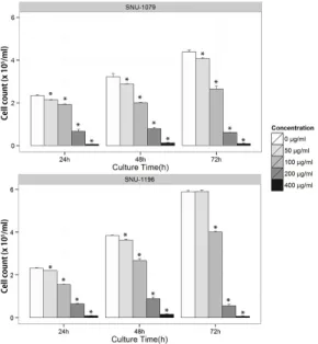
각 0, 50, 100, 200, 400 μg/ml 농도의 漆皮 추출 물을 SNU-1079와 SNU-1196 세포주에 투여하여 24, 48, 72시간까지 배양한 결과, 두 세포주에서 모 두 농도 의존적으로 세포수가 감소하였다(Fig. 1).
Fig. 1. Effects of RVS on cell counting of biliary tract cancer cells.
Biliary tract cancer cells were treated with various concentrations of RVS (0-400 μg/ml) for 24, 48, 72 hours. Cell counting was determined by the ADAM-MC. The data represent the mean
± SD of triplicate samples. *p<0.05 compared to control.
2. 漆皮 추출물이 담도암 세포주 세포증식에 미치는 영향
SNU-1079와 SNU-1196 세포주에 0, 50, 100, 200, 400 μg/ml 농도의 漆皮 추출물을 투여하여 24, 48, 72시간동안 배양한 결과, 전 시간에 걸쳐 농도의존 적으로 세포증식이 억제되는 SNU-1196과 달리, SNU-1079 세포주에서는 72시간에서 농도의존적으 로 유의하게 세포 증식이 억제되었다(Fig. 2).
Fig. 2. Effects of RVS on cell viability of biliary tract cancer cells.
Biliary tract cancer cells were treated with various concentrations of RVS (0-400 μg/ml) for 24, 48, 72 hours. Cell viability was determined by the MTT assay. The data represent the mean ± SD of triplicate samples. *p<0.05 compared to control.
3. 漆皮 추출물이 담도암 세포주 세포자멸에 미치는 영향
SNU-1079와 SNU-1196 세포주에 0, 50, 100, 200, 400
μg/ml 농도의 漆皮 추출물을 투여하고 24, 48, 72시간
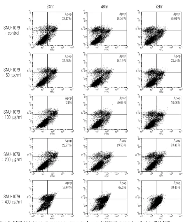
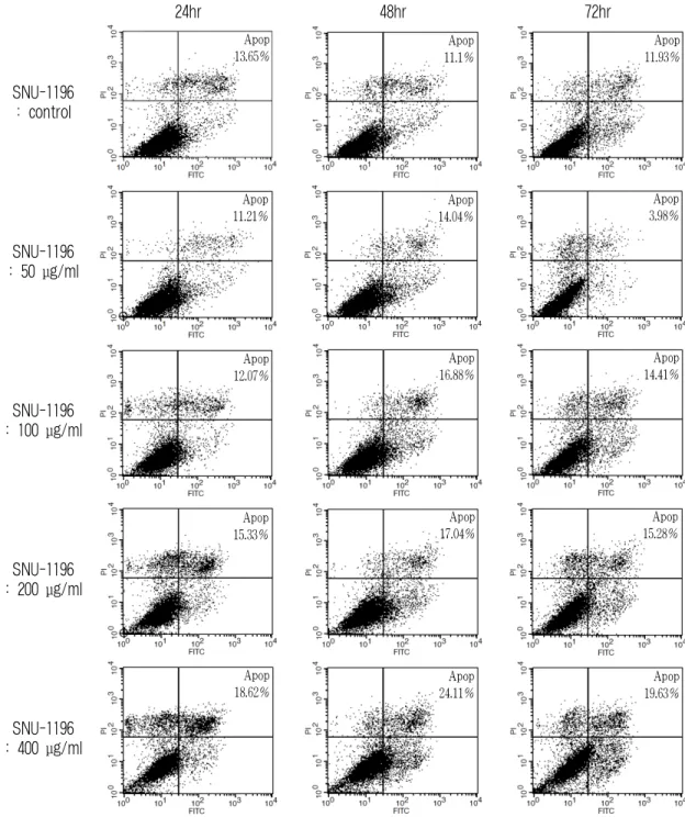
배양하여 세포자멸 효과를 관찰한 결과, SNU-1196
세포주에서 48시간 배양시 농도 의존적으로 세포자멸
이 증가하였고, SNU-1079 세포주에서는 400 μg/ml의
고농도 투여에서 세포자멸이 증가하였다(Fig. 3, Fig. 4).
24hr 48hr 72hr
SNU-1079 : control
Apop 23.17%
Apop 16.53%
Apop 20.01%
SNU-1079 : 50 μg/ml
Apop 25.28%
Apop 18.15%
Apop 21.24%
SNU-1079 : 100 μg/ml
Apop 24%
Apop 20.04%
Apop 19.06%
SNU-1079 : 200 μg/ml
Apop 22.77%
Apop 19.53%
Apop 23.41%
SNU-1079 : 400 μg/ml
Apop 59.67%
Apop 68.3%
Apop 68.46%
Fig. 3. FACS histograms of apoptosis assays by Annexin V-FITC/PI staining method in SNU-1079.
The proportion of early and late stage apoptotic cells which were treated with 400 μg/ml of RVS was increased significantly.
24hr 48hr 72hr
SNU-1196 : control
Apop 13.65%
Apop 11.1%
Apop 11.93%
SNU-1196 : 50 μg/ml
Apop 11.21%
Apop 14.04%
Apop 3.98%
SNU-1196 : 100 μg/ml
Apop 12.07%
Apop 16.88%
Apop 14.41%
SNU-1196 : 200 μg/ml
Apop 15.33%
Apop 17.04%
Apop 15.28%
SNU-1196 : 400 μg/ml
Apop 18.62%
Apop 24.11%
Apop 19.63%
Fig. 4. FACS histograms of apoptosis assays by Annexin V-FITC/PI staining method in SNU-1196.
The proportion of early and late stage apoptotic cells was increased by RVS with dose-dependent manner for 48 hours.
4. 漆皮 추출물에 의한 Bcl-2, Bax, Mcl-1 mRNA 발현의 변화
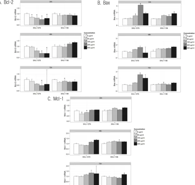
각 세포주에 0, 50, 100, 200, 400 μg/ml 농도의 漆皮 추출물을 투여하여 24, 48, 72시간동안 Bcl-2, Bax, Mcl-1 mRNA 발현을 관찰하였다.
SNU-1079 세포주의 경우 200 μg/ml 투여시 Bax
mRNA 발현이 유의하게 증가하였으며, 같은 농도 에서 Bcl-2 mRNA 발현은 유의하게 감소하였으나, Mcl-1은 mRNA 발현의 차이가 없었다. 한편 SNU-1196 세포주에서는 Bcl-2, Bax, Mcl-1 모두 漆皮 추출물 의 농도와 시간에 따른 유의한 mRNA 발현의 차이 가 없었다(Fig. 5).
A. Bcl-2 B. Bax
C. Mcl-1
Fig. 5. Effects of RVS on the expression of Pro- and Anti-apoptotic mRNAs in SNU-1079 and SNU-1196.
Cells were cultured with 0, 50, 100, 200, 400μg/ml of RVS for 24, 48, 72 hours and RNA levels were measured by RT-PCR.
The crossing point of Bcl-2 (A), Bax (B), Mcl-1 (C) with β-actin was applied to the formula, 2-(targetgene-β-actin), and relative amounts were quantified. The data represent the mean ± SEM of triplicate samples. *p<0.05 compared to control.
5. 漆皮 추출물에 의한 Survivin, Caspase-3 mRNA 발현의 변화
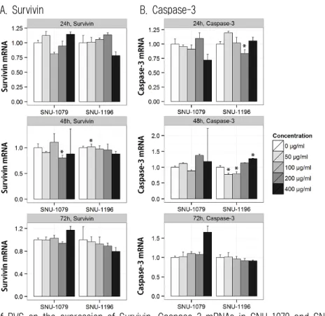
SNU-1079와 SNU-1196 세포주에 0, 50, 100, 200, 400 μg/ml 농도의 漆皮 추출물을 투여하여 24, 48,
72시간동안 Survivin, Caspase-3 mRNA 발현을 관 찰한 결과, 두 세포주 모두에서, 농도와 시간에 따른 유의한 Survivin mRNA 발현의 감소나 Caspase-3 mRNA 발현의 증가는 볼 수 없었다(Fig. 6).
A. Survivin B. Caspase-3
Fig. 6. Effects of RVS on the expression of Survivin, Caspase-3 mRNAs in SNU-1079 and SNU-1196.
Cells were cultured with 0, 50, 100, 200, 400 μg/ml of RVS for 24, 48, 72 hours and RNA levels were measured by RT-PCR. The crossing point of Survivin (A), Caspase-3 (B) with β-actin was applied to the formula, 2-(targetgene-β-actin), and relative amounts were quantified. The data represent the mean ± SEM of triplicate samples. *p<0.05 compared to control.
6. 漆皮 추출물에 의한 Cyclin D1, P21 mRNA 발현의 변화
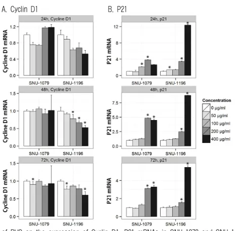
0, 50, 100, 200, 400 μg/ml 농도의 漆皮 추출물을 투여한 뒤 각 세포주에서 24, 48, 72시간동안 Cyclin D1, P21 mRNA 발현을 살펴본 결과, SNU-1079 세포주에서 200 μg/ml 농도의 漆皮 추출물 투여시
P21 mRNA 발현이 유의하게 증가하였다.
SNU-1196 세포주에서는 농도 의존적으로 P21
mRNA 발현이 증가하는 경향으로, 특히 200, 400
μg/ml 농도에서 유의하게 증가하였다. Cyclin D1
mRNA 발현은 48시간 배양에서 유의하게 농도 의
존적으로 감소하였다(Fig. 7).
A. Cyclin D1 B. P21
Fig. 7. Effects of RVS on the expression of Cyclin D1, P21 mRNAs in SNU-1079 and SNU-1196.
Cells were cultured with 0, 50, 100, 200, 400 μg/ml of RVS for 24, 48, 72 hours and RNA levels were measured by RT-PCR. The crossing point of Cyclin D1 (A), P21 (B) with β-actin was applied to the formula, 2-(targetgene-β-actin), and relative amounts were quantified. The data represent the mean ± SEM of triplicate samples. *p<0.05 compared to control.