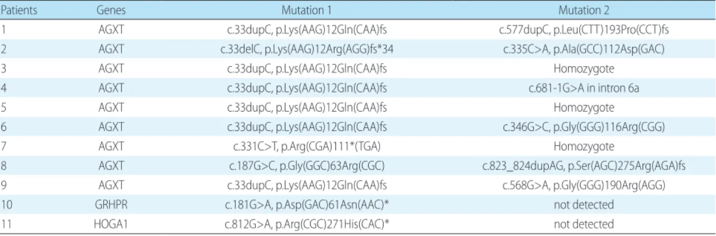
Primary Hyperoxaluria in Korean Pediatric Patients
8
0
0
전체 글
(2)
(3)
(4)
(5)
(6)
(7)
(8)
수치
관련 문서
Seven patients underwent H. pyroli eradication therapy. Three patients showed complete response, but four patients failed... Conclusion
Drug-intoxicated patients were predominantly male, with the highest incidence of intoxication among patients above the seventh decade of age(21.5%) and the
The Incomplete diffuse type PVD found primarily in patients with diabetic retinopathy and retinal detachment and complete diifuse type PVD found primarily in patients
Therefore, nurses should be responsible for the relations to resolve the needs of care between nurses and patients and through communication with the patients,
안 유 석 조선대학교 대학원 Comparative study between resorbable and nonresorbable plate in orthognathic surgery
This showed that the surgical outcome was successfulin 269 patients (98.89%),butthe revision surgery due to surgicalrelapse was performed in three patients.Of 152 patients
The hospital anxiety and depression scale (HADS) and the 9-item patient health questionnaire (PHQ-9) as screening instruments for depression in patients with cancer.. Age
The locations of aneurysms were middle cerebral artery in 15 patients, cerebral artery in 15 patients, cerebral artery in 15 patients, cerebral artery in
Results: In this research, in the group with fibromyalgia patients group, systemic lupus erythematosus patients group and without systemic autoimmune
