Antifungal and Plant Growth Promotion Activities of Recombinant Defensin Proteins from the Seed of Korean
Radish ( Raphanus sativus L.)
Cher-Won Hwang*
Global Leadership School, Department of Environmental Bioscience, Handong Global University (Received November 23, 2009, Accepted December 10, 2009)
Abstract: In the present study, we analyzed the defensin protein deduced from Korean radish (Raphanus sativus L.) seeds.To express the genes in E. coli, we constructed a recombinant expression vector with a defensin gene, named rKRs-AFP gene isolated from Korean radish seeds. Over expressed rKRs-AFP proteins was separated by SDS–PAGE to determine the purity, and protein concentration was determined by the Bradford method. Antifungal activity was assessed by disk assay method against the tested fungi. As a result, when 500 mL of cell culture were disrupted by sonicator, 32.5 mg total proteins were obtained. The purified protein showed a single band on SDS-PAGE with estimated molecular weight about 6 KDa, consistent with the molecular mass calculated from the deduced amino acid sequence. The purified rKRs-AFP protein showed remarkable antifungal activities against several fungi including Aspergillus niger, Botrytis cinerea causing the gray mold disease, and Candida albicans. In field tests using the purified rKRs-AFP protein, the protein showed the reducing activity of disease spot and the mitigating effect of spreading of disease like agrichemicals. The immuno-assay of rKRs-AFP protein showed that the purified protein entirely accumulated at B. cinerea cytoplasm through the hyphal septa shown by fluorescence imaging. There was no fluorescence inside the cell, when the hypha was incubated without the protein.
These all results indicate that the recombinant rKRs-AFP proteins can be utilized as a potential antifungal drug to control harmful plant fungal pathogens.
Key Words: anti-fungal protein, gray mold disease, Raphanus sativus L.
435
*연락저자:
Tel: +82-54-260-1304 Fax: +82-54-261-4603 E-mail: [email protected]
INTRODUCTION
In the world, many fruit, vegetable and plants are damaged by plant fungal pathogens and one of them is Botrytis cinerea which is a casual agent of gray mold. To overcome plant fungal pathogens’ problems, chemical cultivation has been performed in many countries for a long time. This practice causes not only environmental pollution but also harmful effects to human or animal. Due to the undesirable side-effect of synthetic agro-chemicals many biologists have been intensively studying biological control of plant fungal pathogens1-9).
Antimicrobial proteins such as radish antifungal protein in radish seeds, RIP (ribosome inactivating protein) and zeamatin in many plant seeds have been reported in the previous studies1,10-14). Several types of AMPs have been isolated, and their structures have been fully or partially characterized. Until now, defensin, only one conserved class of AMPs was found among plants, invertebrates, and vertebrates15). One type of these AMPs is small (3-5 KDa), basic, and cysteine- rich. Insect defensin and mammalian defensin are the most extensively studied Cys-rich AMPs16).
Plant defensins (45-54aa, 4 disulfide bridges) have been identified as a novel family of Cys-rich peptides17), which implicate in first-line host defense against fungal pathogens. Despite of their structural similarity18-20), plant defensins are highly varied in their primary amino
acid sequences, with exception found in 8 structure- stablilizing Cys residues in common21). The variation in their primary sequences may account for the different biological activities reported for plant defensin, including antifungal activity22), antibacterial activity23). Mutational analysis of the radish (Raphanus sativus) Rs-AFP2 has revealed that the amino acid residues important for antifungal activity are clustered into two adjacent sites. The first site is around the type VI β-turn connecting β-strands 2 and 3, and the second sites is formed by residues on the loop connecting β -strand 1 and the α-helix and the contiguous residues on the α-helix and β-strand 324) When fungal hyphae are treated with Rs-AFP2 or the Dahlia merckii defensin Dm-AMP1, there is a rapid influx of Ca2+, efflux of K+, and alkalinization of the growth medium25). For these reasons, Plant defensin is considered as a hopeful agent of biological control against plant pathogens.
In this study, we described the cloning and purifi- cation of recombinant antifungal protein (rKRs-AFP) derived from Korean radish seeds, and estimated the antifungal efficacy of rKRs-AFP against B. cinerea using immuno-staining analysis and a field study.
MATERIALS AND METHODS
Sample, chemicals, microorganism and media Korean radish seeds (Raphanus sativus L. cv. Beakwoon, Nongwoo Bio Co. Suwon, Korea) were used for the preparation of native antifungal protein1) and total RNA isolation. The total RNA of Korean radish seeds was isolated using easy-BLUETM total RNA extraction kit (iNtRON, Seoul, Korea) and the cDNA of Korean radish seeds was synthesized using Power cDNA synthesis kit (iNtRON, Seoul, Korea). Botrytis cinerea (KACC 40574), Aspergillus flavus (KACC 40250), Fusarium oxyporum (KACC 40053), Aspergillus niger (KACC 40280), and Candida albicans (KACC30050) were used as the test pathogens. The fungal strains were grown in potato dextrose (PD) agar (Difco. Co. Detroit,USA), and spores were harvested and stored as previously described26). C. albicans was cultured overnight in YPD broth/agar (Difco. Co. Detroit, USA) at 30℃, respectively.
E. coli Rosetta (DE3) plac (Novagen, UK.) was cultivated in LB broth (Difco. Co. Detroit,USA) containing appro- priate concentration of antibiotics. Then it was used as a host for the expression of target protein.
Construction of recombinant antifungal protein genes (rKRs-AFP genes)
From the amino acid sequence1,27), we deduced the genetic sequence of the rKRs-AFP genes. In order to amplify the consensus region of coding region form of KRs-AFP genes, the degenerated PCR primer was synthesized described as followings: The sequence of forward primer (mKRs-AFPf-Eco) is 5'-CCG GAA TTC CCA GAA GTT GTG YCA RAG G-3' which contained EcoRI restriction site. The sequence of reverse primer (mKRs-AFPr-Xba-His) is 5'-CCG GTC TAG ATT AGT GGT GGT GGT GGT GAC AWG GRA ART ARC AG-3', which contained XbaI restriction site and 6x His tag sequence. The PCR product amplified by a PCR Pfu DNA polymerase, cDNA of Korean radish seeds as the templates, and the described primer pairs was digested with EcoRI/XbaI and cloned into pHCEIb vector (Bioleaders Co. Seoul, Korea). The recombinant plasmid (rKRs-AFP genes) was sequenced and introduced into E. coli Rosetta (DE3) plac. The transformed E.coli was grown in 500 mL of LB broth (containing 50 ug/mL of ampilcillin and 30 ug/mL of chloramphenicol) at 37℃ with shaking for overnight.
Purification of rKRs-AFP
The fully grown recombinant bacterial cells were harvested by centrifugation at 7,000g for 10 min at 4
℃ and the pellet was re-suspended in 50 mL of 20 mM Tris-HCl (pH 8.0). The cell suspension was sonicated for a total 20 min with a sonicator (Branson Model 250, Branson Ultrasonic, USA). The cracked bacterial cells were centrifuged at 10,000g for 15 min at 4℃. The pellet was re-suspended in 25 mL of 2 M Tris-HCl (pH 12) and 2 M urea and dissolved for 2h at 4℃. The fluid was centrifuged and filtered through a 0.22 µm Acrodisc® syringe filter (Pall Co. USA) to remove insoluble materials28).
A Sephadex G-100 column (16×800 mm) was equili- brated with 1.5 column volumes of 20 mM Tris–HCl (pH 8.0) with a flow rate of 1 mL/min. The solubilized inclusion body was loaded onto a column and the eluted fractions were monitored at 280 nm. Pooled protein fractions were applied onto a metal chelating affinity chromatography column packed with 5 mL Ni-NTA resin (Qiagen Co. Germany) that was pre- charged with 50 mM NiSO4. Before purification, the resin was equilibrated with 20 mM Tris–HCl (pH 8.0),
10 mM imidazole, and 0.5 M NaCl. The protein samples were loaded onto a column and washed with 10 volumes of 20 mM Tris–HCl (pH 8.0), 50 mM imidazole, and 0.5 M NaCl. Finally, the rPhlA protein was eluted with 20 mM Tris–HCl (pH 8.0), 500 mM imidazole, and 0.5 M NaCl, and the final elutant was dialyzed in 20 mM Tris–HCl (pH 8.0), containing 10% glycerol at 4 °C for 12 h. The purification steps were monitored by SDS–PAGE to determine the purity and the concentration of proteins was determined by Bradford method29).
Evaluation of antifungal activity
Antifungal activity was evaluated by disk assay method23). Firstly, B. cinerea KACC49574 was incubated on PDA plate at 25℃ for 1 week. After spore formation, it was diluted in sterile water, centrifuged at 1,000 rpm for 1 min. In the supernatant, hyphae were removed and only spores were collected. Then the spores were added to sterile water, stored at 20℃ and used as required. The obtained spores (100 μl) were mixed on 0.5% soft agar and was added onto a PDA plate.
Afterwards, a sterile 5 mm paper disk (Whatman Ltd.
Maidstone, UK) was placed on the PDA plate and samples were put on the disk. After cultivation at 2 5℃, the antifungal level of sample was determined.
One antifungal unit was defined a minimal inhibitory dose against fungal growth.
Field study of rKRs-AFP
In order to estimate the consequence of rKRs-AFP onto plant infected gray mold rot disease under natural condition, the infected leeks were medicated with 50 mg/L of rKRs-AFP every week for 2 weeks.
One 1 g/L solution of chemical drug Smirex (Dongbang Agro. Seoul, Korea) and distilled water (DW) were applied onto infected plants as a positive control and a negative control, respectively. After application, obser- vations were made to notify the changes in disease points, number of leaves and leaf length for every single plant.
Immunization and immuno-fluorescence microscopy A group of five 6 - 8 week old male Balb/c mice were immunized subcutaneously with 100 µl volumes containing 50 - 60 µg of purified native KRs-AFP in the presence of Freund's incomplete adjuvant. Two
immunizations were performed with a 2-week interval.
A booster dose was given without adjuvant 1 month after the initial administration of the immunogen.
The mice were sacrificed for preparative recovery of serum 2 weeks after the booster. The 2 mL of serum was applied to a column of 5 mL protein A Sepharose® CL-4B (Pharmacia, Sweden). The eluted IgG fraction was evaluated for its quality by SDS-polyacrylamide gel electrophoresis to reveal the presence of heavy and light chains. After dialysis against PBS, the materials was passed through a 0.22 µm syringe filter, quanti- fied by Bradford assay, and then stored frozen in 0.5 mL aliquots at -70℃.
Further, in order to determinate the localization of rKRs-AFP, B. cinerea was incubated on the slide glass.
First, 1 mL of PD agar was onto slide and covered with cover slip to be parallel between medium and slide. After being harden the medium, 1-2×10³ numbers of spores were suspended with 100 µl of 0.5% soft agar then overlaid on slide. Every slide was incubated at 30℃ until the spores which were embedded on slide were germinated sufficiently to observe the elongation of hyphae. 500 µl of 1 mg/mL rKRs-AFP was applied onto slide and incubated at 30℃ for 6 h then the slides were fixed with 2.5% glutaraldehyde/
1% paraformaldehyde mixture at 4℃ for 4h. After fixation, the slides were washed with 0.1% of NaBH4
for 1min to reduce auto fluorescence and then blocked with 2% goat serum / 0.1% Triton X-100 PBS for 1 h.
The slide finished blocking step were treated with 200µl of mouse anti-KRs-AFP IgG diluted with 1%
goat serum / 0.1% Triton X-100 PBS and incubated overnight at 4℃. In order to detect the binding of primary antibody, 500 × diluted Alexa Fluor 488 goat anti-mouse IgG (Molecular ProbesTM USA) with 0.1%
Triton X-100 PBS for 1hr in dark. Every interval of single step, the slide was washed with phosphate buffered saline (PBS) 3 times for 10 min.
RESULTS AND DISCUSSION
Purification of rKRs-AFP
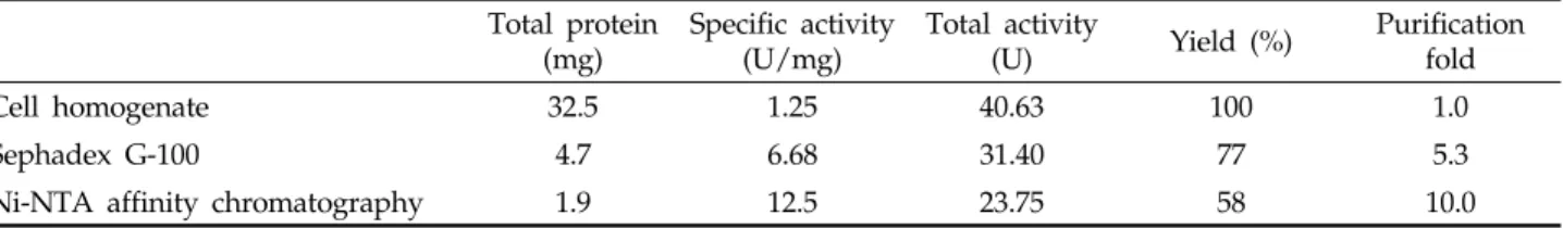
Cells from 500 mL of culture were disrupted by sonicator and 32.5 mg total protein was obtained.
After inclusion body was refolded, the specific activity increased by 5.3 fold while recovery was 77%.
Finally, the rKRs-AFP was purified by metal affinity
Table 1 Purification steps of rKRs-AFP from E. coli Total protein
(mg) Specific activity
(U/mg) Total activity
(U) Yield (%) Purification fold
Cell homogenate 32.5 1.25 40.63 100 1.0
Sephadex G-100 4.7 6.68 31.40 77 5.3
Ni-NTA affinity chromatography 1.9 12.5 23.75 58 10.0
Fig. 1. SDS-PAGE (A) and immunoblotting (B) of recombinant Korean radish antifungal protein from E.
coli Rosetta (DE3) plac. Lane 1, protein molecular weight markers (KDa); lane 2, crude cell extrac lane 3, cytosolic soluble fraction; lane 4, cytosolic insoluble fraction; lane 5, purified recombinant Korean radish antifungal protein from insoluble fraction.
Fig. 2. Determination of antifungal activities of recom- binant Korean radish antifungal protein against pathogenic fungi and yeast by paper disk diffusion assay. 50 µl of diluted samples were applied on each of paper disks. A, Aspergillus niger; B, Botrytis cinerea; C, Candida albicans: 1, 300 µg; 2, 150 µg; 3, 100 µg; 4, 80 µg; 5, 60 µg; 6, 40 µg;
7, 20 µg/ disc.
chromatography and 1.9 mg of protein was obtained.
The specific activity of purified rKRs-AFP was 12.5 U/mg (Table 1). The band was detected with mouse anti-rKRs-AFP IgG on immuno-blotting analysis. The purified protein showed a single band on SDS-PAGE and the size was estimated about 6 KDa (Fig. 1). This value was in good agreement with the molecular mass calculated from the deduced amino acid sequence1,27).
Antifungal activity of rKRs-AFP
Previously we reported the antifungal efficacy of Korean radish against gray mold rot on Chinese chive caused by fungal pathogen B. cinerea30). In the present study, the antifungal activity of rKRs-AFP towards B.
cinerea, A. flavus, F. oxyporum, A. niger, and C. albicans was observed. The antifungal activity was weakest against A. niger and the strongest against B. cinerea, respectively (Fig. 2). The rKRs-AFP revealed moderate antifungal activity against C. albicans. The strength of antifungal activity was positively correlated to the dose of rKRs-AFP. The minimal inhibitory doses of
the antifungal activity of rKRs-AFP towards B. cinerea, A. flavus, F. oxyporum, A. niger, and C. albicans were 80, 120, 100, 150, and 100 ug/disc, respectively. In vitro, the specificity of plant defensins activity was according to the ionic strength of growth medium26) and in high strength growth medium, the spectrum of the activity was narrowed. We thought the difference of minimal inhibitory dose due to high ionic strength PDA growth medium and these purified recombinants rKRs-AFP can be usage for field experiment against gray mold rot.
Leek field study
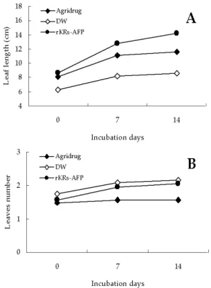
To investigate the potential of rKRs-AFP as an effective antifungal bio-drug, the infected leeks were medicated with 50 mg/L of rKRs-AFP. The protein reduced the disease spot and consequently, mitigated spreading of disease like agrichemicals (Fig. 3). Moreover, the protein showed not only therapeutic efficacy but promotive effect on growth of leek plants. At tow weeks after the treatment, the blades were averagely grown from 8.3 cm to 14 cm, while the plants treated with chemical drug were grown from 8 cm to 10.5 cm, and the numbers of leaves were increased from 1.5 to 2 (Fig. 4). In addition, the width of leaf was broader than positive control. Although the difference on the growth of plants was apparent, further studies
Fig. 3. Antifungal activity of rKRs-AFP against leek (Allium tuberosum Rottler) infected by gray mold rot disease. Every single pot was treated with 200 mL of 50 ug/ mL rRs-AFP, 1 mg/ mL agrichemicals or DW.
Fig. 4. The promotive effect of rKRs-AFP on growth of leek (Allium tuberosum Rottler) A : The leaf length was estimated for every single plant. B : The numbers of leaves was counted for every single plant. The amounts of proteins was used with 40 mg/ L.
Fig. 5. Immunofluorescence microscope images to show the localization of rKRs-AFP in B. cinerea cells. (A) Normal image of B. cinerea cells untreated with rKRs- AFP. (B) rKRs-AFP Untreated cells are immunostained by mouse anti-rKRs-AFP IgG and GFP-labeled goat anti-mouse IgG. There was no detectable fluorescence signal. (C) Merged image of (A) and (B). (D) Normal image of dis- rupted cells (B. cinerea) by rKRs-AFP. (E) The disrupted cells were positively immunostained by mouse anti-rKRs- AFP IgG and GFP-labeled goat anti-mouse IgG (light green). (F) Merged image of (D) and (E).
are required to confirm growth stimulating effects.
Localization of rKRs-AFP in B. cinerea
We have reported that rKRs-AFP belongs to A group (A1, A2, A3 and A4) of defensins families based on its sequence27) and we proposed that rKRs- AFP is included in subfamilies (A3 and A4). To track the movement of antifungal proteins after application to the pathogen, rKRs-AFP was labeled with GFP.
The rKRs-AFP which was treated to B. cinerea was easily detected with GFP-labeled antibody by fluores- cence imaging. The results showed (Fig. 5) that the purified protein entirely accumulated at the cytoplasm through the hyphal septa with affecting pathogenic fungal morphology, but in hyphae incubated without the protein, no binding of primary antibody was observed, resulting in no fluorescence imaging inside the cell.
ACKNOWLEDGEMENT
This study was supported by Sabbatical Leave Grant at Handong Global University (HGU-2008)
REFERENCES
1. Park, J. H., Shin, H. K., and Hwang, C. W. (2001)
New Antimicrobial activity from Korean radish seeds (Raphanus sativus L.) J. Microbiol. Biotechnol., 11(2), 337-341.
2. Boman, H. G. (1995) Peptide antibiotics and their role in innate immunity. Annu. Rev. Immunol. 13, 61-92.
3. Seo, Y. W., K. W. Cho, H. S. Lee, T. M. Yoon, and J. H. Shin (2000) New polyene macrolide antibiotics from Streptomyces sp. M90025. J. Microbiol.
Biotechnol 10, 176-180.
4. Bae, D. W., J. T. Lee, D. Y. Son, E. S. Lee and H.
K. Kim (2000) Isolation of bacteria strain antagonistic to Pyricularia oryzae and its made of antifungal action. J. Microbiol. Biotechnol. 10, 811-816.
5. Bae, D. W., Y. S. Kawk, J. T. Lee, D. Y. Son, S. S.
Chun and H. K. Kim (2000) Purification and characterization of novel antifungal protein from Paenibacillus macerans PMI antagonistic to rice blast fungus Pyricularia oryzae. J. Microbiol. Biotechnol.
10, 805-810.
6. Hwang, C. W., I. C. Park, W. H. Yeh, M. Takagi and J. C. Ryu (1997) A partial nucleotide sequence of chitin synthase(CHS) gene from rice blast fungus Pyricularia oryzae and its cloning. J. Microbiol.
Biotechnol. 7, 157-159.
7. Lacey, L. A. and M. Gottel (1995) Current develop- ment in microbial control of insect past and pros- pects for the 21st century. Entomophage. 40, 3-28.
8. Pfeifer, T. A. and T. A. Grigliatti (1996) Future perspectives on insect pest management: Engineering the pest. J. Invertebr. Pathol. 67, 109-119.
9. Seo, Y. W., K. W. Cho, H. S. Lee, T. M. Yoon and J. H. Shin (200) New polyene macrolide antibiotics from Streptomyces sp. M90025. J. Microbiol. Biotechnol.
10, 176-180.
10. Flant, F., W. Vranken, W. Broekaert and F. Borremans (1998) Determination of the three- dimensional solution structure of Raphanus sativus antifungal protein 1 by 1H NMR. J. Mol. Biol. 279, 257-270.
11. Terras, F. R. G.., H. M. E. Schoofs, M. F. C. De Bolle, F. Van Leuven, S. B. Rees, J. Vanderleyden, B. P. A. Cammue and W. F. Broekaert (1992) Analysis of two novel classes of plant antifungal proteins from radish (Raphanus sativus) seeds. J.
Biol. Chem. 267, 15301-15309.
12. Bohlmann, H., S. Clausen, S. Behnken, H. Giese, H. Hiller, U. Reimann-Philipp, G. Schrader, V.
Barkholt and K. Apel (1988) Leaf-specific thionins of barley-a novel class of cell wall proteins toxic to plant pathogenic fungi and possibly involved in the defence mechanism of plants. EMBO J. 7, 1559-1565.
13. Broekaert , W. F., W. Marien, F. R. G. Terras, M.
F. C. De Bolle, P. Proost, J. Van Damme, L, Dillen, M. Claeys, S. B. Rees, J. Vanderleyden and B. P. A. Cammue (1992) Antimicrobial peptides from Amaranthus cauatus seeds with sequence homology to the cysteine/ glycine-rich domain of chitin–binding proteins. Biochemistry 31, 4308-4314.
14. Cammue, B. P. A., M. F. C. De Bolle, F. R. G.
Terras, P. Proost, J. Van Damme, S. B. Rees, J.
Vanderleyden, and W. F. Broekaert (1992) Isolation and characterization of a novel class of plant antimicrobial peptides from Mirabilis jalapa L. seeds.
J. Biol. Chem. 267, 2228-2233.
15. Thevissen, K., Warnecke, D. C., François, I. E. J.
A., Leipelt, M., Heinz ,E., Ott, C., U., Zähringer, B., Thomma, P. H. J., Ferket, K. K. A. and Cammue, B. P. A. (2004) Defensin from insects and plants interact fungal glucosylceramides, J. Biol. Chem., 279(6), 3900-3905.
16. Hoffmann, J. A. and Hétru, C. (1992) Insect defensins: inducible antibacterial peptides. Immuno.
Today. 13, 411-415.
17. Broekaert, W. F., Terras, F. R. G., Cammue, B. P.
A., and Osborn, R. W. (1995) Synergistic Enhance- ment of the Antifungal Activity of Wheat and Barley Thionins by Radish and Oilseed Rape 2S Albumins and by Barley Trypsin Inhibitors., Plant Physiol, (Bethestda) 108,1353-1358.
18. Bontem F., Roumestand C., Boyot P., Gilquin B., Doljansky Y., Menez A., Toma F. (1991) Three- dimensional structure of natural charybdotoxin in aqueous solution by 1H-NMR. Charybdotoxin possesses a structural motif found in other scopion toxinz.
Eur. J. Biochem., 196, 19-28.
19. Fontecilla-Champs J-C (1989) Three-demensional model of insect-directed scorpion toxin from Androctonus australis Hecter and its implication for the evolution of scorpion toxin in general. J. Mol. Evol. 29, 63-67.
20. Kobayashi Y, Takashima H, Tamaoki H, Kyogoku Y, Lambert P, Kuroda H, Chino N, Watanabe TX, Kimura T, Sakakibara S (1991) The cysteine-stabilized
alpha-helix : a common structural motif of ion- channer blocking neurotoxic peptide. Biopolymers, 31, 1213-1220.
21. Thomma, B. P. H. J., B. P. Cammue, K, Thevissen (2002) Plant defensins., Planta. 216(2), 193-202.
22. Terras F. R., Eggermont K., Kovaleva V., Raikhel N. V., Osborn R. W., Kester A., Rees S. B., Torrekens S., Van Leuven F., Vanderleyden J.
(1995) Small cysteine-rich antifungal proteins from radish: their role in host defense. Plant Cell. 7, 573 –588.
23. Segura A., Moreno M., Molina A., Garcia-Olmedo F. (1998) Novel defensin subfamily from spinach (Spinacia oleracea). FEBS Lett., 435, 159–16.
24. De Samblanx G. W., Goderis I. J., Thevissen K., Raemaekers R., Fant F., Borremans F., Acland D.
P., Osborn R. W., Patel S., Broekaert W. F. (1997) Mutational analysis of a plant defensin from radish (Raphanus sativus L.) reveals two adjacent sites important for antifungal activity. J. Biol.
Chem., 272, 1171–1179.
25. Thevissen K., Ghazi A., De Samblanx G. W., Brownlee C., Osborn R. W., Broekaert W. F. (1996) Fungal membrane responses induced by plant
defensins and thionins. J Biol. Chem., 271, 15018–
1502.
26. Terras, F. R. G., H. M. E. Schoofs, K. Thevissen, R. W. Osborn, J. Vanderleyden, B. P. A. Cammue, and W. F. Broekaert (1993) A new family of basic cysteine-rich plant antifungal proteins from Brassicaceae species. FEBS Lett. 316, 233-24.
27. Hwang, C. W. (2007) Study of distance relation- ships among domestic radish (Raphanus sativus L.) by analyzing its anti-fungal protein gene. J. Life Science. 17, 1294-1297.
28. A. K. Patra, R. Mukhopadhyay, R. Mukhija, A.
Krishnan, L. C. Grag and A. K. Panda, (2000) Optimi- zation of inclusion body solubilization and renatur- ation of recombinant human growth hormone from Escherichia coli. Protein Expr. Purif., 18, pp.
182–192.
29. M. M. Bradford (1976) A rapid and sensitive method for the quantitation of microgram quantities of protein using the principle of protein–dye binding.
Anal. Biochem.72, pp. 248–254.
30. Hwang, C. W. (2003) Antifungal activity of Korean radish (Raphanus sativus L) extracts against pathogenic plant. J. Life Science. 13, 223-229.