branched erect filaments, plurilocular sporangia produced laterally on basal part of erect filaments and occasionally on prostrate filaments, sessile or short pedicellate pluriloc- ular sporangia, and 2 types(big lanceolate and small ovate) of plurilocular sporangia(Saunders 1898; Setchell and Gardner 1925; Abbott and Hollenberg 1976). Feldmannia chitonicola has been reported only from eastern Pacific, in California(Abbott and Hollenberg 1976), Chile(Ramirez and Santelices 1991) and Peru(Acleto 1980).
We collected unidentified tiny filamentous brown algae from west and east coasts of Korea and cultured them. We observed their detailed morphology and developmental pattern in laboratory cultures. We also analysed molecular data based on rbcL and cox1 genes for their phylogenetic relationships with others in their respective genera and a few selected species within the Ectocarpales. We identify them as Feldmannia chitonicola and add it to the Korean marine algal inventory, representing the first report of this species in western Pacific.
https://doi.org/10.11626/KJEB.2019.37.3.278
INTRODUCTION
The filamentous brown algal genus, Feldmannia, was described by G. Hamel(1939) based on F. lebelii. It is char- acterized by having basal meristematic zones in axial fila- ments, branches below meristematic zones and plurilocular sporangia developing from below meristems(Kim and Lee 1994; Abbott and Huisman 2004; Kim 2010). Current- ly, 28 species of Feldmannia have been known around the world(Guiry and Guiry 2019). Of them, six species have been reported in Korea(Kim 2010): F. lebelii(Areschoug ex Crouan and Crouan) Hamel, F. globifera(Montagne) Hamel 1939, F. irregularis(Kützing) Hamel, F. rhizoidea Hollenberg and Abbott, F. indica(Sonder) Womersley and Bailey, and F. mitchelliae(Harvey) Kim.
Feldmannia chitonicola(De A. Saunders) Levring was originally described as Ectocarpus chitonicolus De A. Saun- ders from California based on a small specimen growing on the back of a chiton(Saunders 1898). It is characterized by having small size(1-2mm), epizoic habitat, mostly un-
Original article
A new record of Feldmannia chitonicola from Korea based on laboratory culture and molecular data
Jose Avila-Peltroche, Antony Otinga Oteng’o, So Young Jeong, Boo Yeon Won and Tae Oh Cho*
Department of Life Science, Chosun University, Gwangju 61452, Republic of Korea
Korean J. Environ. Biol.
37(3) : 278-284(2019) ISSN 1226-9999(print) ISSN 2287-7851(online)
* Corresponding author Tae Oh Cho
Tel. 062-230-7161
E-mail. [email protected]
Received: 22 July 2019 Revised: 16 August 2019
Revision accepted: 19 August 2019
Abstract: Feldmannia chitonicola is reported as a new record from Korea based on morphological studies in laboratory-cultured materials and molecular analyses. F.
chitonicola is mainly characterized by a small size(1-2mm), erect filaments mostly unbranched, plurilocular sporangia produced on both prostrate and laterally on the basal part of erect filaments, and 2 types(lanceolate and ovate) of sporangia. In our cultures, sporangia production was slower at 10°C than in 16°C and 20°C. Our molecular analyses of rbcL and cox1 genes supported its independence from other congeners reported for Korea. This is the first report of F. chitonicola for western Pacific.
Keywords: Feldmannia chitonicola, cox1, Ectocarpales, Phaeophyceae, rbcL
MATERIALS AND METHODS
1. Culture and Morphological studies
Two unialgal isolates of filamentous brown algae were es- tablished from samples collected from west and east coasts of Korea in November, 2017 and February, 2019. Young germlines were inoculated in Petri dishes(60mm diam- eter×15mm depth) containing PES medium(Provasoli 1968). Cultures were kept at 10°C, 16°C and 20°C under 20-30μmol photons m-2 s-1 white fluorescent light and 14:10-h light/dark photoperiod. Medium was renewed weekly.
After culture process, thalli were sorted into voucher her- barium specimens, silica gel samples, and formalin samples (4-5% formalin/seawater). Fresh cultured materials were used for morphological analysis. Photomicrographs were taken using an Olympus BX51TRF microscope(Olympus, Tokyo, Japan) and an Olympus DP71 camera. At least 25 individuals were selected for the determination of quanti- tative characters and their means and standard deviations were calculated. For developmental study, a Leica DMi8 inverted microscope(Leica, Wetzlar, Germany) equipped with a Leica DFC450C camera was used. The percentage of fertile plants and different morphologies of sporangia at different temperatures were calculated based on at least 50 plants or sporangia, respectively. Permanent slide speci- mens were mounted by 70% karo syrup and the examined representative specimens were deposited in the herbarium of Chosun University(CUK) and National Institute of Bi- ological Resources(NIBR), Korea.
2. Molecular study
Genomic DNA was manually extracted from silica-gel samples using a NucleoSpin Plant II Kit(Macherey-Na- gel, Düren, Germany). The extracted DNA was stored at -20°C and used to amplify rbcL and cox1. Polymerase chain reaction(PCR) was carried out with a Veriti 96-well Termal cycler(Applied Biosystem). The rbcL gene was am- plified using the primer combinations NDrbcL2-DRL1R and DRL2F-R3A(Kogame et al. 1999; Hwang et al. 2005) with HelixAmp Ready-2x-Go Series(NanoHelix Co., Ltd., Daejeon, Korea). The GAZF2-GAZR2 combination of primers was used for cox1(Saunders 2005; Lane et al.
2007). PCR products were purified using a PCRquick- spinTM PCR product purification kit(iNtRON Biotech- nology, Inc, Seongnam, Korea). Determination of the nu- cleotide sequence or sequencing was performed by Mac-
rogen Inc., Seoul, South Korea. DNA sequence data(rbcL and cox1) were compiled by the present study and ob- tained from GenBank and aligned with ClustalW(Thomp- son et al. 1994). New sequences obtained from Feldmannia chitonicola have been deposited in EMBL/GenBank under the accession numbers MN092346(CUK18833) and MN092347(CUK19719) for rbcL, and MN092344(CUK 18833) and MN092345(CUK19719) for cox1.
Phylogenetic analyses were conducted using MEGA ver- sion 6.06(Tamura et al. 2013). Maximum likelihood anal- yses were conducted using the GTR+G+I model, with 1,000 bootstrap replicates. A bayesian inference was per- formed using MrBayes 3.2.6(Huelsenbeck and Ronguist 2001; Ronguist and Huelsenbeck 2003). Markov chain Monte Carlo runs were conducted for 2 million genera- tions, each with one cold chain and three heated chains using the GTR+Γ+I evolutionary model and sampling and printing every 1,000 generations. Summary trees were generated using a burn-in value of 800.
RESULTS
Feldmannia chitonicola(De A. Saunders) Levring, 1960 전복솜털(신칭)(Figs. 1-3)
Type locality: Pacific Grove, California, USA Habitat: Epiphytic on Sargassum thunbergii.
Material examined: NIBROR0000001611 & CUK188 33(= MBRB0100TC18833), Chaesokgang, Byeonsan- myeon, Buan-gun, Jeollabuk-do, Korea(35°37ʹ27.04ʺN, 126°27ʹ56.88ʺE), November 17, 2017, T. O. Cho and B. Y. Won, at 1m depth by hand; CUK19719(=MBRB 0100TC19719), Guryong Pohang, Guryong Pohang-eup, Pohang-si, Gyeongsangbuk-do, Korea(35°58ʹ48.53ʺN, 129°34ʹ17.33ʺE), February 02, 2019, T. O. Cho and B. Y.
Won, at 1m depth by hand.
World distribution: Korea, USA, Chile, Peru(Guiry and Guiry 2019).
Culture studies
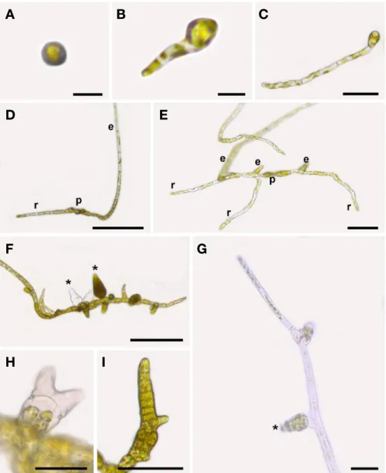
Culture strains produced plurilocular sporangia after 10-12 days in culture. Spores were settled without show- ing any signs of sexuality(Fig. 1A). They developed a “germ tube”(Fig. 1B) which septated by cross-wall and later be- came rhizoid(Fig. 1C). The first erect filament emerged on the opposite side of the rhizoid, forming a heterotrichous thallus after 7 days in culture(Fig. 1D). Additional erect
and rhizoidal filaments arose from the prostrate part during the next days(Fig. 1E). The plants produced plurilocular sporangia in the prostrate part after 10 days in culture(Fig.
1F), and then below the meristem near the base of the erect filament after 12 days in culture. The upper filaments did not show branches in older thalli, but they developed
long pseudohairs and some of them produced secondary meristems on terminal portion with small plurilocular spo- rangia and rhizoids(Fig. 1G).
The developmental pattern of thalli was not affected according to different temperatures but the production of plurilocular sporangia was slower at 10°C than in 16°C and
Fig. 1. Development of Feldmannia chitonicola in laboratory culture. A. Settled spore; B, C. Germination of zoospore showing the devel- opment of rhizoid; D. Heterotrichous thallus with rhizoid(r), prostrate part(p), and erect filament(e) after 7 days in culture; E. Additional erect filaments(e) and rhizoids(r) from prostrate part(p); F. Plurilocular sporangia(asterisks) on prostrate filament after 10 days in culture; G.
Secondary meristem on end portion of erect filament with small plurilocular sporangium(asterisk); H. Branched plurilocular sporangium; I.
Plurilocular sporangium with short outgrowths. Scale bars: A, B=10 μm; C, G, H, I=50 μm; D, F=200 μm; E=100 μm.
A B C
D E
F G
H I
20°C(Table 1). Few irregular morphologies of plurilocular sporangia were observed at 20°C and 16°C(Fig. 1H, I). Di- rect-monophasic life history by plurilocular sporangia was repeated during four generations in our culture. Unilocular sporangia were not found in all the conditions tested.
Morphological observations
Plants are forming small tufts, 1.2-2.1mm tall(Fig. 2A), with numerous uniseriate prostrate filaments, and irreg- ularly branched.(Fig. 2C). Uniseriate erect filaments are mostly unbranched or sometimes sparingly branched near
Table 1. Effect of temperature on percentages of fertile plants and different morphologies of plurilocular sporangia for two strains of Feld- mannia chitonicola from Korea. Percentages were calculated based on at least 50 plants or plurilocular sporangia, respectively.
Strain Temperature(°C) Fertile plants(%) Plurilocular sporangia shape(%)
Ovate Lanceolate Branched or with outgrowths TC18833
(western coast)
20 41.9 16.1 72.6 11.3
16 49.1 90.7 9.3 0
10 PD NS NS NS
TC19719 (eastern coast)
20 81.0 69.3 17.3 13.3
16 73.2 92.9 1.0 6.1
10 42.9 100.0 0.0 0
PD: Poor development; NS: No sporangia
Fig. 2. Development of Feldmannia chitonicola. A. Cultured thallus showing long unbranched filaments; B. Thallus with plurilocular sporan- gia(asterisks) on both prostrate and erect filaments; C. Thallus of numerous prostrate branched filaments; D. Cells of erect filament show- ing discoid chloroplasts; E. Meristematic zones(arrows) composed of small cells on erect filament; F. Pedicellate plurilocular sporangium on prostrate filament; G. Sessile plurilocular sporangium on prostrate filament; H. Elongated plurilocular sporangia on erect filament. Scale bars: A=500μm; B=250μm; C=200μm; D, E=25μm; F, G=50μm; H=100μm.
A B C
D E F G H
base, and slightly attenuated upward or forming long pseudohairs(Fig. 2B). Erect filaments are composed of rec- tangular cells with 19.3±3.1μm in width and with numer- ous discoid chloroplasts and not constricted at the septum (Fig. 2D). Meristematic zones are composed of small-sized cells on erect filaments(Fig. 2E). There are various in the size of cells according to the position: 3.3±0.8 longer than broad in apical and medium parts, 2.1±0.5 longer than broad in basal part, and 1.1±0.3 longer than broad in mer- istematic zone. Plurilocular sporangia are produced as ses- sile and on one(or two)-celled pedicels both on erect and prostrate filaments. Plurilocular sporangia are developed below meristematic zone of erect filament(Fig. 2B). There are 2 types of plurilocular sporangia: large lanceolate(Fig.
2H) and small ovate shapes(Fig. 2F, G). Plurilocular spo- rangia are(42)84-192μm×26-58μm in size. Unilocular sporangia not observed.
Phylogenetic analysis
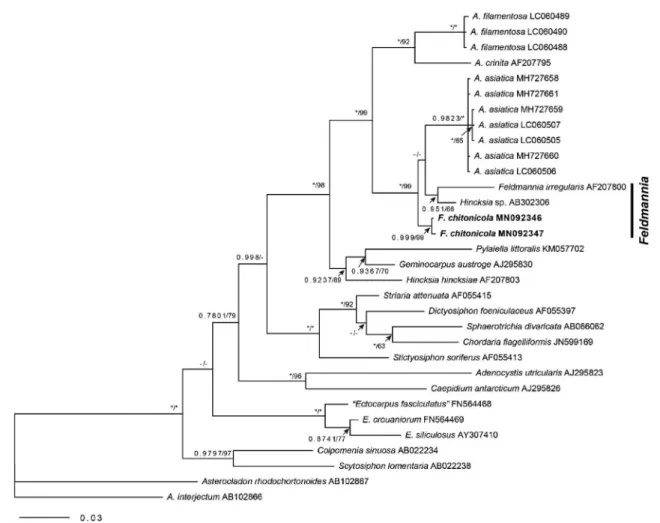
The 1326-nucleotide portion of rbcL and 613-nucleo- tide portion of cox1 was aligned for Feldmannia chitonicola.
The phylogenetic trees were obtained from the alignment of the rbcL sequences newly generated and downloaded from GenBank. Asterocladon rhodochortonoides and A.
interjectum tree were selected as outgroups. Our samples were nested within a clade of Feldmannia(Fig. 3). Gene se- quence divergence differed from F. irregularis by 2.3-2.4%, while from Acinetospora filamentosa and A. crinita by 3.9- 4.0%.
DISCUSSION
Our brown algal collections from Korea matched with
Fig. 3. Phylogenetic tree of Feldmannia species based on Bayesian and ML analysis with rbcL sequences. Value above branches=Bayes- ian posterior probabilities>0.75/Maximum likelihood bootstrap values in %>50. Values lower than BPP 0.75 or BS 50 are indicated by hyphens(-). Values of BPP 1.00 or BS 100 are indicated by asterisks(*).
the diagnosis of Feldmannia chitonicola described from the type locality in thallus size, the lanceolate and ovate shapes of plurilocular sporangia, and the development of plurilocular sporangia on both prostrate and basal part of erect filaments. These samples are nested within a clade of Feldmannia in phylogenetic trees based on rbcL and cox1 genes(Fig. 3). We add F. chitonicola in the list of Korean macroalgal flora. This is also the first record of F. chitonicola in western Pacific.
Although Feldmannia chitonicola closely resembles to F. irregularis, in some morphological features as the size of thalli, unbranched erect filament, and terminal hyaline pseudo-hairs, F. chitonicola always develops plurilocular sporangia first on the prostrate filaments while F. irregularis only on the erect filaments in both wild and culture mate- rials(Kim and Lee 1994). Feldmannia chitonicola may be distinguished from F. irregularis by the presence of repro- ductive organs in prostrate filaments, sporangia shape, and molecular data.
The secondary meristems are the upper part regions producing plurilocular sporangia and rhizoids in erect fil- aments. The presence of a secondary meristem has been known only from culture materials of F. globifera and F. ir- regularis. The upper filaments of Feldmannia chitonicola in older thalli developed the secondary meristems with small plurilocular sporangia and rhizoids. These secondary mer- istems may be attributed to the contact of the filaments with the culture dish, which seems to stimulate the devel- opment of new meristems or rhizoids(Clayton 1974; Kim and Lee 1994).
The temperature has been reported as one of import- ant culture conditions for the production of plurilocular sporangia in filamentous brown algae(Clayton 1974). In this study, the production of plurilocular sporangia was increased under higher temperatures(Table 1) and this matched with the Clayton’s result. Also, our result shows that temperature can determine the morphological types of plurilocular sporangia: ovate plurilocular sporangia in lower temperatures and elongated plurilocular sporangia in higher temperatures(Table 1). These effects of different temperatures on the morphologies of plurilocular sporan- gia have not been reported in Feldmannia species.
ACKNOWLEDGEMENTS
This study was supported by a grant from the National Institute of Biological Resources(NIBR), funded by the
Ministry of Environment(MOE) of the Republic of Korea (NIBR 201801205) and by a grant from the research fund of Chosun University 2018. This research was also sup- ported by Basic Science Research Program through the National Research Foundation of Korea(NRF) funded by the Ministry of Education, Science and Technology (2019R1F1A1060346) and a grant from Marine Bio- technology Program(20170431) funded by Ministry of Oceans and Fisheries of Korean Government to Tae Oh Cho.
REFERENCES
Abbott IA and GJ Hollenberg. 1976. Marine Algae of California.
Stanford University Press, Stanford.
Abbott IA and JM Huisman. 2004. Marine Green and Brown Algae of the Hawaiian Islands. Bishop Museum Press, Ho- nolulu.
Acleto C. 1980. Notas sobre las algas marinas del Perú I. Nuevos registros. Publ. Mus. Hist. Natur. Serie Botánica 30:1-33.
Clayton MN. 1974. Studies on the development, life history and taxonomy of Ectocarpales(Phaeophyta) in southern Austra- lia. Aust. J. Bot. 22:743-813.
Guiry MD and GM Guiry. 2019. AlgaeBase. World-wide electron- ic publication, National University of Ireland, Galway. http://
www.algaebase.org; searched on 18 June 2019.
Hamel G. 1939. Sur la classification des Ectocarpales. Bot. Notis- er 1939:65-70.
Hwang IK, HS Kim and WJ Lee. 2005. Polymorphism in the brown alga Dictyota dichotoma(Dictyotales, Phaeophyceae) from Korea. Mar. Biol. 147:999-1015.
Huelsenbeck JP and F Ronquist. 2001. MrBayes: Bayesian infer- ence of phylogeny. Bioinformatics 17:754-755.
Kim HS and IK Lee. 1994. Morphotaxonomic studies on the Kore- an Ectocarpaceae(Phaeophyta) III. Genus Feldmannia Ham- mel, specially referred to morphogenesis and phylogenetic relationships among related genera. Algae 9:153-168.
Kim HS. 2010. Ectocarpaceae, Acinetopsoraceae, Chordariaceae.
pp. 5-153. In Algal Flora of Korea. Volume 2, Number 1. Het- erokontophyta: Phaeophyceae: Ectocarpales. Marine Brown Algae I.(Kim HS and SM Boo eds.). National Institute of Bio- logical Resources, Incheon.
Kogame K, T Horiguchi and M Masuda. 1999. Phylogeny of the order Scytosiphonales(Phaeophyceae) based on DNA se- quences of rbcL, partial rbcS, and partial LSU nrDNA. Phyco- logia 38:496-502.
Lane CE, SC Lindstrom and GW Saunders. 2007. A molecular
assessment of north east Pacific Alaria species(Laminariales, Phaeophyceae) with reference to the utility of DNA barcod- ing. Mol. Phylogenet. Evol. 44:634-648.
Provasoli L. 1968. Media and prospects for the cultivation of ma- rine algae. pp. 63-75. In Cultures and Collections of Algae (Watanabe A and A Hattori eds.). Proceedings of the U.S.- Japan Conference, Hakone.
Ramirez ME and B Santelices. 1991. Catálogo de las algas ma- rinas bentónicas de la costa temperada del Pacífico de Su- damérica. Monografías Biológicas 5:1-437.
Ronquist F and JP Huelsenbeck. 2003. MrBayes 3: Bayesian phylogenetic inference under mixed models. Bioinformatics 19:1572-1574.
Saunders DA. 1898. Phycological memoirs. Proceedings of the California Academy of Sciences, Series 3. Botany 1:147-168.
Saunders GW. 2005. Applying DNA barcoding to red macroal- gae: a preliminary appraisal holds promise for future applica- tions. Philos. Trans. R. Soc. B-Biol. Sci. 360:1879-1888.
Setchell WA and NL Gardner. 1925. The marine algae of the Pa- cific coast of North America. Part III. Melanophyceae. Univ.
Calif. Publ. Bot. 8:383-898.
Tamura K, G Stecher, D Peterson, A Filipski and S Kumar. 2013.
MEGA6: Molecular Evolutionary Genetics Analysis Version 6.0. Mol. Biol. Evol. 30:2725-2729.
Thompson JD, DG Higgins and TJ Gibson. 1994. Clustal W:
improving the sensitivity of progressive multiple sequence alignment through sequence weighting, position-specific gap penalties and weight matrix choice. Nucleic Acids Res.
22:4673-4680.