운동 강도 차이가 고지방식이 Sprague-Dawley Rat의 골격근 내 PGC-1α, PPAR-γ 및 인슐린 저항에 미치는 영향
정현령․강호율
경북대학교 사범대학 체육교육과 운동대사 실험실
Effects of Exercise Intensity on PGC-1α, PPAR-γ, and Insulin Resistance in Skeletal Muscle of High Fat Diet-fed Sprague-Dawley Rats
Hyun-Lyung Jung and Ho-Youl Kang
Exercise Metabolism Laboratory, Department of Physical Education, Kyungpook National University
ABSTRACT This study investigated the effects of exercise intensity on PGC-1α, PPAR-γ, and insulin resistance in skeletal muscle of high fat diet-fed Sprague-Dawley rats. Forty rats were randomly divided into five groups: sedentary control group (SED), high fat diet group (HF), high fat diet+low-intensity exercise group (HFLE, 22 m/min, 60 min, 6 days/week), high fat diet+moderate-intensity exercise group (HFME, 26 m/min, 51 min), and high fat diet+high-in- tensity exercise group (HFHE, 30 m/min, 46 min). After 4 weeks of high fat diet and endurance exercise training, the lipid profiles, insulin, and glucose concentrations were determined in plasma. PGC-1α, PPAR-γ, and GLUT-4 contents were measured in plantaris muscle. The rate of glucose transport in soleus muscle was determined under submaximal insulin concentration (1,000 μIU/mL insulin, 20 min) during muscle incubation. Plasma glucose during oral glucose tolerance test in HF was significantly greater than that in SED, and plasma glucose levels in the three exercise (EX) groups were significantly lower that those in SED and HF at 30 and 60 min, respectively (P<0.05).
Plasma insulin levels in the EX groups were significantly reduced by 60 min compared to that in HF (P<0.05). The protein expression level of PGC-1α as well as muscle glucose uptake were significantly higher in SED and HF than those in the three EX groups (P<0.05), and HFHE showed significantly higher levels than HFLE and HFME. Expression levels of GLUT-4 and PPAR-γ were significantly higher in the HFLE, HFME, and HFHE groups compared to the SED and HF (P<0.05). Therefore, the results of this study indicate that 4 weeks of high fat diet significantly developed whole body insulin resistance but did not affect PGC-1α, PPAR-γ, or the glucose transport rate in skeletal muscle, and exercise training was able to attenuate deteriorated whole body insulin resistance due to high fat diet. In addition, high intensity training significantly affected PGC-1α expression and the glucose transport rate of skeletal muscle in comparison with low and middle training intensities.
Key words: high fat diet, exercise intensity, insulin resistance, PGC-1α, PPAR-γ
Received 21 April 2014; Accepted 26 June 2014
Corresponding author: Ho-Youl Kang, Exercise Metabolism Labor- atory, Department of Physical Education, Kyungpook National University, Daegu 702-701, Korea
E-mail: [email protected], Phone: +82-53-950-5945
서 론
현대인의 고칼로리, 고지방 섭취 및 신체활동 부족은 비 만 인구의 증가와 함께 고인슐린증・고혈당증을 야기하며 지 속적으로 방치할 경우 제2형 당뇨병 발병률을 증가시킨다 (1-4). 선행연구에서는 제2형 당뇨병의 발병 원인으로 골격 근의 인슐린 저항의 증가가 주된 원인이라고 보고되고 있으 며(5), 운동은 골격근의 인슐린 저항을 완화시킴으로써 비만 인, 2형 당뇨병 환자뿐만 아니라 정상인에서도 인슐린 민감 도를 개선하며 만성질환들을 예방하고 감소시키는 것으로
보고하고 있다(5,6).
운동의 대사적 적응을 촉발하는 중재자인 peroxisome proliferator-activated receptor γ coactivator-1α(PGC -1α)는 PPAR-γ의 보조활성인자로 알려져 있으며, 최근 PGC-1α는 산화적 인산화와 지방 산화를 증가시키는 효소 의 보조활성인자로 에너지 항상성, 체온 조절뿐만 아니라 근육 내 미토콘드리아 생합성과 산화대사, 당대사에 관여하 는 것으로 알려져 있다(7-12). PGC-1α와 인슐린 저항성에 관련된 선행연구를 살펴보면 obese Zucker 쥐와 인슐린 저항이 있는 사람의 근육에서는 PGC-1α가 정상 근육에 비 해 감소한 것으로 나타났으며(13-15), 당뇨환자를 대상으 로 한 연구에서도 정상인에 비해 골격근 내 PGC-1α가 감소 한 것으로 나타났다(15). 그러나 운동은 PGC-1α 증가와 함 께 유전자의 전사를 활성화시켜 근육 내 미토콘드리아 생합 성, 근육 내 지방산 대사, GLUT-4 발현, 인슐린 신호체계
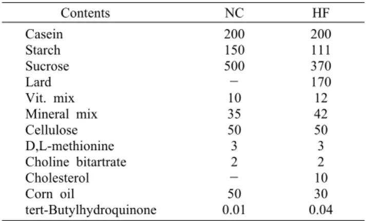
Table 1. Composition of the experimental diet Contents NC HF
Casein Starch Sucrose Lard Vit. mix Mineral mix Cellulose D,L-methionine Choline bitartrate Cholesterol Corn oil
tert-Butylhydroquinone
200 150 500
- 10 35 50 3 2
- 50 0.01
200 111 370 170 12 42 50 3 2 10 30 0.04 NC, normal control diet; HF, high fat diet.
Table 2. Four weeks exercise training protocol Group Exercise intensity HFLE
HFME HFHE
22 m/min, 60 min 26 m/min, 51 min 30 m/min, 46 min
HFLE, high fat diet+low-intensity exercise group; HFME, high fat diet+moderate-intensity exercise group; HFHE, high fat diet+
high-intensity exercise group.
및 근육 내 포도당 운반을 증가시켜 인슐린 저항성을 완화시 킨다고 보고하고 있다(16). 운동과 관련된 선행연구에서 지 구성 트레이닝이 골격근 내 PGC-1α 발현을 증가시키지 않 는다고 보고되고 있으나(17,18), 이러한 트레이닝에 의한 근육 내 PGC-1α 발현에 차이가 나는 것은 운동 형태, 운동 기간 그리고 운동 강도에 영향을 받을 것이라고 사료된다.
이처럼 운동과 PGC-1α에 대한 선행연구는 상당수 이루어 지고 있으나 운동 강도가 PGC-1α 발현과 골격근의 인슐린 저항에 미치는 효과에 대한 선행연구는 미비한 실정이다.
그러므로 장기간의 운동 시 운동 강도 차이가 골격근 내 PGC-1α, PPAR-γ, GLUT-4, mitochondrial enzyme content 및 인슐린 저항에 미치는 효과를 검증할 필요가 있 을 것으로 생각된다. 따라서 본 연구에서는 운동 강도 차이 가 고지방식이 Sprague-Dawley rat의 골격근 내 PGC-1α, PPAR-γ 및 인슐린 저항성에 미치는 영향을 알아보고자 한 다.
재료 및 방법
실험동물
본 연구는 4주령의 SD 수컷 쥐 40마리로 하였으며 쥐는 한 케이지(20.7×35×17 cm)에 1마리씩 넣어 사육하였다.
사육실의 온도는 21°C로 유지하였고 명기와 암기를 각각 12시간으로 조절하였다. 1주간의 환경 적응 기간을 거친 후 4주간 고지방 섭취와 유산소 운동을 실시하였다. 집단은 무 작위로 선정하여 비교집단(sedentary control group, SED), 고지방식이집단(high fat diet group, HF), 고지방식이+저 강도 운동집단(high fat diet+low-intensity exercise group, HFLE), 고지방식이+중강도 운동집단(high fat di- et+moderate-intensity exercise group, HFME), 고지방 식이+고강도 운동집단(high fat diet+high-intensity ex- ercise group, HFHE) 다섯 집단으로 구분하였다.
실험설계
식이는 AIN-76(Harlan Teklad, Indianapolis, IN, USA) 을 바탕으로 하여 제조한 사료를 제공하고 사료와 물은 자유 롭게 섭취하도록 하였다. 식이 조성표는 Table 1과 같다.
운동은 전동식 동물용 트레드밀(Quinton Instrument Co., Seattle, WA, USA)을 이용하여 8% 경사도에서 4주간 주당 6회의 지구성 운동을 실시하였다. 운동집단은 1주 동안 15 m/min 30분으로 운동 적응 훈련을 실시한 후 트레드밀 속도 와 지속시간을 점증적으로 증가시켜 4주 후에는 서로 다른 운동 강도에서 달리도록 하였다(19). 본 연구의 운동 프로토 콜은 Table 2와 같다. 모든 집단은 체중 100 g당 6.5 mg의 sodium pentobarbital(Hanlim Pharm. Co., Ltd., Yongin, Korea)을 복강 내 주사로 마취한 다음 근조직을 적출하였 다. 적출한 근조직은 GLUT-4, PGC-1α, PPAR-γ, GAPDH 분석 전까지 액체질소에 냉각시켜 -70°C(DF-9007, IlShin
Lab. Co., Busan, Korea)에 보관하였다. 가자미근(soleus muscle)은 포도당 운반률을 측정하기 위해 즉시 배양하였 다.
혈당내성검사(oral glucose tolerance test, OGTT) 4주간의 처치 후 12시간의 금식을 거쳐 혈당내성검사를 실시하였다. 안정 시 혈액은 각각의 케이스에 쥐를 넣고 안 정을 취한 후 꼬리 끝을 0.1 cm 이하로 잘라 500 μL 혈액을 채취하였다. 안정 시 혈액 채취한 쥐는 50%의 포도당 용액 을 체중당 1 g/kg으로 경구투여 하고 포도당 투여 30분과 60분 후에 500 μL의 혈액을 채취하였다. 채취된 혈액은 응 고를 방지하기 위해 50 μL의 헤파린(Choonawae Pharma.
Co., Jeonbuk, Korea)을 이용해 처리하였고 원심분리 하여 혈당과 인슐린 분석 전까지 -70°C 초저온 냉장고(VS-550, Vision Scientific Co., Ltd., Bucheon, Korea)에 보관하였 다.
혈중 지질(plasma lipids)
혈중 총콜레스테롤(total cholesterol: TC), 중성지방 (triglyceride: TG) 및 고밀도 지단백 콜레스테롤(high- density lipoprotein cholesterol, HDL-C)은 진단용 시약 (Asan Pharma. Co., Gyeonggi, Korea)을 이용해 효소분석 법으로 측정하였다.
혈당(plasma glucose)과 인슐린(insulin)
혈당은 자동 혈당분석기 YSI-2300(Yellow Springfield Instruments Co., Yellow Springs, OH, USA)를 이용하여
측정하였고, 혈중 인슐린은 진단용 시약(Lincoplex Re- search, Inc., St. Louis, MO, USA)을 이용하여 방사선 면역 측정법(radioimmunoassay)으로 분석하였다.
구연산 합성효소(citrate synthase, CS)
구연산 합성효소 분석을 위해 근육(plantaris muscle)을 차가운 완충액(100 mM Tris-HCl buffer pH 8.1)과 함께 1:30의 비율로 혼합한 다음 polytron homogenizer(Kin- ematica, Littau, Switzerland)로 균질화하였다. 균질화된 근조직은 0.4%의 Triton X-100 buffer(pH 8.1)를 이용하 여 1:9로 희석시켰다. 분광광도계(spectrophotometer)와 큐벳(cuvette)을 37°C 유지한 후 시약(100 mM Tris-HCl pH 8.1, 1 mM DTNB, 0.03 mg acetyl CoA)과 희석한 근조 직을 첨가한 후 1∼3분간 방치한 다음 10 mM ox- aloacetate를 첨가하여 흡광도 412 nm에서 1분 간격으로 5∼9분간 citrate synthase activity를 측정하였다(20).
Muscle homogenization
족척근은 차가운 완충액(20 mM tris-HCl pH 7.4, 1%
Triton X-100, 50 mM NaCl, 250 mM sucrose, 5 mM Na4P2O7, 50 mM NaF, 0.5 mM PMSF, 2 mM DTT, 0.1 mM benzamidine, 4 mg/L leupeptin, 50 mg/L trypsin in- hibitor)과 함께 1:20 비율로 혼합한 다음 polytron homog- enizer(Kinematica)로 균질화하였다. 균질화한 근조직과 기준액은 16,000×g에서 4°C, 40분간 원심분리(VS-550, Vision Scientific Co., Ltd., Bucheon, Korea) 하여 상층액 을 분리하였다.
Western blotting
균질화된 근육(plantaris musle)의 상층액은 DC protein assay(Bio Rad Laboratories, Hercules, CA, USA)를 이 용하여 단백질 정량을 하였다. Laemmli sample buffer (125 mM tris, 20% glycerol, 2% SDS, 0.008% bromo- phenol blue, pH 6.8)로 1:2 희석하여 95°C에서 5분간 중탕 한 후 22,000×g에서 1분간 원심분리 하여 상층액을 분리하 였다. 단백질을 함유한 상층액을 10% SDS-polyacryl- amide gel electrophoresis(PAGE)에 주입한 다음 전기영 동(Mini Trans-Blot Electrophoretic Transfer Cell, Bio Rad Laboratories)한 후 transfer buffer를 이용하여 PVDF membrane으로 이동시켰다. Membrane은 T-TBS (3번×10분)로 세척하고 10% skim milk를 이용하여 60분 간 blocking한 후 1차 항체(sc-1608; goat polyclonal an- ti-GLUT4, sc-13067; rabbit polyclonal anti-PGC-1α, sc-7196; rabbit polyclonal anti-PPAR-γ, sc-20357;
goat polyclonal anti-GAPDH, Santa Cruz Biotechnol- ogy, Santa Cruz, CA, USA, 1:1,000 dilution)에 넣어 배양 (4°C with gentle shaking, overnight)하였다. T-TBS를 이용하여 세척한 후 membrane은 2차 항체(81-1620:
ZYMED Laboratories, South San Francisco, CA, USA;
#7074: Cell Signaling Technology, Beverly, MA, USA, 1:2,000 dilution)와 함께 60분 동안 배양하고 다시 세척하 였다. Protein은 X-ray film에 ECL Western blot de- tection kit(RPN2106, Amersham, Arlington Heights, IL, USA)를 사용하여 시각화하였다. X-ray film 시각화된 밴드 는 스캔 후 Image J(http://imagej.nih.gov/ij/, Image Proc- essing and Analysis in Java) 프로그램을 이용하여 densi- tometry analysis(gel internal density and gel peak analysis) 방법으로 분석하였다.
포도당 운반률(muscle glucose transport rate) 포도당 운반률은 비대사물질인 glucose analog 2-de- oxy-D-glucose(2-DG)(American Radiolabeled Chemicals Inc., St. Louis, MO, USA)를 이용하여 측정하였다(21). 측 정을 위해 좌우측 가자미근을 적출하였고 즉시 근육 내 glu- cose를 제거하기 위해 KHB(32 mM mannitol, 8 mM glu- cose, 0.1% BSA)에 60분간 세척하였다. 세척 후 32 mM mannitol, 8 mM glucose, 0.1% BSA(1,000 μIU/mL in- sulin, absence or presence)가 함유된 KHB에 20분간 배 양하였다. 다시 10분간 36 mM mannitol, 2 mM pyruvate, 0.1% BSA에 세척 후 deoxy-D-glucose, 2-[1,2-3H](2.5 μCi/mL)와 16 mM[14C] mannitol(0.3 μCi/mL)이 함유된 1.5 mL KHB(36 mM mannitol, 4 mM deoxy glucose, 0.1% BSA)(1,000 μIU/mL insulin, absence or presence) 에 30°C, 20분간 하였다. 세포 내외의 2-DG의 활성도는 scintillation counter(Wallac 1220 QUANTULUS, Perkin- Elmer Inc., Waltham, MA, USA)를 이용하여 10분간 측정 하였다.
통계처리
각 측정항목에 대한 결과는 평균과 표준오차(mean±SE) 를 산출하였으며 통계적 분석은 SPSS 18.0 통계프로그램 (SPSS Inc., Chicago, IL, USA)을 이용하였다. 통계처리는 집단 간 처치 후 변화를 알아보기 위해 일원분산분석법 (One-way ANOVA)을 이용하였고, 집단 간 유의성은 Duncan's multiple range test를 실시하여 P<0.05 수준에 서 비교하였다.
결과 및 고찰
체중의 변화
체중의 변화는 1일 1회 운동 2시간 전 저울(METTER PJ6, Metter Toledo International Inc., Seoul, Korea)을 이용하여 쥐의 체중을 측정하였으며 4주간의 실험 처치 후 집단의 체중의 변화는 Table 3과 같다. 체중은 고지방식이 집단(HF)이 비교집단(SED)에 비해 유의하게 증가한 것으 로 나타났으며, 운동집단(HFLE, HFME, HFHE)은 고지방
Table 3. Changes of body weight (g)
Group Pre Post
SED HF HFLE HFME HFHE
88.6±1.8 88.6±1.7 88.4±1.6 88.2±1.6 88.3±1.6
228.2±2.6a 252.6±2.7b 231.6±2.7a 237.0±4.1a 234.1±3.4a SED, sedentary control group; HF, high fat diet group; HFLE, high fat diet+low-intensity exercise group; HFME, high fat diet+
moderate-intensity exercise group; HFHE, high fat diet+high- intensity exercise group.
Values are means±SE (n=8). Values with different superscripts are significantly different (P<0.05).
Table 4. Fasting plasma lipids after 4 weeks exercise (mg/dL)
SED HF HFLE HFME HFHE
TC TG HDL-C
64.6±3.1a 62.1±2.5b 30.8±1.7b
80.6±4.2b 82.4±8.7c 25.0±1.0a
68.8±3.3a 46.2±2.7a 29.6±3.8c
70.3±2.4a 48.8±2.0a 30.4±0.8c
68.8±2.6a 46.8±2.0a 33.3±1.1d SED, sedentary control group; HF, high fat diet group; HFLE, high fat diet+low-intensity exercise group; HFME, high fat diet+moderate-intensity exercise group; HFHE, high fat diet+high-intensity exercise group.
TC, total cholesterol; TG, triglyceride; HDL-C, high-density lipoprotein cholesterol.
Values are means±SE (n=8).
Values with different superscripts in the same row are significantly different (P<0.05).
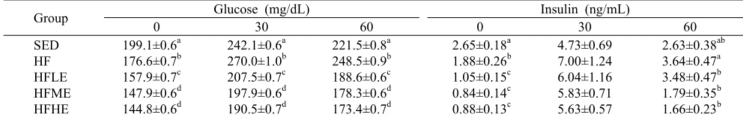
Table 5. Plasma glucose insulin during OGTT
Group Glucose (mg/dL) Insulin (ng/mL)
0 30 60 0 30 60
SED HF HFLE HFME HFHE
199.1±0.6a 176.6±0.7b 157.9±0.7c 147.9±0.6d 144.8±0.6d
242.1±0.6a 270.0±1.0b 207.5±0.7c 197.9±0.6d 190.5±0.7d
221.5±0.8a 248.5±0.9b 188.6±0.6c 178.3±0.6d 173.4±0.7d
2.65±0.18a 1.88±0.26b 1.05±0.15c 0.84±0.14c 0.88±0.13c
4.73±0.69 7.00±1.24 6.04±1.16 5.83±0.71 5.63±0.57
2.63±0.38ab 3.64±0.47a 3.48±0.47b 1.79±0.35b 1.66±0.23b SED, sedentary control group; HF, high fat diet group; HFLE, high fat diet+low-intensity exercise group; HFME, high fat diet+
moderate-intensity exercise group; HFHE, high fat diet+high-intensity exercise group.
Values are means±SE (n=8).
Values with different superscripts in a same column are significantly different (P<0.05).
식이집단(HF)에 비해 유의하게 낮게 나타났다(F=9.112, P=0.000). 본 연구 결과 고지방식이집단이 일반식이집단과 운동집단에 비해 체중이 높은 것으로 보아 고지방식이로 인 하여 비만이 유도된 것을 알 수 있었으며, 지구성 운동이 고지방식이로 증가한 체중을 감소시키는데 효과적인 것으 로 나타났다.
혈중 지질의 농도
4주간의 처치 후 혈중 TC의 농도는 고지방식이집단(HF) 이 비교집단(SED)에 비해 유의하게 증가한 것으로 나타났 으며 운동집단(HFLE, HFME, HFHE)은 고지방식이집단 (HF)에 비해 유의하게 낮게 나타났다(F=3.051, P=0.017).
HDL-C의 농도는 고지방식이집단이 일반식이집단에 비해 유의하게 낮게 나타났으며, 세 운동집단은 고지방식이집단 에 비해 유의하게 증가한 것으로 나타났다(F=5.899, P= 0.001). 혈중 TG의 농도는 고지방식이집단이 비교집단
(SED)에 비해 유의하게 증가한 것으로 나타났으며, 운동집 단(HFLE, HFME, HFHE)은 비교집단과 고지방식이집단 (HF)에 비해 유의하게 낮게 나타났다(F=7.256, P=0.000, Table 4). 본 연구 결과 4주간의 고지방식이와 운동 처치 후 혈중 TC와 TG는 고지방식이에 비해 일반식이와 운동집 단이 유의하게 감소한 것으로 나타났으며, 운동집단의 혈중 TG는 비교집단(SED)에 비해 유의하게 낮은 것으로 나타났 다. 또한 HDL-C는 고지방식이집단에 비해 운동집단이 유 의하게 증가한 것을 알 수 있었다. 이는 고지방식이가 혈중 지질(TC, TG)을 증가시키는 것으로 생각되며 선행연구의 결과처럼 지구성 운동이 혈중 지질을 개선시키는데 효과적 인 것으로 사료된다(22). 최근 임상 실험에서는 고강도 운동 이 저강도와 중강도 운동에 비해 혈중 지질을 감소시키는데 효과적인 것으로 보고하고 있다(23,24). 반면 Jang 등(25) 은 SD 수컷 쥐를 대상으로 일회성 저강도, 고강도 운동을 실시한 결과 운동 강도에서는 혈중 지질의 유의한 차이가 없는 것으로 나타났다. 본 연구에서는 4주간 다른 강도의 지구성 운동을 실시한 결과 운동 집단 간 차이가 없는 것으 로 나타났다(Table 4). 이러한 결과는 선행연구와 일치하며 (25) 혈중 지질 개선은 트레이닝의 강도보다는 운동 기간, 운동량이 중요한 요소라 생각된다.
혈당내성검사
4주간의 처치 후 혈당내성검사의 혈당과 인슐린 농도의 변화는 Table 5와 같다. 포도당 투여 전(0) 공복 시 혈당의 농도는 비교집단에 비해 고지방식이와 운동집단 모두 유의 하게 낮게 나타났다(F=37.890, P=0.000). 또한 세 운동집 단은 HF에 비해 유의하게 낮게 나타났으며, HFHE는 HFLE
0.0 2.0 4.0 6.0 8.0 10.0 μmol/g/min
SED HF HFLE HFME HFHE b
c
a a ab
Fig. 1. Citrate synthase activity in plantaris muscle. SED, seden- tary control group; HF, high fat diet group; HFLE, high fat di- et+low-intensity exercise group; HFME, high fat diet+moder- ate-intensity exercise group; HFHE, high fat diet+high-intensity exercise group. Values are means±SE (n=8). Values with differ- ent letters on bars are significantly different (P<0.05).
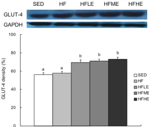
SED HF HFLE HFME HFHE GLUT-4
GAPDH
0 20 40 60 80 100
GLUT-4 density (%) .
SED HF HFLE HFME HFHE
b b b
a a
Fig. 2. Expression of GLUT-4 in plantaris muscle. SED, seden- tary control group; HF, high fat diet group; HFLE, high fat di- et+low-intensity exercise group; HFME, high fat diet+moder- ate-intensity exercise group; HFHE, high fat diet+high-intensity exercise group. Values are means±SE (n=8). Values with differ- ent letters on bars are significantly different (P<0.05).
에 비해서도 유의하게 낮게 나타났다(P<0.05). 그러나 포도 당 투여 후 30분과 60분에서는 SED에 비해 HF가 유의하게 높은 것으로 나타났으나 세 운동집단 모두 SED와 HF에 비 해 낮게 나타났다(P<0.05). 혈중 인슐린의 농도에서는 포도 당 투여 전(0) 비교집단과 비교하여 고지방식이집단과 운동 집단에서 유의하게 낮게 나타났으며 세 운동집단은 HF에 비해서도 유의하게 낮게 나타났다(F=19.462, P=0.000). 포 도당 투여 후 30분에서는 집단 간 유의한 차이가 없는 것으 로 나타났으나 투여 후 60분에서는 HF에 비해 HFME와 HFHE가 유의하게 낮은 것으로 나타났다(P<0.05). 본 연구 에서는 포도당 투여 전 혈당과 인슐린 농도에서 고지방식이 가 일반식이집단에 비해 유의하게 감소한 것으로 나타났다.
이것은 고지방식이집단의 공복 시 인슐린과 혈당이 낮으므 로 인슐린 저항이 개선된 것으로 생각할 수 있으나 실제 포 도당 투여 후 30분과 60분에서는 고지방식이가 일반식이집 단보다 유의하게 높은 것을 알 수 있었다. 그러므로 본 연구 의 4주간의 고지방식이 처치는 실제 쥐의 whole body 인슐 린 저항을 증가시키는 것으로 사료되며 이러한 결과는 본 연구와 실험방법이 유사한 최근 2013년 발표된 Bielohu- by 등(26)의 연구 결과와 일치한다. 본 연구 결과 세 운동집 단(HFLE, HFME, HFHE)에서 식이집단(SED, HF)에 비해 인슐린 그리고 혈당이 유의하게 감소하는 것을 알 수 있었 다. 이 결과는 선행연구에서처럼 지구성 운동이 골격근의 인슐린 민감도를 증가시켜 인슐린 저항성을 개선시키는 결 과로 사료된다.
구연산 합성효소의 활성도(citrate synthase activity) 4주간의 처치 후 구연산 합성효소의 활성도는 SED(5.7±
0.2 μmol/g/min), HF(5.9±0.2 μmol/g/min)에 비해 HFME (6.7±0.2 μmol/g/min)에서 유의하게 높게 나타났으며, HFHE (8.4±0.5 μmol/g/min)는 다른 네 집단에 비해 유의하게 높 게 나타났다(F=16.532, P=0.000, Fig. 1). 그러나 SED, HF,
HFLE에서는 집단 간 유의한 차이가 없는 것으로 나타났다.
본 연구 결과는 장기간의 트레이닝과 높은 강도의 운동은 트레이닝 적응(skeletal muscle adaptation) 현상이 나타난 다는 선행연구와 일치하는 것으로(27,28) 본 연구에 사용된 운동 강도가 연구목적에 합당한 것으로 생각된다.
골격근의 GLUT-4의 발현량
4주간의 실험 처치 후 GLUT-4의 발현량은 비교집단과 비교하여 고지방식이집단에서는 유의한 차이가 없는 것으로 나타났으나(P=0.980) 운동집단(HFLE: 69.4±3.0%, HFME:
71.2±1.8%, HFHE: 73.1±2.1%)은 두 식이집단(SED: 56.1
±2.1%, HF: 57.8±1.8%)에 비해 유의하게 높게 나타났다 (F=12.973, P=0.000, Fig. 2). 지구성 운동은 인슐린 수용기 의 활성도와 GLUT-4를 증가시키고 인슐린의 민감도와 골 격근 내 혈당 흡수를 개선시켜 골격근 내 인슐린 저항을 감 소시킨다(29-31). 본 연구 결과 운동집단에서는 골격의 GLUT-4 발현이 증가한 것을 알 수 있었다. 이는 장기간의 지속적인 지구성 운동이 근육의 인슐린 저항성을 감소시킨 다는 선행연구들과 일치하였으며(29-31) 장기간의 지구성 운동이 세포막의 GLUT-4 이동(translocation)과 골격근 내 GLUT-4 발현을 증가(overexpression)시킨 것으로 생각 된다(31,32). Koshinaka 등(33)은 일회성 고강도 운동과 저 강도 운동을 실시한 결과 total GLUT-4 content는 서로 다른 운동 강도에서 유의한 차이가 없는 것으로 나타났다.
반면 Hansen 등(34)은 수영 운동(acute exercise) 후 근육 내 total GLUT-4 contents에는 비교집단과 유의한 차이가 없었으나 세포표면의 GLUT-4 이동에서는 비교집단과 비 교하여 운동집단이 유의하게 증가한 것으로 보고하였다. 본 연구 결과 4주간의 지구성 운동은 근육 내 total GLUT-4
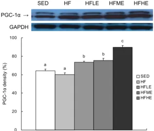
SED HF HFLE HFME HFHE PGC-1α →
GAPDH
0 20 40 60 80 100
PGC-1α density (%) .
SED HF HFLE HFME HFHE
b b
c
a a
Fig. 3. Expression of PGC-1α in plantaris muscle. SED, seden- tary control group; HF, high fat diet group; HFLE, high fat di- et+low-intensity exercise group; HFME, high fat diet+moder- ate-intensity exercise group; HFHE, high fat diet+high-intensity exercise group. Values are means±SE (n=8). Values with differ- ent letters on bars are significantly different (P<0.05).
발현을 증가시키는 것으로 나타났으나 운동 강도의 차이에 서는 유의한 차이가 없는 것을 알 수 있었다. 이는 선행연구 가 뒷받침해주나(34) 일회성 운동으로 운동 강도, 운동량, 운동 기간에서 본 연구와 차이가 있는 것을 알 수 있다. 그러 므로 추후 본 연구 결과를 바탕으로 고지방식이와 지구성 트레이닝 방법(운동 강도, 운동량, 운동 기간 등)을 고려하여 세포표면의 GLUT-4 이동에 대한 연구가 필요할 것으로 생 각된다.
골격근의 PGC-1α의 발현량
4주간의 실험 처치 후 PGC-1α의 발현량은 비교집단 (SED)과 비교하여 고지방식이집단(HF)에서는 유의한 차이 가 없는 것으로 나타났다(P=0.554). 운동집단(HFLE: 73.6
±1.0%, HFME: 75.4±2.3%, HFHE: 89.7±2.0%)에서는 SED(64.2±1.9%)와 HF(60.3±1.7%)에 비해 유의하게 높 게 나타났으며 고강도 운동집단(HFHE)은 저강도(HFLE)와 중강도 운동집단(HFME)에 비해서도 유의하게 높게 나타났 다(F=38.800, P=0.000, Fig. 3). 선행연구에 의하면 영양과 잉, 신체활동 부족 및 비만은 골격근 내 PGC-1α의 발현을 감소시키고 미토콘드리아의 기능을 약화시킨다고 보고하고 있다(6,7,35). 또한 골격근의 PGC-1α의 감소는 미토콘드리 아 유전자의 전사를 감소시켜 탄수화물과 지방 산화 감소, 근육 내 지방 축적과 함께 골격근의 인슐린 저항성과 제2형 당뇨병을 증가시킨다고 보고하고 있다(6,36). 본 연구 결과 4주의 고지방 섭취 후 골격근 내 PGC-1α는 일반식이에 비 해 고지방식이집단이 6.2% 감소한 것으로 나타났으나 통계 적인 유의한 차이가 없는 것으로 나타났다. 이는 본 연구의 혈당내성검사에서 알 수 있듯이 4주간 고지방 섭취는 whole body 인슐린 저항을 발생시키는 것으로 생각되나 골격근
내 인슐린 저항과 PGC-1α 발현에 영향을 미치기에는 부족 한 것으로 생각되며 추후 좀 더 장기간의 연구가 필요할 것 으로 생각된다.
지구성 운동은 구연산 합성효소를 증가시키고 미토콘드 리아의 수와 크기를 증가시킨다(37,38). 또한 트레이닝에 의한 골격근의 적응은 PGC-1α mRNA와 근육 내 PGC-1α 단백질 발현에 영향을 미치며(38-41), Terada 등(42)은 고 강도 운동과 저강도 운동이 골격근의 citrate synthase(CS) 를 증가시키며, 지속적인 운동은 PGC-1α 발현을 증가시킨 다고 보고하였다. Goto 등(41)은 3일간 2시간의 수영을 시 킨 결과 PGC-1α mRNA를 증가시킨다고 보고하였다. 또한 21∼87세 남녀를 대상으로 16주간 유산소 운동을 실시한 결과 citrate synthase, PGC-1α mRNA가 증가하였다고 보 고하였다(39). 본 연구 결과 고지방을 섭취시킨 운동집단에 서는 일반식이와 고지방식이집단에 비해 PGC-1α가 유의하 게 증가한 것으로 나타났다. 이는 트레이닝에 의한 골격근 내 미토콘드리아의 증가가 PGC-1α 발현을 증가시킨다는 연구와 일치한다. 또한 본 연구에서는 고강도 운동집단은 다른 운동집단에 비해 유의하게 증가한 것을 알 수 있었다.
이는 본 연구의 citrate synthase의 결과(Fig. 1)와 선행연 구의 결과에서 알 수 있듯이(41) 4주간의 높은 강도의 지구 성 운동이 저강도 운동에 비해 미토콘드리아(mitochondrial enzyme content)를 증가시켜 골격근 내 PGC-1α를 발현을 증가시킨 것으로 생각된다.
골격근의 PPAR-γ의 발현량
4주간의 실험 처치 후 PPAR-γ 발현량은 비교집단과 비 교하여 고지방식이집단에서는 유의한 차이가 없는 것으로 나타났으나(P=0.966) 운동집단(HFLE: 147.4±5.6%, HFME:
149.3±5.2%, HFHE: 146.3±3.0%)에서는 비교집단(137.7
±3.2%)과 고지방식이집단(129.7±4.3%)에 비해 유의하게 높게 나타났다(F=4.159, P=0.007, Fig. 4). PPAR-γ는 탄 수화물과 지방산 산화대사 및 지방산 합성에 관여하는 것으 로 알려져 있다(43). 선행연구에 의하면 근육 내 PPAR-γ 활 성화는 골격근의 유리지방산 흡수와 근육 내 지방을 감소시 키고 결과적으로 인슐린 민감도를 증가시킨다고 보고하고 있으며(6), 장기간의 지구성 운동은 혈중 유리지방산과 근육 내 지질 축적을 감소시킨다고 보고하고 있다(22). Jo 등(44) 은 일회성 운동과 12주간 지구성 운동을 실시한 결과 골격 근의 PPAR-γ 발현이 증가한 것으로 나타났으며, 두 운동집 단에서는 유의한 차이가 없는 것으로 나타났다. 본 연구에서 는 4주간의 고지방 섭취와 지구성 운동을 실험한 결과 식이 집단에서는 통계적으로 유의한 차이가 없는 것으로 나타났 으나 운동집단에서는 식이집단에 비해 세 집단 모두 유의하 게 증가한 것을 알 수 있었다. 이는 선행연구의 결과와 일치 하며(44), 장기간의 지구성 운동이 골격근의 PPAR-γ 발현 을 증가시켜 지방대사를 증가시키고 골격근의 인슐린 저항 성을 감소시킨 것으로 생각된다. 이 결과는 본 연구의 혈당내
0.0 2.0 4.0 6.0 8.0
SED HF HFLE HFME HFHE SED HF HFLE HFME HFHE Submaximal insulin concentration Non insulin condition μmol/mL/hr
b b
c
a a
Fig. 5. Glucose transport rate under submaximal insulin concen- trations and non-insulin concentrations in soleus muscle. SED, sedentary control group; HF, high fat diet group; HFLE, high fat diet+low-intensity exercise group; HFME, high fat diet+mod- erate-intensity exercise group; HFHE, high fat diet+high-in- tensity exercise group. Values are means±SE (n=8). Values with different letters on bars are significantly different (P<0.05).
SED HF HFLE HFME HFHE PPAR-γ
GAPDH
0 40 80 120 160 200
PPAR-γ density (%) .
SED HF HFLE HFME HFHE
b b
b
a a
Fig. 4. Expression of PPAR-γ in plantaris muscle. SED, seden- tary control group; HF, high fat diet group; HFLE, high fat di- et+low-intensity exercise group; HFME, high fat diet+moder- ate-intensity exercise group; HFHE, high fat diet+high-intensity exercise group. Values are means±SE (n=8). Values with differ- ent letters on bars are significantly different (P<0.05).
성검사의 혈당과 인슐린의 결과가 뒷받침해준다(Table 5).
골격근 포도당 운반률의 변화
4주간의 실험 처치 후 non-insulin condition과 sub- maximal 인슐린 농도(1,000 μIU/mL)에서 골격근 내 포도 당 운반률을 측정하였다(45,46). Submaximal condition에 서 근육 내 포도당 운반률은 비교집단(SED)과 비교하여 고 지방식이집단(HF)에서는 유의한 차이가 없는 것으로 나타 났다(P=0.966). 운동집단(HFLE: 4.7±0.3 μmol/mL/hr, HFME: 5.0±0.3 μmol/mL/hr, HFHE: 5.5±0.3 μmol/mL/
hr)은 SED(4.1±0.2 μmol/mL/hr)와 HF(3.7±0.2 μmol/
mL/hr)에 비해 유의하게 높게 나타났으며, HFHE가 HFLE
와 HFME에 비해서도 유의하게 높게 나타났다(F=8.329, P=0.000, Fig. 5). 그러나 non-insulin condition에서는 집 단 간 유의한 차이가 없는 것으로 나타났다(F=1.372, P= 0.264). 고지방 섭취는 혈중 유리지방산을 증가시키고 근육 의 지질 축적을 증가시킨다(22). 근육 내 지질 축적은 세포 내 glucose-fatty acid를 통해 지방 산화를 증가시키고 근육 내 혈당 이동을 감소시켜 인슐린 저항성을 증가시킨다고 보 고하고 있다(47). 본 연구 결과 4주간의 고지방식이 섭취에 서는 일반식이와 비교하여 유의한 차이가 없는 것으로 나타 났다. 그러므로 본 연구의 4주간 고지방식이는 whole body 의 인슐린 저항성을 발생시켰으나 근육 내 혈당 이동(glucose uptake)에는 영향을 미치는 못한 것으로 사료된다. 그러므 로 추후 고지방식이의 함량, 섭취 기간 등을 고려한 연구가 필요하다고 사료된다. 선행연구에 의하면 고강도 운동이 저 강도 지구성 운동에 비해 근육 내 혈당 이동을 증가시킨다고 보고하고 있다(33,46,48,49). Koshinaka 등(49)은 Wistar rat을 대상으로 일회성 간헐적 지구성 운동(swimming)을 실시한 결과 운동 후 고강도 운동에서 저강도 운동에 비해 근육 내 AMP-activated protein kinase(AMPK), acetyl coenzyme A protein kinase(ACC), p-38 mitogen-acti- vated protein kinase의 활성화를 증가시키고 근육 내 혈당 이동을 증가시킨다고 보고하였으며, 비만 쥐를 대상으로 한 연구에서는 7주간의 지구성 운동(treadmill)을 실시한 결과 고강도 운동이 저강도 운동에 비해 근육 내 포도당 운반률이 증가한 것으로 보고하고 있다(46). 반면 Terada 등(50)은 일회성 수영 운동(intermittent exercise and intermittent prolonged exercise)을 실시한 결과 운동 강도에서는 유의 한 차이가 없다고 보고하였다. 본 연구 결과 세 운동집단의 포도당 운반률이 증가한 것으로 나타났으며 운동 강도가 높 을수록 근육 내 혈당 이동을 유의하게 증가시킨 것을 알 수 있었다. 이는 선행연구의 결과처럼 지구성 고강도 운동이 저강도 운동에 비해 근육 내 AMPK와 ACC phosporylation 을 증가시키고 AMPK 활성화를 증가시켜 insulin inde- pendent glucose uptake를 향상시키는데 더 효과적인 것 으로 생각된다(32,48). 그러므로 본 연구 결과를 바탕으로 운동 강도에 따른 골격근 내 인슐린 신호체계(down stream signaling) 및 혈당이동에 대한 세부적인 연구가 필요할 것 으로 생각된다.
요 약
본 연구에서는 4주간의 고지방식이와 지구성 운동이 골격근 의 PGC-1α, PPAR-γ 및 인슐린 저항성(glucose uptake, GLUT-4)에 미치는 영향을 분석하였다. 인슐린 민감도 지 표인 혈당내성검사에서는 일반식이와 비교하여 고지방식이 에서 포도당 투여 후 30분과 60분에서 유의하게 증가하였으 며, 운동집단에서는 일반식이와 고지방식이집단에 비해 유 의하게 감소한 것을 알 수 있었다. 골격근의 포도당 운반률,
PGC-1α, GLUT-4, PPAR-γ의 결과에서는 일반식이에 비 해 고지방식이집단에서 감소하는 경향이 나타났으나 통계 적으로 유의한 차이가 없는 것으로 나타났다. 그러나 운동집 단(저/중/고강도운동)에서는 일반/고지방식이집단과 비교 하여 유의하게 증가한 것으로 나타났다. 운동집단의 운동 강도 차이에서 GLUT-4와 PPAR-γ는 집단 간 유의한 차이 가 없는 것으로 나타났다. 그러나 골격근의 포도당 운반률과 PGC-1α 단백질 발현은 저/중강도 운동과 비교하여 고강도 운동이 유의하게 증가한 것을 알 수 있었다. 이상의 결과를 종합해 볼 때 4주간의 고지방식이는 whole body의 인슐린 저항성을 발생시켰으나 근육 내 인슐린 저항에는 영향을 미 치는 못한 것으로 사료된다. 그러므로 추후 고지방식이의 함량, 섭취 기간 등을 고려한 연구가 필요할 것으로 생각된 다. 또한 4주간의 지속적인 지구성 운동이 고지방식이로 인 해 발생된 골격근 인슐린 저항성을 감소시키는데 효과적인 으로 나타났으나 운동 강도에 따른 골격근의 포도당 운반률, PGC-1α, GLUT-4, PPAR-γ의 변화가 인슐린 저항성이 개 선시켰다고 설명하기는 부족한 것으로 판단된다. 그러므로 추후 본 연구의 결과를 바탕으로 운동 형태(운동 기간, 운동 강도)에 따른 골격근의 PGC-1α와 insulin signalling path- way에 대한 세부적인 연구가 필요할 것으로 생각된다.
감사의 글
이 논문은 2011년도 정부(교육부)의 재원으로 한국연구재단 의 지원을 받아 연구되었으며(NRF-2011-327-G00117) 지원에 감사드립니다.
REFERENCES
1. Lee JS, Pinnamaneni SK, Eo SJ, Cho IH, Pyo JH, Kim CK, Sinclair AJ, Febbraio MA, Watt MJ. 1985. Saturated, but not n-6 polyunsaturated, fatty acids induce insulin resist- ance: role of intramuscular accumulation of lipid metabo- lites. J Appl Physiol 100: 1467-1474.
2. Mullen KL, Pritchard J, Ritchie I, Snook LA, Chabowski A, Bonen A, Wright D, Dyck DJ. 2009. Adiponectin resistance precedes the accumulation of skeletal muscle lipids and in- sulin resistance in high-fat-fed rats. Am J Physiol Regul Integr Comp Physiol 296: R243-251.
3. Mullen KL, Smith AC, Junkin KA, Dyck DJ. 2007. Globular adiponectin resistance develops independently of impaired insulin-stimulated glucose transport in soleus muscle from high-fat-fed rats. Am J Physiol Endocrinol Metab 293: E83- 90.
4. Zderic TW, Davidson CJ, Schenk S, Byerley LO, Coyle EF.
2004. High-fat diet elevates resting intramuscular triglyceride concentration and whole body lipolysis during exercise. Am J Physiol Endocrinol Metab 286: E217-225.
5. Benton CR, Wright DC, Bonen A. 2008. PGC-1alpha-medi- ated regulation of gene expression and metabolism: im- plications for nutrition and exercise prescriptions. Appl Physiol Nutr Metab 33: 843-862.
6. Handschin C, Spiegelman BM. 2008. The role of exercise
and PGC1α in inflammation and chronic disease. Nature 454: 463-469.
7. Russell AP. 2005. PGC-1alpha and exercise: important part- ners in combating insulin resistance. Curr Diabetes Rev 1:
175-181.
8. Russell AP, Hesselink MK, Lo SK, Schrauwen P. 2005.
Regulation of metabolic transcriptional co-activators and transcription factors with acute exercise. FASEB J 19: 986- 988.
9. Kelly DP, Scarpulla RC. 2004. Transcriptional regulatory circuits controlling mitochondrial biogenesis and function.
Genes Dev 18: 357-368.
10. Knutti D, Kralli A. 2001. PGC-1, a versatile coactivator.
Trends Endocrinol Metab 12: 360-365.
11. Puigserver P, Spiegelman BM. 2003. Peroxisome prolifer- ator-activated receptor-gamma coactivator 1 alpha (PGC-1 alpha): transcriptional coactivator and metabolic regulator.
Endocr Rev 24: 78-90.
12. Rhee J, Inoue Y, Yoon JC, Puigserver P, Fan M, Gonzalez FJ. 2003. Regulation of hepatic fasting response by PPAR gamma coactivator-1alpha (PGC-1): requirement for hepato- cyte nuclear factor 4alpha in gluconeogenesis. Proc Natl Acad Sci 100: 4012-4017.
13. Sriwijitkamol A, Ivy JL, Christ-Roberts C, DeFronzo RA, Mandarino LJ, Musi N. 2006. LKB1-AMPK signaling in muscle from obese insulin-resistant Zucker rats and effects of training. Am J Physiol Endocrinol Metab 290: E925-932.
14. De Filippis E, Alvarez G, Berria R, Cusi K, Everman S, Meyer C. 2008. Insulin-resistant muscle is exercise resistant:
evidence for reduced response of nuclear-encoded mitochon- drial genes to exercise. Am J Physiol Endocrinol Metab 294:
E607-614.
15. Patti ME, Butte AJ, Crunkhorn S, Cusi K, Berria R, Kashyap S, Miyazaki Y, Kohane I, Costello M, Saccone R, Landaker EJ, Goldfine AB, Mun E, DeFronzo R, Finlayson J, Kahn CR, Mandarino LJ. 2003. Coordinated reduction of genes of oxidative metabolism in humans with insulin resistance and diabetes: Potential role of PGC1 and NRF1. Proc Natl Acad Sci U S A 100: 8466-8471.
16. Matiello R, Fukui RT, Silva ME, Rocha DM, Wajchenberg BL, Azhar S, Santos RF. 2010. Differential regulation of PGC-1alpha expression in rat liver and skeletal muscle in response to voluntary running. Nutr Metab (Lond) doi:
10.1186/1743-7075-7-36.
17. Akimoto T, Pohnert SC, Li P, Zhang M, Gumbs C, Rosen- berg PB, Williams RS, Yan Z. 2005. Exercise stimulates Pgc-1alpha transcription in skeletal muscle through activa- tion of the p38 MAPK pathway. J Biol Chem 280: 19587- 19593.
18. Tunstall RJ, Mehan KA, Wadley GD, Collier GR, Bonen A, Hargreaves M, Cameron-Smith D. 2002. Exercise training increases lipid metabolism gene expression in human skel- etal muscle. Am J Physiol Endocrinol Metab 283: E66-72.
19. Garekani ET, Mohebbi H, Kraemer RR, Fathi R. 2011.
Exercise training intensity/volume affects plasma and tissue adiponectin concentrations in the male rat. Peptides 32:
1008-1012.
20. Srere PA. 1969. Citrate synthase. Methods Enzymol 13: 3-5.
21. Young DA, Uhl JJ, Cartee GD, Holloszy JO. 1986. Activa- tion of glucose transport in muscle by prolonged exposure to insulin. Effects of glucose and insulin concentrations. J Biol Chem 261: 16049-16053.
22. Jung HL, Kang HY. 2010. Effects of endurance exercise and high-fat diet on insulin resistance and ceramide contents of
skeletal muscle in Sprague-Dawley rats. J Korean Diabetes 34: 244-252.
23. Paoli A, Pacelli QF, Moro T, Marcolin G, Neri M, Battaglia G, Sergi G, Bolzetta F, Bianco A. 2013. Effects of high-in- tensity circuit training, low-intensity circuit training and en- durance training on blood pressure and lipoproteins in mid- dle-aged overweight men. Lipids Health Dis 12: 131.
24. Racil G, Ben Ounis O, Hammouda O, Kallel A, Zouhal H, Chamari K, Amri M. 2013. Effects of high vs. moderate exercise intensity during interval training on lipids and adi- ponectin levels in obese young females. Eur J Appl Physiol 113: 2531-2540.
25. Jang YS, Chae HW, Kang HY. 1999. Effects of low and high exercise training intensities on body composition of growing male rats. Korean J Obesity 8: 89-101.
26. Bielohuby M, Sisley S, Sandoval D, Herbach N, Zengin A, Fischereder M, Menhofer D, Stoehr BJ, Stemmer K, Wanke R, Tschöp MH, Seeley RJ, Bidlingmaier M. 2013. Impaired glucose tolerance in rats fed low-carbohydrate, high-fat diets. Am J Physiol Endocrinol Metab 305: E1059-1070.
27. Leek BT, Mudaliar SR, Henry R, Mathieu-Costello O, Richardson RS. 2001. Effect of acute exercise on citrate synthase activity in untrained and trained human skeletal muscle. Am J Physiol Regul Integr Comp Physiol 280: R441- 447.
28. Dudley GA, Abraham WM, Terjung RL. 1982. Influence of exercise intensity and duration on biochemical adapta- tions in skeletal muscle. J Appl Physiol Respir Environ Exerc Physiol 53: 844-850.
29. Gan SK, Kriketos AD, Ellis BA, Thompson CH, Kraegen EW, Chisholm DJ. 2003. Changes in aerobic capacity and visceral fat but not myocyte lipid levels predict increased insulin action after exercise in overweight and obese men.
Diabetes Care 26: 1706-1713.
30. Goodpaster BH, Katsiaras A, Kelley DE. 2003. Enhanced fat oxidation through physical activity is associated with im- provements in insulin sensitivity in obesity. Diabetes 52:
2191-2197.
31. Tanaka S, Hayashi T, Toyoda T, Hamada T, Shimizu Y, Hirata M, Ebihara K, Masuzaki H, Hosoda K, Fushiki T, Nakao K. 2007. High-fat diet impairs the effects of a single bout of endurance exercise on glucose transport and insulin sensitivity in rat skeletal muscle. Metabolism 56: 1719- 1728.
32. Ivy JL. 2004. Muscle insulin resistance amended with ex- ercise training: role of GLUT4 expression. Med Sci Sports Exerc 36: 1207-1211.
33. Koshinaka K, Kawasaki E, Hokari F, Kawanaka K. 2009.
Effect of acute high-intensity intermittent swimming on post-exercise insulin responsiveness in epitrochlearis muscle of fed rats. Metabolism 58: 246-253.
34. Hansen PA, Nolte LA, Chen MM, Holloszy JO. 1985.
Increased GLUT-4 translocation mediates enhanced insulin sensitivity of muscle glucose transport after exercise. J Appl Physiol 85: 1218-1222.
35. Holloway GP, Perry CG, Thrush AB, Heigenhauser GJ, Dyck DJ, Bonen A, Spriet LL. 2008. PGC-1alpha's relation- ship with skeletal muscle palmitate oxidation is not present
with obesity despite maintained PGC-1alpha and PGC-1beta protein. Am J Physiol Endocrinol Metab 294: E1060-1069.
36. Liang H, Ward WF. 2006. PGC-1alpha: a key regulator of energy metabolism. Adv Physiol Educ 30: 145-151.
37. Holloszy JO, Coyle EF. 1984. Adaptations of skeletal mus- cle to endurance exercise and their metabolic consequences.
J Appl Physiol Respir Environ Exerc Physiol 56: 831-838.
38. Baar K, Wende AR, Jones TE, Marison M, Nolte LA, Chen M, Kelly DP, Holloszy JO. 2002. Adaptations of skeletal muscle to exercise: rapid increase in the transcriptional co- activator PGC-1. FASEB J 16: 1879-1886.
39. Short KR, Vittone JL, Bigelow ML, Proctor DN, Rizza RA, Coenen-Schimke JM. 2003. Impact of aerobic exercise train- ing on age-related changes in insulin sensitivity and muscle oxidative capacity. Diabetes 52: 1888-1896.
40. Terada S, Goto M, Kato M, Kawanaka K, Shimokawa T, Tabata I. 2002. Effects of low-intensity prolonged exercise on PGC-1 mRNA expression in rat epitrochlearis muscle.
Biochem Biophys Res Commun 296: 350-354.
41. Goto M, Terada S, Kato M, Katoh M, Yokozeki T, Tabata I, Shimokawa T. 2000. cDNA cloning and mRNA analysis of PGC-1 in epitrochlearis muscle in swimming-exercised rats. Biochem Biophys Res Commun 274: 350-354.
42. Terada S, Tabata I, Higuchi M. 2004. Effect of high-intensity intermittent swimming training on fatty acid oxidation en- zyme activity in rat skeletal muscle. Jpn J Physiol 54: 47- 52.
43. Lin J, Puigserver P, Donovan J, Tarr P, Spiegelman BM.
2002. Peroxisome proliferator-activated receptor gamma co- activator 1beta (PGC-1beta ), a novel PGC-1-related tran- scription coactivator associated with host cell factor. J Biol Chem 277: 1645-1648.
44. Jo JS, Jang HS, Yoon CS, Kim JC. 2006. Endurance train- ing-induced expressions of PPAR-α, PPAR-γ and PGC-1α protein in skeletal muscles of rats. Korean J Exercise Nutrition 10: 289-296.
45. Sherman WM, Katz AL, Cutler CL, Withers RT, Ivy JL.
1988. Glucose transport: locus of muscle insulin resistance in obese Zucker rats. Am J Physiol 255: E374-382.
46. Willems ME, Brozinick JT Jr, Torgan CE, Cortez MY, Ivy JL. 1985. Muscle glucose uptake of obese Zucker rats trained at two different intensities. J Appl Physiol 70: 36-42.
47. Roden M, Price TB, Perseghin G, Petersen KF, Rothman DL, Cline GW, Shulman GI. 1996. Mechanism of free fatty acid-induced insulin resistance in humans. J Clin Invest 97:
2859-2865.
48. Cortez MY, Torgan CE, Brozinick JT Jr, Ivy JL. 1991.
Insulin resistance of obese Zucker rats exercise trained at two different intensities. Am J Physiol 261: E613-619.
49. Koshinaka K, Sano A, Howlett KF, Yamazaki T, Sasaki M, Sakamoto K, Kawanaka K. 2008. Effect of high-in- tensity intermittent swimming on postexercise insulin sensi- tivity in rat epitrochlearis muscle. Metabolism 57: 749-756.
50. Terada S, Yokozeki T, Kawanaka K, Ogawa K, Higuchi M, Ezaki O, Tabata I. 1985. Effects of high-intensity swim- ming training on GLUT-4 and glucose transport activity in rat skeletal muscle. J Appl Physiol 90: 2019-2024.