INTRODUCTION
Helicobacter pylori (H. pylori) infection is related to the devel- opment of gastritis, peptic ulcer, and gastric adenocarcinoma.
1A hallmark of H. pylori-associated gastric change is hyperprolif- eration of gastric epithelial cells.
2,3Oncogenes, such as β-catenin
and c-myc, stimulate cell proliferation and promote malignant changes. H. pylori infection leads to an increase of nuclear β-catenin in gastric epithelial NCI-N87 cells.
4In the nucleus, β-catenin serves as a transcriptional regulator.
5,6c-Myc is one of target genes that are regulated by β-catenin.
7As a proto-onco- gene, c-myc stimulates the expression of genes, which are in- volved in cell proliferation.
8Reactive oxygen species (ROS) are one of the potential toxic factors in H. pylori-induced gastric injury.
9The levels of ROS were increased in the gastric mucosa of H. pylori-infected pa- tients.
10Previously, we and others showed that nicotinamide adenine dinucleotide phosphate oxidase produces ROS in H.
pylori-infected gastric epithelial cells
11and gastric mucosa of humans and mice.
12,13H. pylori-induced ROS production stimulates the expression of various genes by activating NF-κB and AP-1.
14Since these redox-sensitive transcription factors
Activation of NF-κB and AP-1 Mediates
Hyperproliferation by Inducing β-Catenin and c-Myc in Helicobacter pylori -Infected Gastric Epithelial Cells
Eunyoung Byun
1, Bohye Park
1, Joo Weon Lim
1, and Hyeyoung Kim
1,21Department of Food and Nutrition, Brain Korea 21 PLUS Project, College of Human Ecology, Yonsei University, Seoul;
2Department of Pharmacology, Yonsei University College of Medicine, Seoul, Korea.
Purpose: In the gastric mucosa of Helicobacter pylori (H. pylori)-infected patients with gastritis or adenocarcinoma, proliferation of
gastric epithelial cells is increased. Hyperproliferation is related to induction of oncogenes, such as β-catenin and c-myc. Even though transcription factors NF-κB and AP-1 are activated in H. pylori-infected cells, whether NF-κB or AP-1 regulates the expression of β-catenein or c-myc in H. pylori-infected cells has not been clarified. The present study was undertaken to investigate whether H.
pylori-induced activation of NF-κB and AP-1 mediates the expression of oncogenes and hyperproliferation of gastric epithelial cells.
Materials and Methods: Gastric epithelial AGS cells were transiently transfected with mutant genes for IκBα (MAD3) and c-Jun
(TAM67) or treated with a specific NF-κB inhibitor caffeic acid phenethyl ester (CAPE) or a selective AP-1 inhibitor SR-11302 to sup- press activation of NF-κB or AP-1, respecively. As reference cells, the control vector pcDNA was transfected to the cells. Wild-type cells or transfected cells were cultured with or without H. pylori.
Results: H. pylori induced activation of NF-κB and AP-1, cell proliferation, and expression of oncogenes (β-catenein, c-myc) in AGS
cells, which was inhibited by transfection of MAD3 and TAM67. Wild-type cells and the cells transfected with pcDNA showed similar activities of NF-κB and AP-1, proliferation, and oncogene expression regardless of treatment with H. pylori. Both CAPE and SR-11302 inhibited cell proliferation and expression of oncogenes in H. pylori-infected cells.
Conclusion: H. pylori-induced activation of NF-κB and AP-1 regulates transcription of oncogenes and mediates hyperprolifera-
tion in gastric epithelial cells.
Key Words: Helicobacter pylori, NF-κB, AP-1, oncogenes, hyperproliferation Yonsei Med J 2016 May;57(3):647-651
http://dx.doi.org/10.3349/ymj.2016.57.3.647 pISSN: 0513-5796 · eISSN: 1976-2437
Received: January 14, 2015 Revised: July 28, 2015 Accepted: September 3, 2015
Corresponding author: Dr. Hyeyoung Kim, Department of Food and Nutrition, Yonsei University, 50 Yonsei-ro, Seodaemun-gu, Seoul 03722, Korea.
Tel: 82-2-2123-3125, Fax: 82-2-364-5781, E-mail: [email protected]
•The authors have no financial conflicts of interest.
© Copyright: Yonsei University College of Medicine 2016
This is an Open Access article distributed under the terms of the Creative Com- mons Attribution Non-Commercial License (http://creativecommons.org/licenses/
by-nc/3.0) which permits unrestricted non-commercial use, distribution, and repro- duction in any medium, provided the original work is properly cited.
are activated by ROS,
15ROS may trigger the activation of these transcription factors in H. pylori-infected gastric epithelial cells. NF-κB regulates immune response, inflammatory reac- tions, cell proliferation, and apoptosis. IκB-α acts as the cyto- plasmic inhibitory protein of NF-κB.
16Activation of NF-κB and AP-1 is shown in H. pylori-infected human gastric epithelial AGS cells.
17Nollet, et al.
18reported that NF-κB and AP-1 play a role in β-catenin expression in certain situations. However, whether NF-κB and AP-1 directly regulate the expression of β-catenin or c-myc in H. pylori-infected cells has not been clari- fied. The purpose of this study is to investigate whether H. py- lori-induced activation of NF-κB and AP-1 mediates the expres- sion of oncogenes (β-catenin, c-myc) and hyperproliferation of gastric epithelial cells.
MATERIALS AND METHODS
A human gastric epithelial cell line AGS (adenocarcinoma gas- tric, ATCC CRL 1739) and H. pylori (strain NCTC 11637) were obtained from the American Type Culture Collection (Manas- sas, VA, USA). AGS cells were cultured as previously described.
14H. pylori was inoculated onto chocolate agar plates at 37°C un- der microaerophilic conditions using GasPak
TMEZ Gas Gen- erating Pouch Systems (BD Biosciences, San Jose, CA, USA).
14Prior to infection, H. pylori were harvested and then suspend- ed in antibiotic-free cell culture medium. H. pylori was added to cultured cells at a bacterium/cell ratio 50:1. In the ratio of bacterium/cell (50:1), H. pylori did not induce apoptotic cell death, which was reported in our previous study.
19A mutated IκBα gene, called MAD3 double-point mutant was prepared as described previously
14to inhibit activation of NF- κB. A dominant negative mutant of c-Jun, called TAM67, was a kind gift from Dr. Andreas von Knethen (University of Erlan- gen, Erlangen, Germany) and transfected to AGS cells to inhibit AP-1 activation. The control vector pcDNA (Invitrogen Corp., Carlsbad, CA, USA) was transfected to the cells instead of mu- tant genes for IκBα (MAD3) and c-Jun (TAM67). Subconfluent AGS cells were transfected with DOTAP {N-[1-(2,3-dioleoyloxy) propyl]-N,N,N trimethyl ammonium methylsulfate} (Boeh- ringer-Mannheim, Pentzberg, Germany) for 16 h.
11The trans- fected cells were cultured with or without H. pylori.
11Wild-type cells were cultured with or without H. pylori and expressed as control and none cells. The cells transfected with pcDNA, MAD3, and TAM67 were expressed as pcDNA, MAD3, and TAM67 cells. In the other set of experiment, AGS cells were treated with a specific NF-κB inhibitors caffeic acid phenethyl ester (CAPE) (40 μM) (Sigma-Aldrich, St. Louis, MO, USA) or selective AP-1 inhibitor SR-11302 (2 μM) (Santa Cruz Biotech- nology, Santa Cruz, CA, USA) for 2 h before H. pylori infection and cultured for 24 h (protein levels of oncogenes) and 48 h (vi- able cell numbers).
The cells were infected with H. pylori for 1 h (for NF-κB and
AP-1), 24 h (for thymidine incorporation and oncogene expres- sion), and for 72 h (viable cell numbers). The time points for determining NF-κB and AP-1, thymidine incorporation, onco- gene expression, and viable cell numbers were adapted from a previous study.
11Viable cell numbers was determined by direct counting with a hemocytometer using the trypan blue exclusion test. For thy- midine incorporation, 1 μCi/mL [
3H] thymidine (Amersham Biosciences, Piscataway, NJ, USA) was added to the cells and cultured with or without H. pylori for 24 h.
20The cells were washed, incubated in 10% trichloroacetic acid and a solution consisting of 0.3 M NaOH and 1% sodium dodecyl sulfate (SDS) as described.
20The radioactivity of cell extracts was measured by liquid scintillation counting. The relative amount of [
3H] thy- midine incorporation, which reflected the extent of DNA syn- thesis, was expressed as a percentage of wild-type cells cultured without H. pylori (none).
Nuclear extracts were prepared for electrophoretic mobility shift assay.
14DNA binding activity of NF-κB or AP-1 was deter- mined by the previously described method.
14For real-time PCR analysis, total RNA in cells were isolated and converted into cDNA by reverse transcription process us- ing a random hexamer and virus reverse transcriptase (Pro- mega, Madison, WI, USA). RNA expression levels of β-catenin, c-myc and β-actin were determined by the method described previously.
11For Western blot analysis, proteins in whole cell extracts were subjected to 8–12% SDS-polyacrylamide gel electropho- resis and transferred to nitrocellulose membranes. Membranes were blocked using 3–5% nonfat dry milk in Tris-buffered sa- line and 0.2% Tween 20 (TBS-T), incubated with antibodies for β-catenin, c-myc, or actin (Santa Cruz Biotechnology, Dal- las, TX, USA) diluted in TBS-T containing 3% dry milk, and washed with TBS-T. Primary antibodies were detected using horseradish peroxidase-conjugated secondary antibodies and visualized by the enhanced chemiluminescence detection sys- tem (Santa Cruz Biotechnology).
11To measure the density ra- tios among protein bands, the blots were scanned using a Bio- Rad laser densitometer (GS-700). The Scion image program (Scion Corporation, Frederick, MD, USA) was used to measure band intensities. The Western blot results presented in each fig- ure are representative of four independent experiments. The protein level was compared to that of the loading control actin and expressed as the percentage ratio of the band densities.
One-way ANOVA and Newman-keul’s test were used for de- termining the statistical differences. All values are expressed as mean±SE of four different experiments. A value of p<0.05 was considered statistically significant.
RESULTS
To determine the role of NF-κB and AP-1 in cell proliferation
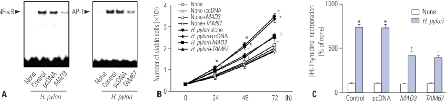
and oncogene expression, the cells were transiently transfect- ed with mutant genes for IκBα (MAD3) and c-Jun (TAM67) and then cultured with H. pylori. As shown Fig. 1A, H. pylori-infec- tion induced activation of NF-κB and AP-1 in wild-type cells (control) and the cells transfected with pcDNA (pcDNA) at 1 h-culture. H. pylori-induced activation of NF-κB and AP-1 were inhibited by transfection of MAD3 and TAM67. Similarly, H. pylori-stimulated cell proliferation time-dependently (Fig.
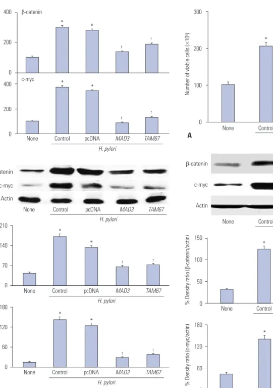
1B), and DNA synthesis at 24 h-culture (Fig. 1C) was inhibited by transfection with the mutants. However, transfection of MAD3 and TAM67 had no effect on the cells cultured without H. pylori. It may be explained that NF-κB and AP-1 were not activated in the cells cultured without H. pylori. As shown in Fig. 2, the mRNA and protein levels of β-catenin and c-myc were lower in the cells transfected with MAD3 and TAM67 than those transfected with pcDNA or wild-type cells cultured with H. pylori at 24 h-culture.
CAPE, one of the active compounds of propolis, has been shown to inhibit NF-κB activation.
21,22SR-11302 is a retinoid that transrepresses AP-1 without transactivating the retinoic acid re- sponse element.
23,24To investigate the involvement of NF-κB and AP-1 activities on H. pylori-induced oncogene expression and hyperproliferation, the cells were pretreated with CAPE or SR- 11302 and then cultured with H. pylori. As shown in Fig. 3, CAPE and SR-11302 inhibited H. pylori-induced cell proliferation (de- termined by viable cell numbers at 48 h-culture) in AGS cells (Fig. 3A). H. pylori-induced expression of β-catenin and c-myc was inhibited by CAPE and SR-11302 in AGS cells at 24 h-culture (Fig. 3B). Taken together, H. pylori-induced oncogene expres- sion and hyperproliferation are mediated by activation of NF-κB and AP-1 in gastric epithelial AGS cells.
DISCUSSION
In the present study, we found that activation of NF-κB and
AP-1 transcriptionally regulate cell proliferation and expres- sion of β-catenin and c-myc in H. pylori-infected gastric epi- thelial AGS cells. Hyper-proliferation of gastric epithelial cells and up-regulation of several gene expressions are reported to be associated with ROS under H. pylori infection. Even though ROS are considered to be responsible for the proliferation and oncogene expression, how ROS mediate cell proliferation in H.
pylori-infected cells has not been clarified. We demonstrated that redox-sensitive transcription factors NF-κB and AP-1 me- diate cell proliferation by inducing important oncogenes β-ca- tenin and c-myc in H. pylori-infected AGS cells. However, this study is limited in proving the hypothesis since AGS cells are already transformed cancer cells. Further study should be per- formed to establish the role of NF-κB and AP-1 on oncogene expression using primary gastric epithelial cells isolated from normal gastric tissues and in vivo animal models.
For the signaling mechanism for cell proliferation, β-catenin inactivates glycogen synthase kinase 3β and migrates to the nucleus, which induces the expression of cyclin D1. Bandapal- li, et al.
25reported that overexpression of β-catenin increases its nuclear level and carcinogenesis including metastasis. In the present study, inhibition of NF-κB and AP-1 upon trans- fection of MAD3 or TAM67 suppressed the expression of β-catenin in H. pylori-infected cells. These results suggest that H. pylori may activate β-catenin through modulation of NF-κB and AP-1 activities in gastric epithelial cells. Even though c- myc expression is reported to be regulated by β-catenin,
7,8there has been no studies to determine the role of NF-κB and AP-1 on c-myc expression in gastric epithelial cells. In the present study, we found that NF-κB and AP-1 regulate the ex- pression of both β-catenin and c-myc at the transcription lev- el, which was determined using transfection of MAD3 (a mu- tated IκBα gene) or TAM67 (a dominant negative mutant of c- Jun) or treatment with CAPE (a specific NF-κB inhibitor) or SR-11302 (a selective AP-1 inhibitor). Therefore, targeting transcription factor NF-κB and AP-1 may be beneficial for
Fig. 1. Activation of NF-κB and AP-1, viable cell numbers, and thymidine incorporation of H. pylori-infected cells with or without transfection of MAD3 or TAM67. (A) The cells were cultured with H. pylori for 1 h. The DNA binding activities of NF-κB and AP-1 were determined by EMSA. (B) Viable cell numbers were determined by the trypan blue exclusion assay for indicated time period. *p<0.05 vs. 0 h, †p<0.05 vs. H. pylori (the cells without trans- fection of MAD3 and TAM67 and cultured with H. pylori) or H. pylori+pcDNA (the cells with transfection of pcDNA and cultured with H. pylori). (C) DNA synthesis was determined by thymidine incorporation. [3H] Thymidine was added to the cells and cultured with H. pylori for 24 h. *p<0.05 vs. cor- responding none (the cells cultured without H. pylori), †p<0.05 vs. corresponding H. pylori (the cells without transfection of MAD3 and TAM67 and cul- tured with H. pylori) or H. pylori pcDNA (the cells with transfection of pcDNA and cultured with H. pylori). AGS, adenocarcinoma gastric; EMSA, lec- trophoretic mobility shift assay; H. pylori, Helicobacter pylori.
4
3
2
1
0
1000
500
0 Control pcDNA MAD3 TAM67 NoneControlpcDNAMAD3 NoneControlpcDNATAM67
NoneNone+pcDNA None+MAD3 None+TAM67 H. pylori alone H. pylori+pcDNA H. pylori+MAD3 H. pylori+TAM67
0 24 48 72 (h)
Number of viable cells (×105) [3H]-Thymidine incorporation (% of none)
None H. pylori
B C
A
*
* *
*
*
**
*
†
† †
†
†
†
NF-κB AP-1
H. pylori H. pylori
Fig. 2. Expression of β-catenin and c-myc of H. pylori-infected AGS cells with or without transfection of MAD3 or TAM67. The cells were cultured in with or without H. pylori for 24 h. (A) mRNA expression of β-catenin and c-myc were measured by real-time PCR analysis. *p<0.05 vs. none (the cells cultured without H. pylori), †p<0.05 vs. H. pylori control (the cells without transfection of MAD3 and TAM67 and cultured in with H. pylori) or H. pylori pcDNA (the cells transfected with pcDNA and cultured with H.
pylori). (B) Protein levels of β-catenin and c-myc were determined by Western blot analysis. Actin served as a loading control. The protein level was compared to that of the loading control actin and expressed as the percentage ratio of the band densities. All data are presented as the mean±SE of four independent experiments. *p<0.05 vs. none (the cells cultured without H. pylori), †p<0.05 vs. H. pylori control (the cells without transfection of MAD3 and TAM67 and cultured in with H. pylori) or H. py- lori pcDNA (the cells transfected with pcDNA and cultured with H. pylori).
H. pylori, Helicobacter pylori.
400
200
0 400
200
0 None pcDNA MAD3 TAM67
β-catenin
c-myc
Relative amount of mRNA (% of none)
A
*
*
*
*
†
†
†
†
H. pylori Control
β-catenin c-myc Actin
None pcDNA MAD3 TAM67
H. pylori Control
210
140
70
0
180
120
60
0
None
None
Control
Control
pcDNA
pcDNA
MAD3
MAD3
TAM67
TAM67
% Density ratio (β-catenin/actin)% Density ratio (c-myc/actin)
*
*
*
*
†
†
†
†
H. pylori
H. pylori B
Fig. 3. Viable cell numbers and expression of β-catenin and c-myc of H.
pylori-infected AGS cells with treatment of CAPE or SR-11302. (A) The cells were pretreated with CAPE or SR-11302 and cultured with H. pylori for 48 h. Viable cell numbers were determined by the trypan blue exclu- sion assay. *p<0.05 vs. none, †p<0.05 vs. H. pylori control (the cells with- out treatment of CAPE or SR-11302 and cultured with H. pylori). (B) Pro- tein levels of β-catenin and c-myc were determined by Western blot analysis. Actin served as a loading control. The protein level was com- pared to that of the loading control actin and expressed as the percent- age ratio of the band densities. All data are presented as the mean±SE of four independent experiments. *p<0.05 vs. none (the cells cultured without H. pylori), †p<0.05 vs. H. pylori control (the cells with H. pylori). H.
pylori, Helicobacter pylori.
300
200
100
0 None Control CAPE SR
Number of viable cells (×105)
A
*
† †
H. pylori
None Control CAPE SR
H. pylori β-catenin
c-myc
Actin
150
100
50
0
180
120
60
0
None
None Control CAPE SR
Control CAPE SR
% Density ratio (β-catenin/actin)% Density ratio (c-myc/actin)
*
†
†
*
† †
B
H. pylori
H. pylori
preventing progression of H. pylori-associated carcinogenesis by suppressing expression of β-catenin and c-myc, as well as hyper-proliferation of gastric epithelial cells.
ACKNOWLEDGEMENTS
This study was supported by a grant from the NRF of Korea, funded by the Korean government (MSIP) (NRF-2012R1A1A2 043423).
REFERENCES
1. Negrini R, Savio A, Poiesi C, Appelmelk BJ, Buffoli F, Paterlini A, et al. Antigenic mimicry between Helicobacter pylori and gastric mucosa in the pathogenesis of body atrophic gastritis. Gastroen- terology 1996;111:655-65.
2. Nagy TA, Wroblewski LE, Wang D, Piazuelo MB, Delgado A, Rome- ro-Gallo J, et al. β-Catenin and p120 mediate PPARδ-dependent proliferation induced by Helicobacter pylori in human and rodent epithelia. Gastroenterology 2011;141:553-64.
3. Peek RM Jr, Moss SF, Tham KT, Pérez-Pérez GI, Wang S, Miller GG, et al. Helicobacter pylori cagA+strains and dissociation of gastric epithelial cell proliferation from apoptosis. J Natl Cancer Inst 1997;89:863-8.
4. Gnad T, Feoktistova M, Leverkus M, Lendeckel U, Naumann M.
Helicobacter pylori-induced activation of beta-catenin involves low density lipoprotein receptor-related protein 6 and Dishev- elled. Mol Cancer 2010;9:31.
5. Behrens J, Jerchow BA, Würtele M, Grimm J, Asbrand C, Wirtz R, et al. Functional interaction of an axin homolog, conductin, with beta-catenin, APC, and GSK3beta. Science 1998;280:596-9.
6. Behrens J, von Kries JP, Kühl M, Bruhn L, Wedlich D, Grosschedl R, et al. Functional interaction of beta-catenin with the transcrip- tion factor LEF-1. Nature 1996;382:638-42.
7. Shih IM, Yu J, He TC, Vogelstein B, Kinzler KW. The beta-catenin binding domain of adenomatous polyposis coli is sufficient for tumor suppression. Cancer Res 2000;60:1671-6.
8. Dang CV. c-Myc target genes involved in cell growth, apoptosis, and metabolism. Mol Cell Biol 1999;19:1-11.
9. Shimoyama T, Fukuda S, Liu Q, Nakaji S, Fukuda Y, Sugawara K.
Production of chemokines and reactive oxygen species by human neutrophils stimulated by Helicobacter pylori. Helicobacter 2002;
7:170-4.
10. Davies GR, Simmonds NJ, Stevens TR, Sheaff MT, Banatvala N, Laurenson IF, et al. Helicobacter pylori stimulates antral mucosal reactive oxygen metabolite production in vivo. Gut 1994;35:179-85.
11. Byun E, Lim JW, Kim JM, Kim H. α-Lipoic acid inhibits Helico- bacter pylori-induced oncogene expression and hyperprolifera-
tion by suppressing the activation of NADPH oxidase in gastric epithelial cells. Mediators Inflamm 2014;2014:380830.
12. Keenan JI, Peterson RA 2nd, Hampton MB. NADPH oxidase in- volvement in the pathology of Helicobacter pylori infection. Free Radic Biol Med 2005;38:1188-96.
13. Tominaga K, Kawahara T, Sano T, Toida K, Kuwano Y, Sasaki H, et al. Evidence for cancer-associated expression of NADPH oxidase 1 (Nox1)-based oxidase system in the human stomach. Free Rad- ic Biol Med 2007;43:1627-38.
14. Seo JH, Lim JW, Kim H, Kim KH. Helicobacter pylori in a Korean isolate activates mitogen-activated protein kinases, AP-1, and NF- kappaB and induces chemokine expression in gastric epithelial AGS cells. Lab Invest 2004;84:49-62.
15. Müller JM, Rupec RA, Baeuerle PA. Study of gene regulation by NF-kappa B and AP-1 in response to reactive oxygen intermedi- ates. Methods 1997;11:301-12.
16. Ghosh S, Karin M. Missing pieces in the NF-kappaB puzzle. Cell 2002;109 Suppl:S81-96.
17. Chu SH, Kim H, Seo JY, Lim JW, Mukaida N, Kim KH. Role of NF- kappaB and AP-1 on Helicobater pylori-induced IL-8 expression in AGS cells. Dig Dis Sci 2003;48:257-65.
18. Nollet F, Berx G, Molemans F, van Roy F. Genomic organization of the human beta-catenin gene (CTNNB1). Genomics 1996;32:413- 24.
19. Lim JW, Kim H, Kim KH. NF-kappaB, inducible nitric oxide syn- thase and apoptosis by Helicobacter pylori infection. Free Radic Biol Med 2001;31:355-66.
20. Coward L, Smith M, Kirk M, Barnes S. Chemical modification of isoflavones in soyfoods during cooking and processing. Am J Clin Nutr 1998;68(6 Suppl):1486S-91S.
21. Chen MF, Keng PC, Lin PY, Yang CT, Liao SK, Chen WC. Caffeic acid phenethyl ester decreases acute pneumonitis after irradia- tion in vitro and in vivo. BMC Cancer 2005;5:158.
22. Jung WK, Choi I, Lee DY, Yea SS, Choi YH, Kim MM, et al. Caffeic acid phenethyl ester protects mice from lethal endotoxin shock and inhibits lipopolysaccharide-induced cyclooxygenase-2 and inducible nitric oxide synthase expression in RAW 264.7 macro- phages via the p38/ERK and NF-kappaB pathways. Int J Biochem Cell Biol 2008;40:2572-82.
23. Fanjul A, Dawson MI, Hobbs PD, Jong L, Cameron JF, Harlev E, et al. A new class of retinoids with selective inhibition of AP-1 inhib- its proliferation. Nature 1994;372:107-11.
24. Huang C, Ma WY, Dawson MI, Rincon M, Flavell RA, Dong Z.
Blocking activator protein-1 activity, but not activating retinoic acid response element, is required for the antitumor promotion effect of retinoic acid. Proc Natl Acad Sci U S A 1997;94:5826-30.
25. Bandapalli OR, Dihlmann S, Helwa R, Macher-Goeppinger S, Weitz J, Schirmacher P, et al. Transcriptional activation of the beta- catenin gene at the invasion front of colorectal liver metastases. J Pathol 2009;218:370-9.