Molecular identification of Mycoplasma cynos from laboratory beagle dogs with respiratory disease
Sunhwa Hong1,2, Okjin Kim1,3*
1Center for Animal Resources Development, Wonkwang University, Iksan, Korea
2Institute of Animal Experiment & Efficacy Evaluation, Wonkwang University, Iksan, Korea
3Digestive Disease Research Institute, Wonkwang University, Iksan, Korea
In this study, we examined a colony of 20 beagle dogs in a laboratory animal facility. Mycoplasma was detected by consensus PCR assay in 1 dog with respiratory and constitutional symptoms. None of the other dogs were affected. The dog was euthanized and necropsied. In postmortem examinations, gray or plum-colored gross lesions were found on the lung, most commonly in the apical and cardiac lobes.
Some lesions showed clear demarcation and consolidation. Microscopic examination showed peribronchiolar lymphoid hyperplasia and interstitial thickening, lesions pathognomonic for mycoplasma pneumonia. To identify canine Mycoplasma species, we used species-specific PCR reactions for M. arginini, M. canis, M.
cynos, M. edwardii, M. felis, M. gateae, M. maculosum, M. molare, M. opalescens, M. spumans, Mycoplasma sp. HRC 689, and M. collis. As the result, we identified Mycoplasma cynos by amplification of DNA extracted from lung tissue of the laboratory beagle dog with respiratory disease.
Keywords: Mycoplasma, PCR, consensus, Mycoplasma cynos, Beagle, dog
Received 6 February 2012; Revised version received 1 March 2012; Accepted 4 March 2012
Mycoplasmas belong to the class Mollicutes. They are among the smallest free-living microorganisms capable of autoreplication. They are highly fastidious, difficult to culture, and slow growing [1]. Many species are important veterinary pathogens and cause respiratory infections, mastitis, conjunctivitis, arthritis, and infrequently, abortion [1]. It has been reported that mycoplasma infections are highly contagious and are very common in the field of laboratory animal medicine [2]. It is necessary to determine the extent of Mycoplasma contamination in laboratory animal colonies because these organisms are prevalent in commercial and research animal facilities [3], and mycoplasma infections in laboratory animals interfere with the results of biomedical research [4].
Mycoplasmas are thought to be part of the normal bacterial flora in the upper respiratory tract in dogs [5], but there have been conflicting reports about the
presence of mycoplasmas in the lower respiratory tract of healthy dogs. Randolph et al. [6] found that the lungs of up to 27% of healthy dogs were colonized, but other authors have failed to replicate those findings [5]. The role of individual Mycoplasma species in respiratory infections of dogs is not well understood. [5] M.
bovigenitalium, M. canis, M. cynos, M. edwardii, M.
feliminutum, M. gateae, and M. spumans have been isolated from dogs with respiratory disease [7,8].
Pneumonia in dogs has been reproduced by experimental endobronchial inoculation with an isolate of M. cynos that was isolated from a dog with pneumonia, and by exposure of non-infected dogs to infected dogs [9,10]. In addition, several cases have been described in which pure cultures of mycoplasmas were isolated from dogs with respiratory disease, but species-level typing was not performed [6,11]. The overall importance and distribution
Letter
http://dx.doi.org/10.5625/lar.2012.28.1.61
*Corresponding author: Okjin Kim, Center for Animal Resources Development, Wonkwang University, 460 Iksandaero, Iksan, Jeonbuk 570-749, Korea
Tel: +82-63-850-6668; Fax: +82-63-850-7308; E-mail: [email protected]
This is an Open Access article distributed under the terms of the Creative Commons Attribution Non-Commercial License (http://creativecommons.org/licenses/
by-nc/3.0) which permits unrestricted non-commercial use, distribution, and reproduction in any medium, provided the original work is properly cited.
In this study, we detected mycoplasma in an infected laboratory dog by genus-specific consensus PCR using 16S ribosomal DNA. Thereafter, Mycoplasma cynos was identified by a species-specific PCR.
Twenty male Beagle dogs (age, 1 year) were obtained from the Animal Facilities of the Center for Animal Resources Development, Wonkwang University, Korea.
The animal experiments in this study were conducted according to the ethical procedures of Wonkwang University IACUC. Over the previous 7 days, 1 dog (No.
8) had developed a dry cough, anorexia, vomiting, and depression. None of the other 19 dogs had symptoms.
All dogs, including No. 8, underwent physical and laboratory examinations. Abdominal sonography and radiography did not demonstrate any abnormal lesions.
Nasal swabs were collected from all 20 dogs. Samplings were performed from the nares with dry unmoistened swabs. The tip of the swab was inserted into the nares and rolled 5 times in each nostril. Specimens were transported and stored at room temperature. Specimens were processed for PCR analysis within 24 h of collection. Each swab was put into 2 mL of 0.1 M PBS buffer, vortexed, and discarded, and the PBS was used to extract genomic DNA for Mycoplasma consensus PCR assay.
Dog No. 8 was euthanized and necropsied. During the necropsy, tissue specimens were collected from the lungs, and gross and microscopic observations of pathological lesions were recorded. For Mycoplasma identification, fresh tissues were submitted for species- specific PCR analysis.
For histopathological observation, lung tissues were fixed in 10% neutral buffered formalin. After fixation, the tissues were embedded in paraffin in the usual manner. Sections were cut with a thickness of 4 µm, floated on a water bath, mounted on slides, and stained with hematoxylin & eosin. After staining, microscopic examination was conducted.
The lung tissue of dog No. 8 was homogenized and
To screen for mycoplasma infection, we conducted Mycoplasma consensus PCR with the DNA extracted from the nasal swab samples, as described previously [14]. In brief, amplification of the V3 region of the 16S ribosomal DNA was performed with consensus primers GC-341F (5'-CGCCCGCCGCGCGCGGCGGGCGGG GCGGGGGCACGGGGGGCCTACGGGAGGCAGC AG-3') and 534R (5'-ATTACCGCGGCTGCTGG-3') [1].
The template DNA (50 ng) and 20 pmol of each primer were added to a PCR mixture tube (AccuPower PCR PreMix; Bioneer Corporation) containing 2.5 U of Taq DNA polymerase, 250 µM of each deoxynucleoside triphosphate, 10 mM Tris-HCl (pH 8.3), 40 mM KCl, 1.5 mM MgCl
2, and the gel loading dye. The final volume was adjusted to 20 µL with distilled water. The reaction mixture was subjected to denaturation at 94
oC for 5 min, followed by 30 cycles of 95
oC for 1 min, 55
oC for 45 s, and 72
oC for 1 min and a final extension step of 72vC for 10 min, and samples were stored at 4
oC until analysis. Reactions were conducted using My Genie 32 Thermal Block PCR (Bioneer Corporation). Eight microliters of each sample were mixed with 2 µL of loading buffer, and electrophoretically separated on 1.2%
agarose gels stained with 0.5 µg/mL ethidium bromide.
DNA bands were observed under ultraviolet light.
To identify canine Mycoplasma species, we used species-specific PCR reactions for M. arginini, M. canis, M. cynos, M. edwardii, M. felis, M. gateae, M.
maculosum, M. molare, M. opalescens, M. spumans, and Mycoplasma sp. HRC 689. Due to the high similarity of the 16S rRNA genes of canine Mycoplasma species [15], species-specific PCR tests for regions identified in the 16S/23S rRNA intergenic spacer region had been developed for the Mycoplasma species [16]. PCR reactions were performed with forward primer (Myc1;
5'-CACCGCCCGTCACACCA-3'), and following reverse
primers: M. arginini (5'-GTTGTATGACCTATTGTTGT
C-3'), M. canis (5'-CTGTCGGGGTTATCTCGAC-3'),
M. cynos (5'-GATACATAAACACAACATTATAATAT
TG-3'), M. edwardii (5'-CTGTCGGGTTATCATGCGA C-3'), M. felis (5'-GGACTATTATCAAAAGCACATAA C-3'), M. gateae (5'-GTTGTATGACCTATTGTTGTC- 3'), M. maculosum (5'-CCTATGATTGTTACAGATG- 3'), M. molare (5'-AGCCTATTGTTTTTGATTTG-3'), M. opalescens (5'-TAAGCTTTGTAGACCATAA-3'), M. spumans (5'-GTTGTATGACCTATTGTTGTC-3'), and Mycoplasma sp. HRC 689 (5'-CTTGCGACCTA ACAAGTCC-3') [16]. Also, we conducted species- specific PCR to detect M. collis with Primer 1 (5'- AAAAGAAGCTTGAATTATAG-3') and Primer 2 (5'- ATTAAGAGTCATTTCCTACT-3') [17]. All PCRs commenced with an initial denaturation at 95
oC for 5 min and were followed by the specific annealing step and then by final extension at 72
oC for 5 min. PCR reactions were performed with each 25 pmol of forward and reverse primers, and template DNA (50 ng) added to a PCR mixture tube (AccuPower PCR PreMix; Bioneer Corporation) containing 2.5 U of Taq DNA polymerase, 250 µM of each deoxynucleoside triphosphate, 10 mM Tris-HCl (pH 8.3), 40 mM KCl, 1.5 mM MgCl
2, and the gel loading dye. The final volume was adjusted to 50 µL with distilled water. Reactions were conducted using My Genie 32 Thermal Block PCR (Bioneer Corporation).
Eight microliters of each sample were mixed with 2 µL of loading buffer, and electrophoretically separated on 1.2% agarose gels stained with 0.5 µg/mL ethidium bromide. DNA bands were observed under ultraviolet light.
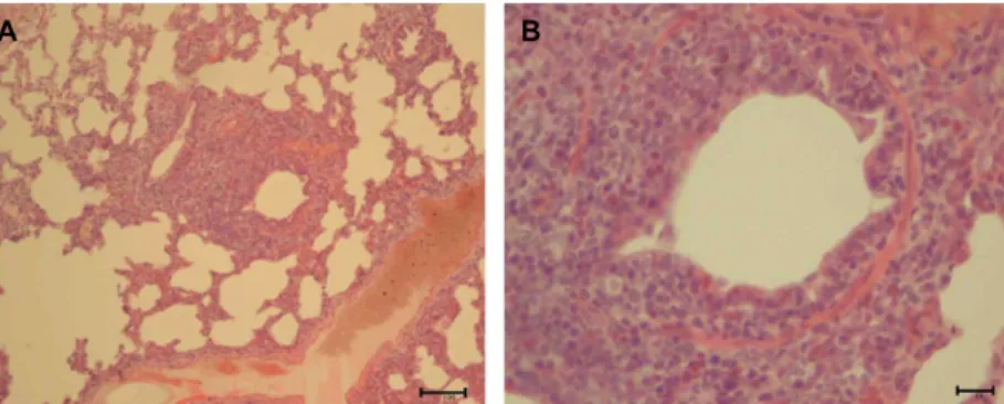
Gross lesions that were gray or plum colored were found on the lung of dog No. 8, most commonly in the apical and cardiac lobes. Some lesions showed clear demarcation and consolidation. The microscopic lesions were confined to the apical and cardiac lobes of lung.
The plum colored and consolidated areas of the infected
lungs corresponded to mild to severe characteristic perivascular and peribronchiolar lymphomononuclear nodules of infiltration, often compressing the lumen of the bronchioles (Figure 1). The bronchial and bronchiolar epithelium showed a loss of cilia on the epithelial cells and sloughing of epithelial cells into the airway lumina.
Infiltrating lymphocytes and mononuclear cells were frequently seen interspersed between epithelial cells.
Edema fluid, neutrophils, and large mononuclear cells were seen in the lumina of the airways. The lymphoid nodules were associated with a narrowing of the lumina of the airways. Peribronchiolar lymphoid hyperplasia and interstitial thickening, the pathognomonic lesions of mycoplasma pneumonia, were observed (Figure 1).
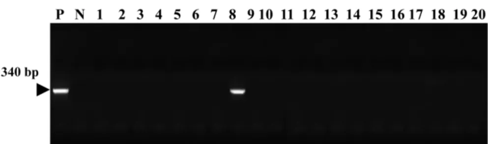
A consensus PCR analysis method using 16S ribosomal DNA was employed to detect mycoplasmas. The 16S rDNA gene (340 bp) was specifically amplified by PCR with the Mycoplasma genus-specific primers (GC-341F and 534R). The target nucleic acid fragments were specifically amplified by consensus PCR with 16S ribosomal DNA primers. As a result, mycoplasma was detected in the nasal swab sample from dog No. 8 by PCR (Figure 2). We did not detect any mycoplasmas in the healthy dogs.
To identify the canine Mycoplasma species from the lung tissue of dog No. 8, Mycoplasma species-specific PCR reactions were conducted. We identified M. cynos by positive amplification of a 227-bp DNA strand (Figure 3). We did not detect amplification during PCR assays for M. arginini, M. canis, M. edwardii, M. felis, M. gateae, M. maculosum, M. molared, M. opalescens, M. spumans, Mycoplasma sp. HRC 689 or M. collis.
Mycoplasmas are recognized agents of respiratory disease in a wide range of animal species, including man. Generally, the disease is mild, but it has been
Figure 1. Histopathological findings in the lung of dog No. 8. (A) Peribronchiolar lymphoid hyperplasia and interstitial thickening lesions. H&E stain, Bar=100µm, (B) Infiltrating lymphocytes and mononuclear cells in peribronchiolar hyperplasia lesion. H&E stain, Bar=20µm
shown to predispose the host to more severe secondary infection and to exacerbate concurrent infections [18].
Naturally occurring mycoplasmosis is an insidious disease that may significantly impact research in a variety of disciplines in the field of laboratory animal medicine, as the organisms may be disseminated widely in the hosts [2]. Imperceptible infection is known to affect physiological mechanisms and the immune system, thereby influencing experimental results obtained with contaminated animals [19]. It alters both the number and subpopulation distribution of lymphocytes in the lung [4] and it induces elevations in NK cell activity, suppressing humoral antibody response [20] and enhancing production of TNF, IL-1, IL-6, and interferons [21].
Mycoplasma infection in laboratory animals interferes with the results of biomedical research and can threaten the heath of personnel conducting animal experiments.
A zoonotic M. pneumoniae outbreak was reported in a group of students who had an infected Syrian gold hamster family in their classroom [22]. Recently, Mycoplasma species have been accepted as important emerging and reemerging pathogens in human and animal diseases [23]. There are many questions to be answered concerning Mycoplasma infection in humans and animals. In the case of laboratory animals, a number of Mycoplasma species have been identified and found to
be highly prevalent in animal facilities [2]. The monitoring and control of these mycoplasmas is necessary to ensure optimal experimental results in laboratory animals.
Bacterial culture has been the best method for diagnosing a bacterial infection. However, the sensitivity of the culture-isolation method is low [12]. Therefore, a culture is not considered the most practical diagnostic tool. PCR is a specific and sensitive molecular method for detecting mycoplasma DNA, and can supplement other methods.
However, PCR using species-specific primers may require multiple assays because of the presence of a number of Mycoplasma species [1]. In this study, consensus PCR successfully detected mycoplasma DNA in a nasal swab specimen. Thus, consensus PCR may be recommended for monitoring Mycoplasma species in laboratory animals.
Mycoplasmas have been implicated as potential agents of canine respiratory disease [8,24]. Some authors also describe Mycoplasma collis as a canine mycoplasma [25]. However, we cannot find any report of M. collis infection in dogs and reports indicate that this species was originally isolated from rodents [26]; therefore, this species may have been mistakenly identified as being of canine origin. In addition, other publications describe the isolation of untyped Mycoplasma spp. from dogs [11, 27]. In the past 20 years, work on canine mycoplasmas has been extremely limited, with only a dozen publications
Figure 3. Amplification of DNA extracted from the lung tissue of dog No. 8 with canine Mycoplasma species-specific primers was confirmed on 1.2% agarose gel electrophoresis. Lane P: positive control, N: distilled water, 1: M. arginini, 2: M. canis, 3: M. cynos, 4: M. edwardii, 5: M. felis, 6: M. gateae, 7: M. maculosum, 8: M. molare, 9: M. opalescens, 10: M. spumans, 11: Mycoplasma sp.
HRC 689, 12: M. collis
on mycoplasmas in dogs. M. cynos was isolated from 2 kenneled dogs with pneumonia [9] and was shown to induce pneumonia with severe inflammation of bronchi and respiratory tract tissue [8]. To date, there has been limited information regarding the etiological role of mycoplasmas in natural canine disease [5,6,8,9].
However, an association between M. cynos isolated from the lower respiratory tract and the severity of canine infectious respiratory disease in kenneled dogs has now been demonstrated [15]. Only M. cynos was significantly associated with respiratory disease. Mycoplasmas have been known for some time to modulate host susceptibility to secondary infections [18,28]. Thus, the presence of other initiating agents could be suspected and mycoplasma agents could be involved. M. cynos has been isolated from dogs with pneumonia [8,9,29] and was particularly abundant in the most necrotic areas of the lung [29].
Furthermore, M. cynos was the only agent detected in an outbreak of severe bronchopneumonia in a litter of young puppies that resulted in several deaths, but was resolved in the surviving littermates after the administration of appropriate antibiotics [30]. Experimental endobronchial inoculation of dogs with M. cynos has produced localized pneumonia with destruction and loss of the bronchial epithelial cilia and alveolar infiltration with neutrophils and macrophages [10], and in our study, M.
cynos was detected in the lung tissue by PCR. It is known that M. cynos can persist in the lung for up to 3 weeks following infection [10], and that it can also be isolated from the trachea, conjunctiva, tonsils, and even kennel aerosols [16,31]. The capacity of M. cynos to persist in the environment is unknown, but other Mycoplasma species can survive for weeks to months outside the host, and the environment could therefore be a source of infection [32]. Recently, it has been shown that biofilm formation is important for persistence of mycoplasmas and may aid environmental survival [33], and it seems feasible that M. cynos may be able to persist in the kennel environment as an adherent biofilm layer.
M. cynos was originally isolated from an outbreak of enzootic pneumonia in kenneled dogs in which 2 healthy dogs were exposed to an infected litter [9]. In our study, M. cynos was identified from the lung of dog with respiratory disease. Infection with M. cynos may have been preceded or superseded by infection with another microorganism. A previous study found that a greater number of bacteria were isolated from dogs with more
severe clinical disease [34], and this may have made isolation of M. cynos more difficult due to the increased number of other bacterial colonies growing on the medium.
In such cases, using Mycoplasma species-specific PCR directly on clinical samples or filtering samples through a 0.2- µm filter prior to plating may improve the detection of M. cynos.
In this study, we encountered a laboratory Beagle dog with respiratory disease and conducted health examinations on a colony of 20 of Beagle dogs in the laboratory animal facilities. Mycoplasma was detected in the dog with respiratory symptoms by consensus PCR assay, and no mycoplasma was detected in the healthy dogs. The affected dog was euthanized. The postmortem examination revealed pathological lesions on the lung that were gray or plum colored and most commonly present in the apical and cardiac lobes. Some lesions showed clear demarcation and consolidation. Microscopic lesions were confined to the apical and cardiac lobes of the lung.
The plum-colored and consolidated areas of the infected lungs corresponded to mild to severe characteristic perivascular and peribronchiolar lymphomononuclear nodules of infiltration, often compressing the lumen of the bronchioles. Peribronchiolar lymphoid hyperplasia and interstitial thickening lesions, which are known as pathognomonic findings for mycoplasma pneumonia, were observed. For Mycoplasma species identification, fresh tissues were submitted for species-specific PCR analysis, and M. cynos was identified.
Consensus PCR can successfully detect mycoplasma and has been recommended for monitoring Mycoplasma species in laboratory animals [14]. In this study, consensus PCR was used to detect mycoplasma in laboratory beagle dogs. The results indicate that consensus PCR may be useful and effective for monitoring Mycoplasma species in various kinds of laboratory animals, including rodents and dogs. The application of consensus PCR before species-specific PCR would be the best strategy for the detection and identification of Mycoplasma species.
Acknowledgments
This research was supported by Technology
Development Program for Agriculture and Forestry
(grant No. 610004-3), Ministry for Food, Agriculture,
Forestry and Fisheries, Republic of Korea.
pulmonis. Am J Pathol 1971; 64(3): 675–708.
4. Davis JK, Thorp RB, Maddox PA, Brown MB, Cassell GH.
Murine respiratory mycoplasmosis in F344 and LEW rats:
evolution of lesions and lung lymphoid cell populations. Infect Immun 1982; 36(2): 720–729.
5. Rosendal S. Canine mycoplasmas: their ecologic niche and role in disease. J Am Vet Med Assoc 1982; 180(10): 1212–1214.
6. Randolph JF, Moise NS, Scarlett JM, Shin SJ, Blue JT, Bookbinder PR. Prevalence of mycoplasmal and ureaplasmal recovery from tracheobronchial lavages and prevalence of mycoplasmal recovery from pharyngeal swab specimens in dogs with or without pulmonary disease. Am J Vet Res 1993; 54(3):
387–391.
7. Armstrong D, Morton V, Friedman MH, Steger L, Tully JG.
Canine pneumonia associated with mycoplasma infection. Am J Vet Res 1972; 33(7): 1471–1478.
8. Rosendal S. Canine mycoplasmas: pathogenicity of mycoplasmas associated with distemper pneumonia. J Infect Dis 1978; 138(2):
203–210.
9. Rosendal S. Mycoplasmas as a possible cause of enzootic pneumonia in dogs. Acta Vet Scand 1972; 13(1): 137–139.
10. Rosendal S, Vinther O. Experimental mycoplasmal pneumonia in dogs: electron microscopy of infected tissue. Acta Pathol Microbiol Scand B 1977; 85B(6): 462–465.
11. Chandler JC, Lappin MR. Mycoplasmal respiratory infections in small animals: 17 cases (1988-1999). J Am Anim Hosp Assoc 2002; 38(2): 111–119.
12. Harasawa R, Koshimizu K, Takeda O, Uemori T, Asada K, Kato I. Detection of Mycoplasma hyopneumoniae DNA by the polymerase chain reaction. Mol Cell Probes 1991; 5(2): 103–109.
13. Cho SJ, Lee HA, Hong S, Kim O. Uterine adenocarcinoma with feline leukemia virus infection. Lab Anim Res 2011; 27(4): 347–
351.
14. Hong S, Lee HA, Park SH, Kim O. Sensitive and Specific Detection of Mycoplasma species by Consensus Polymerase Chain Reaction and Dot Blot Hybridization. Lab Anim Res 2011;
27(2): 141–145.
15. Chalker VJ, Brownlie J. Taxonomy of the canine Mollicutes by 16S rRNA gene and 16S/23S rRNA intergenic spacer region sequence comparison. Int J Syst Evol Microbiol 2004; 54(Pt 2):
537–542.
16. Chalker VJ, Owen WM, Paterson C, Barker E, Brooks H, Rycroft AN, Brownlie J. Mycoplasmas associated with canine infectious respiratory disease. Microbiology 2004; 150(Pt 10): 3491–3497.
17. van Kuppeveld FJ, van der Logt JT, Angulo AF, van Zoest MJ, Quint WG, Niesters HG, Galama JM, Melchers WJ. Genus- and species-specific identification of mycoplasmas by 16S rRNA amplification. Appl Environ Microbiol 1992; 58(8): 2606-2615.
18. Thacker EL, Halbur PG, Ross RF, Thanawongnuwech R, Thacker
gamma interferon in C3H/HeN and C57BL/6N mice in acute Mycoplasma pulmonis disease. Infect Immun 1995; 63(10):
4084–4090.
22. Mikola I, Balogh G, Nagy A, Mátyás M, Glávits R, Stipkovits L.
Mycoplasma pneumoniae epidemic as zoonosis. Orv Hetil 1997;
138(46): 2933–2935.
23. Rosengarten R, Citti C, Much P, Spergser J, Droesse M, Hewicker-Trautwein M. The changing image of mycoplasmas:
from innocent bystanders to emerging and reemerging pathogens in human and animal diseases. Contrib Microbiol 2001; 8: 166–
185.
24. Greig AS. The Significance Of A Pleuropneumonia-Like Organism In Kennel Cough. Can J Comp Med Vet Sci 1954;
18(8): 275–279.
25. Johansson K-E, Pettersson B. Taxonomy of Mollicutes. In:
Molecular Biology and Pathogenicity of Mycoplasmas (Razin S, Herrmann R, ed), : Kluwer, New York, 2002; pp1–27.
26. Hill AC. Mycoplasma collis, a new species isolated from rats and mice. Int J Syst Bacteriol 1983; 33(4): 847–851.
27. Kirchner BK, Port CD, Magoc TJ, Sidor MA, Ruben Z.
Spontaneous bronchopneumonia in laboratory dogs infected with untyped Mycoplasma spp. Lab Anim Sci 1990; 40(6): 625–628.
28. Kaklamanis E, Pavlatos M. The immunosuppressive effect of mycoplasma infection. I. Effect on the humoral and cellular response. Immunology 1972; 22(4): 695–702.
29. Chvala S, Benetka V, Mostl K, Zeugswetter F, Spergser J, Weissenbock H. Simultaneous canine distemper virus, canine adenovirus type 2, and Mycoplasma cynos infection in a dog with pneumonia. Vet Pathol 2007; 44(4): 508–512.
30. Zeugswetter F, Weissenbock H, Shibly S, Hassan J, Spergser J.
Lethal bronchopneumonia caused by Mycoplasma cynos in a litter of golden retriever puppies. Vet Rec 2007; 161(18): 626–
627.
31. Rosendal S. Canine mycoplasmas. I. Cultivation from conjunctivae, respiratory- and genital tracts. Acta Pathol Microbiol Scand B Microbiol Immunol 1973; 81(4): 441–445.
32. Nagatomo H, Takegahara Y, Sonoda T, Yamaguchi A, Uemura R, Hagiwara S, Sueyoshi M. Comparative studies of the persistence of animal mycoplasmas under different environmental conditions.
Vet Microbiol 2001; 82(3): 223–232.
33. McAuliffe L, Ellis RJ, Miles K, Ayling RD, Nicholas RA. Biofilm formation by mycoplasma species and its role in environmental persistence and survival. Microbiology 2006; 152(Pt 4): 913–922.
34. Chalker VJ, Toomey C, Opperman S, Brooks HW, Ibuoye MA, Brownlie J, Rycroft AN. Respiratory disease in kennelled dogs:
serological responses to Bordetella bronchiseptica lipopolysaccharide do not correlate with bacterial isolation or clinical respiratory symptoms. Clin Diagn Lab Immunol 2003; 10(3): 352–356.