aResearch Assistant, bAssociate Professor, Department of Ortho- dontics, Gazi University Faculty of Dentistry.
Corresponding author: Çağrı Türköz.
Department of Orthodontics, Gazi University Faculty of Dentistry, 06510, Emek, Ankara, Turkey.
+903122034289; e-mail, [email protected].
Received February 25, 2010; Last Revision May 5, 2010;
Accepted May 11, 2010.
DOI:10.4041/kjod.2010.40.4.260
Bond strength of different bonding systems to the lingual surface enamel of mandibular incisors
Çağrı Türköz, DDS, PhD,a Burcu Baloş Tuncer, DDS, PhD,b Mehmet Çağrı Ulusoy, DDS, PhD,b Cumhur Tuncer, DDS, PhDb
Objective: The aim of this study was to determine whether different types of adhesive systems and enam- el-protective agents will affect the tensile bond strength of lingual brackets. Methods: A total of 75 extracted mandibular incisors were randomly divided into 5 groups and lingual brackets were bonded. Group 1 speci- mens received Transbond XT (3M Unitek, Monrovia, CA, USA), Group 2 required the application of a fluo- ride-releasing resin (Ortho-coat, Pulpdent, Watertown, MA, USA) with Transbond XT, Group 3 specimens received a chlorhexidine varnish (Cervitec Plus, Ivoclar Vivadent, Schaan, Lichtenstein) with Transbond XT.
In Group 4, a light-cured orthodontic adhesive (Aegis Ortho, Bosworth, Skokie, USA) was applied and in Group 5, an antimicrobial self-etching primer (Clearfil Protect Bond, Kuraray, Osaka, Japan) was used.
Results: There were no significant differences in bond strength whether fluoride-releasing resin or chlorhex- idine varnish were used or not. Group 5 had significantly higher bond strength and adhesive remnant index (ARI) values than other groups (p < 0.001). The application of enamel-protective products did not have an adverse affect on the bond strength of lingual brackets. Conclusions: These products might provide ben- efits both for the patient and the clinician, by supporting the oral hygiene during lingual orthodontic treatment. The higher ARI score may be beneficial for Clearfil Protect Bond but its excessive bond strength should be considered in clinical practice, especially where the enamel is thin. (Korean J Orthod 2010;40(4):260-266)
Key words: Lingual, Bonding, Resin, Adhesive
INTRODUCTION
Esthetic materials have appeared in the market, since the number of adult patients seeking orthodontic treat- ment has increased in recent years. During the last two decades, the problem of esthetics during orthodontic
treatment has been solved by attaching fixed appli- ances to the lingual surfaces of teeth.1 Despite the es- thetic advantages, disadvantages such as patient dis- comfort have also been mentioned in several studies.1-3 While some studies have focused on tongue irritations, pain, speech and eating problems,1,3 other studies dealt with difficulties with oral hygiene.2,4
Fixed orthodontic appliances induce changes in the oral environment, such as increased retention sites for food particles and dental plaque, a low-pH environ- ment and increased proportions of Streptococcus mutans.5-7 Consequently, the risk of decalcification and dental caries increases in some cases.8,9 Attempts have been made to maintain proper oral hygiene around brackets and bands in orthodontic patients. For this
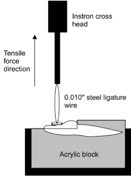
Fig 1. Schematic drawing of tensile stress testing.
purpose, fluoride releasing and/or antimicrobial agents have been applied in orthodontic patients.9 New mate- rials have recently appeared in the market which in- cludes fluoride-releasing resins,10 chlorhexidine var- nishes,11 amorphous calcium phosphate (ACP) contain- ing orthodontic adhesive,12 and anti-microbial agents.13,14 These products claim to prevent demineralization and white spot lesions around orthodontic brackets, and maintain plaque control throughout the treatment.
Although Gorelick et al.15 emphasized that flow of saliva is a major factor in avoiding decalcification of enamel on the lingual surfaces of the mandibular in- cisors, maintaining oral hygiene is more difficult for patients with lingual brackets than with buccal brack- ets, since perfect oral hygiene is particularly important in these patients. Also the small tooth surface from the bracket to gingival margin is conducive to the retention of dental plaque. The clinical application of afore- mentioned agents can be useful, but they must first provide an acceptable level of bond strength. Morpho- logical differences exist between labial and lingual tooth surfaces especially in anterior teeth, as well as the etching surface areas.16 Besides, the base surface area of lingual brackets show differences due to the lingual surface anatomy of incisors.
Therefore, the aim of this study was to evaluate whether different orthodontic adhesives provide satis- factory bond strength for lingual orthodontic brackets in the mandibular anterior region. For this purpose, the effect of a fluoride-releasing resin (Ortho-Coat; Pulp- dent Co, Watertown, MA, USA), a chlorhexidine var- nish (Cervitec Plus; Ivoclar Vivadent, Schaan, Lichten- stein), a light-cured orthodontic adhesive containing Amorphous Calcium Phosphate (ACP) (Aegis Ortho;
Bosworth, Skokie, Ill, USA), and an antimicrobial self-etching primer (Clearfil Protect Bond; Kuraray Medical Inc., Okayama, Japan) were compared with a commonly used orthodontic adhesive, Transbond XT (3M Unitek, Monrovia, CA, USA).
MATERIAL AND METHODS
A total of seventy-five caries-free human mandibular incisors, extracted with orthodontic and periodontal in- dications, were used in this study. The teeth were stor-
ed in distilled water with thymol crystals (%1 wt/vol) added to inhibit bacterial growth at room temperature after extraction. The criteria for tooth selection in- cluded intact enamel that had not been pretreated with chemical agents and no visible cracks or enamel irregularities.
The teeth were cleaned and polished with a fluo- ride-free pumice slurry and rubber cups for 10 seconds and thoroughly washed and dried with an oil-free air stream. They were examined under a light stereo- microscope at × 10 magnification to ensure the ab- sence of caries and enamel cracks. The sample was randomly divided in five groups of 15 specimens each.
All were embedded horizontally in cold-curing acrylic (Orthocryl, Dentaurum, Ispringen, Germany) using metal ring moulds (Fig 1).
Lower incisor metal lingual orthodontic brackets (American Orthodontics, Sheboygan, Wis, USA) were used and placed by the same researcher in order to standardize the pressure. The dimensions of the lingual brackets were derived from the manufacturer and mean base surface area of the brackets were calculated as 9.16 mm2.
Group 1 utilized the conventional bonding with Trans- bond XT primer (3M Unitek, Monrovia, CA, USA) ac- cording to the manufacturer’s guidelines. The lingual enamel was etched for 30 seconds with 37% phos-
phoric acid gel (Gel Etch, 3M Unitek, Monrovia, CA, USA) and washed with water spray for 20 seconds and air-dried with an oil-free air force. Primer was rubbed with pressure onto the enamel surface of each tooth for 5 seconds and dried with air. The brackets were coated with Transbond XT adhesive paste and positioned at the center of the lingual surface. Excess adhesive was removed from the margins of the bracket base with a scaler before polymerization. All brackets were light- cured for a total of 40 seconds (10 seconds each from the mesial, distal, gingival and occlusal margins) with a halogen curing unit (Hilux Ultra Plus, 600 mW/cm2, Benlioğlu Dental, Ankara, Turkey). The light intensity of the halogen curing unit was checked before each testing procedure with a curing radiometer (Demetron Kerr, Danbury, CT, USA). There was no measurable reduction in intensity for any light during the ex- periment.
In Group 2, the same bonding procedure was per- formed as in Group 1. After the bonding of the brack- ets, a fluoride-releasing resin, Orthocoat (Orthocoat, Pulpdent, Watertown, MA, USA) was applied around the bracket-enamel margin and light cured for a total of 40 seconds (10 seconds each from the mesial, distal, gingival and occlusal margins) in accordance with the manufacturer’s guidelines.
In Group 3, Cervitec Plus (Ivoclar Vivadent, Schaan, Liechtenstein) which is a varnish that contains a com- bination of chlorhexidine and thymol, was applied to the bracket-enamel margin after bonding the brackets as in the first group.
The brackets were bonded by following the manu- facturer’s instructions with Aegis Ortho (Bosworth, Skokie, Ill, USA), a light-cure orthodontic adhesive containing amorphous calcium phosphate (ACP) in Group 4. First, the enamel was etched for 30 seconds with 35% phosphoric acid, washed with water for 20 seconds and air-dried with an oil-free air stream. The primer was rubbed with pressure onto the enamel sur- face of each tooth for 5 seconds and dried with air.
The adhesive paste was applied to the bracket base and the brackets were positioned at the center of the lin- gual surface. After the removal of excess adhesive, the teeth were light-cured for a total of 40 seconds, 10 seconds each from the mesial, distal, gingival and oc-
clusal margins.
In Group 5, an anti-bacterial self-etching primer, Clearfil Protect Bond (Kuraray Medical, Osaka, Japan) was applied as suggested by the manufacturer. Clearfil Protect Bond was manufactured to provide adequate bond strength for bonding to dentine and prepared en- amel surfaces. As brackets were bonded to uncut en- amel surfaces, the enamel surface was etched for 10 seconds with 37% phosphoric acid gel prior to bonding. Second, the teeth were washed with water for 20 seconds and air-dried. Third, the Clearfil Protect Bond primer was applied with a brush on the etched enamel surfaces in a thin uniform layer, left for 20 seconds and sprayed with an air stream to evaporate the solvent. Later, the bonding agent was applied and light cured for 10 seconds. Brackets were bonded with Transbond XT adhesive paste, placed on the center of the lingual surfaces and after removal of excess adhe- sive, they were light cured for 40 seconds.
All specimens were stored in distilled water at room temperature for 24 hours. Tensile debonding test was performed with a Universal testing machine (Instron, Canton, MA, USA). The specimens were stressed in a vertical direction to the bracket base with a crosshead speed of 1 mm/min. The maximum tensile force neces- sary to debond each bracket was recorded in Newton and then converted into Megapascal (MPa).
The debonded enamel surfaces were examined under a stereomicroscope (Nikon, Osaka, Japan) at 20 × magnification to assess the residual adhesive remaining on the tooth surface by a blinded examiner (CU). A modified adhesive remnant index (ARI) was used to quantify the amount of remaining adhesive on the tooth surface. The following scale was used: 1, all ad- hesive remaining on the tooth; 2, more than 90% of the adhesive remaining on the tooth; 3, between 10 - 90% of adhesive remaining on the tooth; 4, less than 10% of the adhesive remaining on the tooth; 5, no ad- hesive remaining on the tooth.
Statistical evaluation was performed, using SPSS for Windows version 11.0 (Chicago, IL, USA). Descriptive statistics including the mean, standard deviation, mini- mum and maximum values were calculated for each group. The distribution of the variables was evaluated by Kolmogorov-Smirnov test. Since the measurements
95% Confidence interval
Mann-Whitney N Mean (MPa) SD Minimum Maximum ‐‐‐‐‐‐‐‐‐‐‐‐‐‐‐‐‐‐‐‐‐‐‐‐‐‐‐‐‐‐‐‐‐‐‐‐‐‐‐‐‐‐‐‐‐‐‐‐‐‐‐
U test Lower bound Upper bound
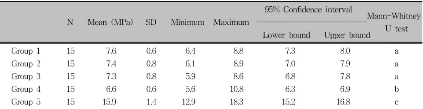
Group 1 15 7.6 0.6 6.4 8.8 7.3 8.0 a
Group 2 15 7.4 0.8 6.1 8.9 7.0 7.9 a
Group 3 15 7.3 0.8 5.9 8.6 6.8 7.8 a
Group 4 15 6.6 0.6 5.6 10.8 6.3 6.9 b
Group 5 15 15.9 1.4 12.9 18.3 15.2 16.8 c
Group 1, Transbond XT; Group 2, Transbond XT + Ortho-Coat; Group 3, Transbond XT + Cervitec Plus; Group 4, Aegis Ortho; Group 5, Transbond XT Paste + Clearfil Protect Bond; n, sample size; MPa, Megapascal; SD, standard deviation. aSame letters indicate lack of statistically significant difference.
Table 1. Descriptive statistics related to shear bond strength data and statistical comparisons
Groups Mean SD Median Minimum Maximum
Group 1 2.4a 0.9 2 1 4
Group 2 2.1a 0.9 2 1 3
Group 3 2.1a 0.9 2 1 4
Group 4 1.9a 0.8 2 1 3
Group 5 4.1b 0.9 4 3 5
Group 1, Transbond XT; Group 2, Transbond XT + Ortho-Coat; Group 3, Transbond XT + Cervitec Plus;
Group 4, Aegis Ortho; Group 5, Transbond XT Paste + Clearfil Protect Bond; SD, standard deviation. Groups with same subscripts are not significantly different; p
< 0.001.
Table 2. Descriptive statistics of adhesive remnant in- dex (ARI) scores
were not normally distributed and because of the small sample size, nonparametric tests were used. Kruskal- Wallis one-way ANOVA was used to determine differ- ences in bond strength data and ARI values. As the p-values of Kruskal-Wallis tests were less than 0.001, pairwise comparisons were performed using Mann- Whitney U-test adjusted according to Bonferoni at a significance level of p < 0.005.
RESULTS
The bond strength values and the statistical compar- isons are presented in Table 1. Kruskal-Wallis test re- vealed significant differences among groups (p < 0.005). Pairwise comparison of the groups with the Mann-Whitney U test showed no significant differ- ences between groups 1, 2 and 3, whereas groups 4 and 5 showed statistically significant differences when compared with each other and with the other groups (p
< 0.005). Group 5 had the highest bond strength, whereas group 4 had the lowest bond strength values (Table 1).
The ARI scores are displayed in Table 2. No enam- el fractures were detected in any of the specimens.
Statistically significant differences were found with the Kruskal-Wallis test (p < 0.001). The Mann-Whitney U test showed that group 5 displayed a significantly greater ARI score when compared to the other groups (p < 0.001).
DISCUSSION
In the current study the main aim was to determine whether different adhesive systems and enamel pro- tective agents would affect the tensile bond strength of lingual brackets bonded to the enamel of lower incisor teeth. Different from the labial surface, the lingual side of lower anterior teeth have a convex surface top- ography such as a cingulum, which may exaggerate the results of commonly used shear bond strength tests.
For this reason, the tensile bond strength test was pre- ferred in order to eliminate the effect of lingual morphology.
The use of orthodontic fixed appliances creates sig- nificant oral hygiene challenges for the patient and the
clinician throughout the long treatment periods. The re- sults of studies have suggested using enamel protective methods which reduce the dependence on patient com- pliance in preventing the development of white spot le- sions around brackets and at the gingival margins.17 Proper oral hygiene is more difficult to maintain in pa- tients with lingual brackets, because hygiene control is harder from the lingual than from the buccal side, therefore plaque accumulations, gingivitis and demine- ralization cannot be detected by the patient.2 The prob- lems in maintaining oral hygiene in lingual orthodontic patients can be handled by using special prophylactic procedures.18
The recently developed light-cured reinforced resin, Ortho Coat claims to prevent decalcification, micro leakage, and discoloration of teeth under orthodontic brackets. It coats the enamel around the brackets and prevents food and bacteria from collecting around and under the brackets. Another product used in this study, Cervitec Plus is a further development of Cervitec, a protective varnish containing chlorhexidine. The caries preventive effect of Cervitec during orthodontic thera- pies has been investigated in two studies.19,20 Applicati- on of these products to the enamel surface could pro- vide protection throughout the orthodontic treatment but they could also have an adverse effect on the bond strength of brackets, depending on the application me- thod.5 The effects of similar products on the bond strength of lingual orthodontic brackets have not been elucidated yet. The differences of the etching surface and the anatomical surfaces might have an effect on bond strength quality and quantity of mandibular incisors.
Both of the products were applied around the brack- et periphery in the present study, and the results re- vealed that neither the fluoride-releasing light cure res- in nor the chlorhexidine varnish had a significant ad- verse effect on the enamel bond strength. This was in accordance with previous studies.21-23 In contrast to our findings, Meng et al.24 found that the application of acidulated phosphate fluoride after acid etching enamel had an adverse effect on orthodontic bond strength of human enamel. Additionally, Karaman and Uysal22 stated that different forms of antimicrobial varnishes have some effect on bond strength of orthodontic
brackets.
Aegis Ortho which is a light-cured orthodontic adhe- sive containing Amorphous Calcium Phosphate (ACP), promotes the enhancement of the tooth's natural repair mechanism by releasing calcium and phosphate ions. A recent report has shown that the ACP-containing adhe- sive demonstrated a bond strength of 6.6 ± 1.5 MPa, and declared that although this adhesive demonstrated a low bond strength, it produced a consistent bond.12 Similarly we found a mean bond strength of 6.6 ± 0.6 MPa, which was significantly lower than the other groups. Consistent with previous studies, the anti- microbial self-etching system, Clearfil Protect Bond demonstrated the highest bond strength among the oth- er groups.25,26 The mean bond strength value in this study was 15.9 ± 1.4 MPa, which was similar to the results of a previous study.25 The present bond strength values were found to be higher than Reynolds’27 mini- mal bond strength values that are clinically acceptable (5.9 - 7.8 MPa). However, lower incisors have thinner layers of enamel than other teeth which was claimed to limit their load dissipation potential resulting in en- amel fracture. A bond strength greater than 8 - 9 MPa has been argued to exceed the clinical requirements.16 These results demonstrate that the use of Clearfil Protect Bond, especially where the enamel is likely to be thin may cause a safety issue for orthodontic purposes. On the other hand, current results depend on in-vitro evaluations, and none of the materials have been subjected to different features of an oral environ- ment.
When the ARI scores were considered, a significant difference was observed for group 5, showing less composite remaining on the tooth after debonding when compared with the other groups (Table 2). The majority of the mean scores ranged around 2, indicat- ing that most of the adhesive remained on the tooth surface after debonding in groups 1, 2, 3 and 4. The lowest ARI scores were found in group 4, indicating that almost all the adhesive remained on the tooth, which suggests that the bond between the bracket and the adhesive was weak. Although group 5 had the stat- istically highest bond strength value, ARI scores re- vealed that less adhesive resin remained on the enamel surface in this group. This suggests that the predom-
inant mode of bracket failure was at the enamel-adhe- sive resin interface in Clearfil Protect Bond, which was in accordance with a previous report.28 There are con- flicting results about the amount of residual adhesive on teeth which might have emanated from the different bonding systems, differences in the bracket sizes and designs, or the classification system of ARI.29,30 In our conception, high bond strength during orthodontic treatment and shorter chair time for residual resin re- moval during debonding would be beneficial in clinical performance. One of the major problems in lingual or- thodontics is the clear up of composite from lingual tooth surfaces which is time consuming and un- comfortable for the patient. However, excessive bond strength and bracket failure at the enamel composite interface should be balanced in order to avoid enamel fractures.
CONCLUSION
The findings indicate that application of fluoride-re- leasing resin or chlorhexidine products did not sig- nificantly affect bond strength of lingual orthodontic brackets. Although acceptable bond strengths were ob- served in all tested groups, the excessive bond strength achieved with Clearfil Protect Bond should be taken into consideration especially where the enamel is thin.
- 국문초록 -
하악 전치 설측면에 대한 다양한 접착시스템의 접착강도
Çağrı Türköz, Burcu Baloş Tuncer, Mehmet Çağrı Ulusoy, Cumhur Tuncer
본 연구의 목적은 서로 다른 종류의 접착시스템과 법랑질 보호제 등이 설측 브라켓의 인장강도에 영향을 미치는지 알 아보는데 있다. 75개의 발치된 하악 전치를 5개의 그룹으로 임의로 나눈 후 설측 브라켓을 부착하였다. Group 1은 Transbond XT를, Group 2는 Transbond XT와 함께 불소 유 리레진(Ortho-coat)을 Group 3는 Transbond XT와 Chloro- hexidine varnish (Cervitec Plus)를 Group 4는 광중합 접착 제(Aegis Ortho)를 Group 5는 antimicrobial self-etching pri- mer (Clearfil Protect Bond)을 사용하였다. 불소유리 레진이
나 Chlorohexidine varnish의 사용유무는 접착력에 영향을 미치지 못하였다. Group 5이 다른 그룹에 비해 접착력과 접 착제 잔류지수(adhesive remnant index, ARI)가 통계적으로 유의하게 높았다 (p < 0.001). 법랑질 보호제 적용 시 설측 브라켓의 접착력에 부정적인 영향을 미치지 못하였다. 이상 의 결과로 이번에 사용한 제품들은 설측 교정치료 기간 동안 환자의 구강위생을 개선시킬 수 있어 환자와 술자에게 도움 을 줄 수 있을 것이다. 그러나 에나멜 층이 얇은 경우 Clearfil Protect Bond의 과도한 접착강도에 대해 고려해야 할 것이다.
주요 단어: 설측면, 접착제, 레진, 법랑질 보호제
REFERENCES
1. Caniklioglu C, Oztürk Y. Patient discomfort: a comparison be- tween lingual and labial fixed appliances. Angle Orthod 2005;75:86-91.
2. Hohoff A, Stamm T, Kühne N, Wiechmann D, Haufe S, Lippold C, et al. Effects of a mechanical interdental cleaning device on oral hygiene in patients with lingual brackets. Angle Orthod 2003;73:579-87.
3. Miyawaki S, Yasuhara M, Koh Y. Discomfort caused by bonded lingual orthodontic appliances in adult patients as ex- amined by retrospective questionnaire. Am J Orthod Dentofa- cial Orthop 1999;115:83-8.
4. Sinclair PM, Cannito MF, Goates LJ, Solomos LF, Alexander CM. Patient responses to lingual appliances. J Clin Orthod 1986;20:396-404.
5. Bishara SE, Vonwald L, Zamtua J, Damon PL. Effects of vari- ous methods of chlorhexidine application on shear bond strength. Am J Orthod Dentofacial Orthop 1998;114:150-3.
6. Mattingly JA, Sauer GJ, Yancey JM, Arnold RR. Enhancement of Streptococcus mutans colonization by direct bonded ortho- dontic appliances. J Dent Res 1983;62:1209-11.
7. Scheie AA, Arneberg P, Krogstad O. Effect of orthodontic treatment on prevalence of Streptococcus mutans in plaque and saliva. Scand J Dent Res 1984;92:211-7.
8. Corbett JA, Brown LR, Keene HJ, Horton IM. Comparison of Streptococcus mutans concentrations in non-banded and band- ed orthodontic patients. J Dent Res 1981;60:1936-42.
9. Lundström F, Krasse B. Streptococcus mutans and lactobacilli frequency in orthodontic patients; the effect of chlorhexidine treatments. Eur J Orthod 1987;9:109-16.
10. El Bokle D, Munir H. An in vitro study of the effect of Pro Seal varnish on the shear bond strength of orthodontic brackets. World J Orthod 2008;9:141-6.
11. Sköld-Larsson K, Borgström MK, Twetman S. Effect of an antibacterial varnish on lactic acid production in plaque ad- jacent to fixed orthodontic appliances. Clin Oral Investig 2001;5:118-21.
12. Foster JA, Berzins DW, Bradley TG. Bond strength of an amorphous calcium phosphate-containing orthodontic adhesive.
Angle Orthod 2008;78:339-44.
13. Imazato S, Kinomoto Y, Tarumi H, Torii M, Russell RR, McCabe JF. Incorporation of antibacterial monomer MDPB in- to dentin primer. J Dent Res 1997;76:768-72.
14. Imazato S, Kuramoto A, Takahashi Y, Ebisu S, Peters MC. In vitro antibacterial effects of the dentin primer of Clearfil Protect Bond. Dent Mater 2006;22:527-32.
15. Gorelick L, Geiger AM, Gwinnett AJ. Incidence of white spot formation after bonding and banding. Am J Orthod 1982;
81:93-8.
16. Hobson RS, McCabe JF, Hogg SD. Bond strength to surface enamel for different tooth types. Dent Mater 2001;17:184-9.
17. Geiger AM, Gorelick L, Gwinnett AJ, Benson BJ. Reducing white spot lesions in orthodontic populations with fluoride rinsing. Am J Orthod Dentofacial Orthop 1992;101:403-7.
18. Hohoff A, Fillion D, Stamm T, Goder G, Sauerland C, Ehmer U. Oral comfort, function and hygiene in patients with lingual brackets. A prospective longitudinal study. J Orofac Orthop 2003;64:359-71.
19. Ogaard B, Larsson E, Glans R, Henriksson T, Birkhed D.
Antimicrobial effect of a chlorhexidine-thymol varnish (Cervitec) in orthodontic patients. A prospective, randomized clinical trial. J Orofac Orthop 1997;58:206-13.
20. Øgaard B, Larsson E, Henriksson T, Birkhed D, Bishara SE.
Effects of combined application of antimicrobial and fluoride varnishes in orthodontic patients. Am J Orthod Dentofacial Orthop 2001;120:28-35.
21. Bishara SE, Damon PL, Olsen ME, Jakobsen JR. Effect of ap- plying chlorhexidine antibacterial agent on the shear bond strength of orthodontic brackets. Angle Orthod 1996;66:313-6.
22. Karaman AI, Uysal T. Effectiveness of a hydrophilic primer
when different antimicrobial agents are mixed. Angle Orthod 2004;74:414-9.
23. Wang WN, Sheen DH. The effect of pretreatment with fluo- ride on the tensile strength of orthodontic bonding. Angle Orthod 1991;61:31-4.
24. Meng CL, Li CH, Wang WN. Bond strength with APF applied after acid etching. Am J Orthod Dentofacial Orthop 1998;114:
510-3.
25. Arhun N, Arman A, Sesen C, Karabulut E, Korkmaz Y, Gokalp S. Shear bond strength of orthodontic brackets with 3 self-etch adhesives. Am J Orthod Dentofacial Orthop 2006;
129:547-50.
26. Bishara SE, Soliman M, Laffoon J, Warren JJ. Effect of anti- microbial monomer-containing adhesive on shear bond strength of orthodontic brackets. Angle Orthod 2005;75:397-9.
27. Reynolds IR. A review of direct orthodontic bonding. Br J Orthod 1975;2:171-8.
28. Korbmacher HM, Huck L, Kahl-Nieke B. Fluoride-releasing adhesive and antimicrobial self-etching primer effects on shear bond strength of orthodontic brackets. Angle Orthod 2006;
76:845-50.
29. Bishara SE, Gordan VV, VonWald L, Jakobsen JR. Shear bond strength of composite, glass ionomer, and acidic primer adhesive systems. Am J Orthod Dentofacial Orthop 1999;115:
24-8.
30. Bishara SE, VonWald L, Laffoon JF, Warren JJ. Effect of a self-etch primer/adhesive on the shear bond strength of ortho- dontic brackets. Am J Orthod Dentofacial Orthop 2001;119:
621-4.