Denture due to Multiple Dental Implant Loss of the Fixed Implant Supported Prosthesis
다수의 임플란트발거로 임플란트 고정성 보철이 실패한 환자에서의 잔존 임플란트를 이용한 부분 가철성 국소의치 수복증례
Jeong Kyung Kang,DDS,MSD, Gi Hoon Nam, DDS VHS Medical Center,Dental Hospital,Dept.Prosthodontics 강정경, 남기훈 / 보훈공단 중앙보훈병원 치과보철과
There are several treatment options for rehabilitation of partial edentulism including the use of conventional or implant- retained fixed prostheses. However, such prosthetic options cannot always be possible because of compromised general and oral health (i.e. loss of supporting tissues, medical reasons , extensive surgical protocol and osseointegration failure of dental implant) as well as the affordability of patients. In some cases, removable partial denture provides easier access for oral hygiene procedures and the ability to correct discrepancies in dental arch relationships than implant fixed prosthesis. Recently, Implant Supported Removable Partial Denture (ISRPD) where to place dental implant in strategic position has been suggested to improve the limitation and shortcomings of conventional RPD. ISPRD can overcome mechanical limition of conventional RPD by placing implant in a favorable position and can be cost-effective, prosthetic solution for partially edentulous patients who are not immediate candidates for extensive, fixed implant supported restorations. Incorporation of dental implants to improve the RPD support and retention and to enhance patient acceptance should be considered when treatment planning for RPD.
In this case, 59 years old male patient who received dental treatment of implant fixed prosthesis on both side of the upper jaw and implant overdenture on lower jaw showed implant abutment screw fracture on #15i and osseointegration failure on multiple number of implants. After removing failed implants, we planned ISRPD using #15i,24i,25i,26i and #23 natural tooth for RPD abutment.
We fabricated #23 surveyed crown,#24i=25i=26i surveyed bridge and #15i gold coping for support,retention and stability for RPD.
Periodic follow up check for 2years has been performed since the ISRPD delivery to the patient. No sign of screw loosening, fracture or bone resorption around abutment implants were detected. Keywords: Implant supported RPD, dental implant (J Korean Acad Esthet Dent 2014;23(1):34-40)
부분 무치악을 수복하는 데 있어서 선택할 수 있는 치료의 옵션으로는 전통적인 국소의치와 임플란트 지지-고정성 보철물 등이 있다. 하지만, 환자의 전신적 또는 구강의 상태(수술적인 술식이 제한되는 전신병력, 지지조직의 부족 그리고 골유착에 실 패한 임플란트)와 치료비용에 대한 허용 정도에 따라 모든 옵션이 항상 가능한 것은 아니다. 가철성 국소의치는 임플란트 고정 성 보철물에 비해 구강위생 관리 및 상,하악 악간관계의 부조화를 수정하기에 편리한 장점이 있다. 최근에는 전략적 위치에 임 플란트를 식립하여 기존 악궁 형태에서는 제한되는 국소의치 디자인의 한계를 개선할 수 있는 임플란트지지형 RPD(Implant Supported Removable Partial Denture)가 새로운 방안으로 대두되고 있다. ISRPD는 전략적 위치에 임플란트를 식립하여 역학적 인 한계를 극복할 수 있을 뿐 만 아니라 전악의 임플란트지지형 고정성 보철이 제한되는 환자에서 보다 경제적이고 현실적인 보
로 대두되고 있다. ISRPD는 전략적 위치에 임플란트를 식립하여 역학적인 한계를 극복할 수 있을 뿐 만 아니라 전악 의 임플란트지지형 고정성 보철이 제한되는 환자에서 보다 경제적이고 현실적인 보철적 해결책이 될 수 있다. 따라서, RPD를 이용한 보철계획 수립시 전략적 위치에서의 임플란트 사용은 고전적인 가철성 국소의치에서보다 유지력과 안 정성을 증진시키고 구강위생관리 또한 용이하여 환자의 적응도를 높이는 방안으로 고려될 수 있다.
임상증례
1. Patient Information
● Age/Sex :59Y/M
● C.C :Mobility of #12i,35i Implant
● PMH :Non-Specific Findings
● Present Illness: #12i, 35i Implant mobility(++). Peri-implantitis 2. Past Dental History
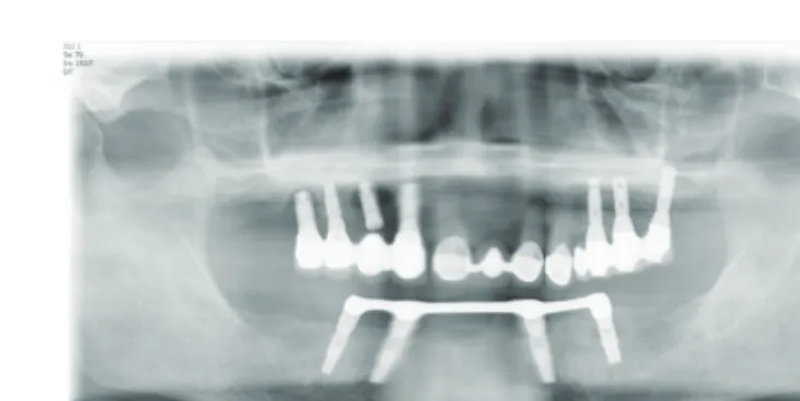
Fig. 1. 2008년 파노라마 사진 Fig. 2. 2009년 파노라마 사진
2008년 환자가 본원에 최초 내원했을 당시에 임상검사 및 방사선검사 결과 상악은#12i=15i,16i(#13i는 sleeping fixture state), #24i=25i=26i 임플란트 고정성 보철물이였고 #11=22 PFM Bridge는 2도 동요도 상태였다. 하악에는
#33i,35i,43i,45i를 이용한 Bar-type overdenture를 사용중이였고 #34i 임플란트는 주변에 골소실 소견이 보였다. 하악 denture 내면에는 fracture line 존재하였다. 진행한 치료로는 #11=22 발치 시행 후 flipper 제작하였고 Peri-implantitis 진행중이었던 #13i 와 #16i는 제거하였다. (Fig. 1)
파절된 하악 overdenture는 재제작하였으며 #11=22 부위는 임플란트 식립 후 임플란트 고정성 보철물 계획하였으 나 환자분이 flipper 당분간 더 쓰신다고 하셔서 더 이상 치료진행을 못하였다. (Fig. 2)
이후에 환자분이 2012년 본원에 재내원했을 때, #35i 임플란트는 bone loss가 진행되어 동요도를 보였으며 #12i 임 플란트 역시 동요도를 보였다.(Fig. 3) 환자분의 추가 수술에 대한 거부와, 현재 잔존치, 잔존 임플란트 상태를 고려하 여 치료계획을 수립하였다.
3. Treatment plan
1. Removal of #12i,35i Implant
2. Mn. Hader Bar cutting & Mn. Overdenture repair
3. #23 single surveyed crown,, #24i,25i,26i Implant PFM Surveyed bridge 4. #15i Gold Coping
5. Mx. Implant Supported Removable Partial Denture (ISRPD)
4. Treatment Sequence
먼저 하악에 대한 치료로는 임플란트 주위염으로인한 골소실로 동요도 보이는 #35i 임플란트 제거하였다. 그리고
#33i,35i 부위 bar를 cutting 하고 기존에 사용중이었던 overdenture는 repair 시행하였다. (Fig. 4, 5) Fig. 3. 2012년 파노라마 사진
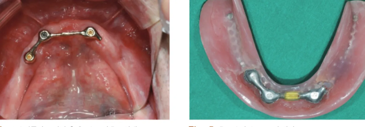
골유착 실패로 동요도 보인 #12i 임플란트는 제거하고 상악 좌측 보철물을 철거하였다. 상악의 잔존 지대치와 잔 존 임플란트는 #23과 #15i,24i,25i,26i가 남게 되었다.(Fig. 6) #23는 surveyed crown 재제작하였고, #24i,25i,26i는 surveyed bridge 제작하였다. #23 surveyed crown은 cingulum rest seat와 mesial guiding plane을, #24i,25i,26i Surveyed Bridge는 distal rest seat와 palatal측에 ledge를, 그리고 #26i distal guiding plane을 부여하였다.(Fig. 7)
#15i 임플란트는 보철 진행 중에 Healing abutment의 screw가 파절되었다. Fixture 내면의 파절된 screw를 round bur 로 삭제 후에 casting post와 같은 방법으로 gold coping 제작 후 final setting하였다. #23 surveyed crown,#24i,25i,26i surveyed bridge 완성 후 시적하였다. (Fig. 8)
Fig. 4. #35i 임플란트 발거 후 Cutting 된 Bar 상태 Fig. 5. Repair된 denture의 내면
Fig. 6. #12i 임플란트 발거하였고 상부 보철물 철거후 구강내 사진
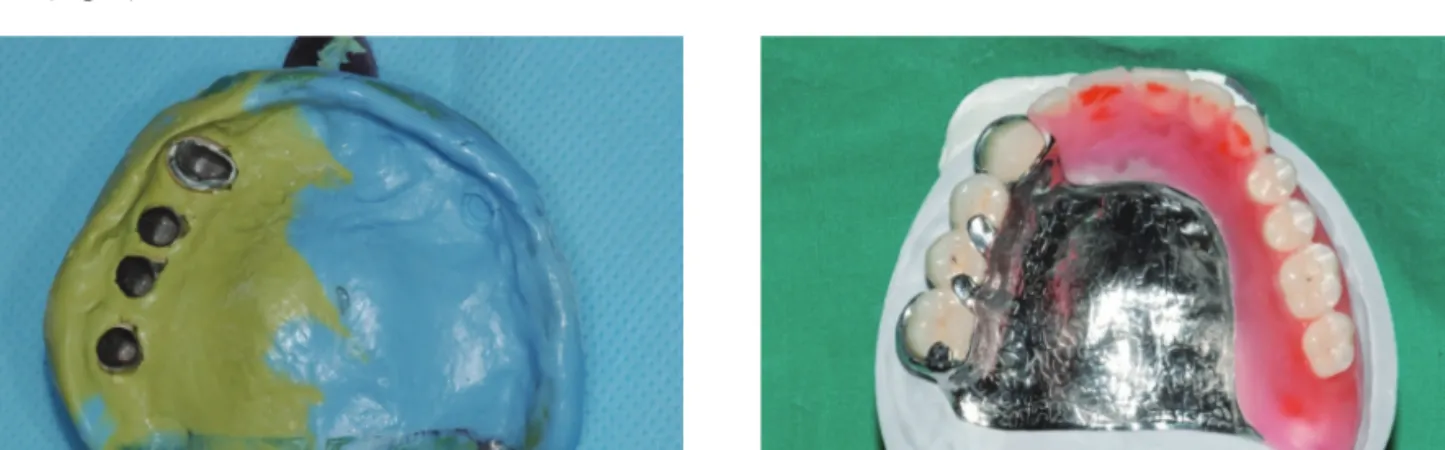
제작된 Surveyed Crown을 이용한 상악 RPD framework 제작을 위해 통법대로 PVS (Polyvinyl siloxane)인상재료로 pick-up impression 채득하였다. (Fig. 9)
[상악 RPD design]
1. Palatal plate-type major connector
2. #23 cingulum rest +proximal plate +akers clasp 3. #24i,25i distal rest
4. #26i distal rest+proximal plate +akers clasp
임플란트로의 과도한 부하를 방지하고 저작력의 분산을 위해 major connector로 full palatal plate를 선택하였고 모 든 지대치에 rest 및 ledge를 형성하였다. 그리고 적절한 lip support 및 심미성 고려하고 납의치 제작하고 시적 후 평가 하였다. (Fig10)
Fig. 7. #23과 #24i,25i,26i Surveyed Crown을 제작
Fig. 7. Functional Impression 채득
Fig. 8. 제작한 surveyed crown과 #14i coping의 시적
Fig. 8. Framework 주조와 납의치 제작
결론
본 증례는 기존의 다수 임플란트 발거로 고정성 임플란트 보철이 불가능한 상태에서 잔존 임플란트를 이용한 임플 란트지지형 가철성 국소의치(ISRPD)로써 환자만족도와 저작능력, 심미성의 회복면에서 만족할 만한 임상결과를 얻 었기에 소개하고자 한다. 반복된 임플란트의 실패로 인한 환자의 수술에 대한 거부와 고정성 보철의 예후가 의심스러 울 경우 잔존 임플란트를 활용한 RPD는 부분 무치악 치료증례에서 훌륭한 대안이 될 수 있을 것으로 보인다. 의치 장 착 후 2년간 F/U 중이며 지대치로 사용한 임플란트에서 screw loosening이나 파절, 골흡수 등의 증상은 현재까지 관찰 되지 않았다.
References
1. Grossmann Y, Nissan J, Levin L. Clinical Effectiveness of implant-supported removable partial dentures. J Oral Maxillofac Surg 2009;67(9):1941-6
2. Kuzmanovic DV, Alan GT, Purton DG. Distal implants to modify the Kennedy classification of a removable partial denture: A clinical report. J Prosthet Dent 2004;92:8-11
3. Uludag B, Celik G. Fabrication of a maxillary implant-supported removable partial denture: A clinical report. J Fig. 11. 의치 완성 후 시적
distal extension: a systematic review. J Oral Rehabil 2012;39:791-8
6. Shahmiri A, Atieh MA. Mandibular Kennedy Class I implant-tooth-borne removable partial denture: a systematic review. J Oral Rehabil 2010;37:225–34