https://doi.org/10.5468/ogs.2018.61.1.38 pISSN 2287-8572 · eISSN 2287-8580
Introduction
Vaginal microbiome composition changes when women be- come pregnant. Pregnancy is accompanied by a shift in the community vaginal bacteria to a composition that is typically dominated by Lactobacillus [1]. This change is believed to in- hibit pathogen growth through secretion of antibacterial bac- teriocins, such as lactic acid that can maintain an acid pH [2,3].
Disturbed vaginal environment is associated with complica- tions of pregnancy, particularly an increased risk of preterm birth [4].
Prevalence of vaginal microorganisms among pregnant women according to trimester and association with preterm birth
Kyung-A Son, Minji Kim, Yoo Min Kim, Soo Hyun Kim, Suk-Joo Choi, Soo-young Oh, Cheong-Rae Roh, Jong-Hwa Kim
Department of Obstetrics and Gynecology, Samsung Medical Center, Sungkyunkwan University School of Medicine, Seoul, Korea
Objective
The aim of this study was to investigate the prevalence of abnormal vaginal microorganisms in pregnant women according to trimester, and to determine whether the presence of abnormal vaginal colonization is associated with higher risk of miscarriage or preterm delivery. Furthermore, we analyzed delivery outcomes according to individual microorganism species.
Methods
We included pregnant women who underwent vaginal culture during routine prenatal check-up between January 2011 and June 2016. We compared delivery outcomes according to the presence or absence of abnormal vaginal flora grouped by trimester.
Results
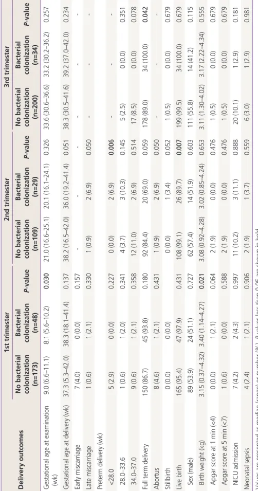
This study included 593 singleton pregnancies. We classified participants into 3 groups, according to the trimester in which vaginal culture was performed; 1st trimester (n=221), 2nd trimester (n=138), and 3rd trimester (n=234). Abnormal vaginal colonization rate significantly decreased with advancing trimester of pregnancy (21.7% for 1st, 21.0% for 2nd, 14.5% for 3rd; P=0.048). Abnormal vaginal colonization detected in the 2nd trimester but not in 1st trimester was associated with a significant increase in preterm delivery before 28 weeks of gestation (6.9% vs. 0%; P=0.006).
Among abnormal vaginal flora isolated in the 2nd trimester, the presence of Klebsiella pneumonia was identified as significant microorganism associated with preterm delivery before 28 weeks of gestation (50% vs. 0.7% for K.
pneumonia; P=0.029).
Conclusion
There is an association between abnormal vaginal colonization detected in the 2nd trimester and preterm delivery before 28 weeks. K. pneumonia has been identified as the likely causative microorganisms.
Keywords: Microbiota; Pregnancy; Premature birth; Klebsiella
Articles published in Obstet Gynecol Sci are open-access, distributed under the terms of the Creative Commons Attribution Non-Commercial License (http://creativecommons.
org/licenses/by-nc/3.0/) which permits unrestricted non-commercial use, distribution, and reproduction in any medium, provided the original work is properly cited.
Copyright © 2018 Korean Society of Obstetrics and Gynecology
Received: 2017.03.29. Revised: 2017.07.06. Accepted: 2017.07.19.
Corresponding author: Soo-young Oh
Department of Obstetrics and Gynecology, Samsung Medical Center, Sungkyunkwan University School of Medicine, 81 Irwon- ro, Gangnam-gu, Seoul 06351, Korea
E-mail: [email protected]
https://orcid.org/0000-0003-3002-0048
Abnormal vaginal colonization, which replaces normal lac- tobacilli during pregnancy, includes bacterial vaginosis or aerobic vaginitis. Whereas bacterial vaginosis is dominated by anaerobic overgrowth, aerobic vaginitis is characterized by microorganisms such as Escherichia coli, group B streptococci, and Enterococci [5,6].
The association between bacterial vaginosis and preterm delivery has been extensively studied, while there is limited research on the clinical significance of abnormal vaginal colo- nization by aerobic bacteria, particularly in relation to preterm delivery. The exact prevalence of each vaginal microorganism throughout gestation has rarely been reported. Ascending infection by abnormal vaginal microorganisms from the lower genital tract is a well-known route of intra-amniotic infection associated with preterm birth. Furthermore, microorganisms isolated from infected amniotic fluid are mostly aerobic bac- teria, rather than anaerobe causing bacterial vaginosis, a de- tailed analysis of the change in vaginal microorganism during pregnancy is critically important [4,7,8].
The aim of this study is to investigate the prevalence of ab- normal vaginal microorganisms, detected by Gram staining and culture in pregnant women, classified by trimester, and to determine if the presence of abnormal vaginal colonization is associated with a higher risk of miscarriage or preterm de- livery. Furthermore, we tried to analyze delivery outcomes ac- cording to individual microorganism species.
Materials and methods
We included pregnant women who underwent vaginal culture during their routine prenatal check-up in our institution be- tween January 2011 and June 2016. Those included were pa- tients who visited our institution for prenatal check-up for the first time. Women with multiple pregnancies were excluded.
All vaginal sampling for Gram staining and culture was con- secutively done regardless of symptoms by a single physician who performed routine vaginal screening upon his principle.
After insertion of a water-lubricated sterile speculum, a smear was taken from vaginal posterior fornix using a sterile cotton swab. All vaginal culture was incubated primarily under an aerobic condition.
By retrospective review of medical records, we collected data on the maternal baseline characteristics including age, par- ity, pre-pregnancy body mass index (BMI), smoking, history
of spontaneous preterm delivery, and comorbidity. Maternal hypertension, diabetes, and thyroid disease were classified as comorbidities. Gestational age at vaginal culture examination and the results were also examined. We divided the study population into 3 groups according to the trimester in which vaginal culture was performed. Delivery outcomes including gestational age at delivery, mode of delivery, birth weight, sex, Apgar score, neonatal intensive care unit (NICU) admission and early neonatal sepsis were investigated. Miscarriage was divided into early miscarriage (<14 weeks) and late miscar- riage (14.0–21.6 weeks) as previously indicated [9]. Preterm delivery was further divided into 3 groups (22.0–27.6 weeks;
28.0–33.6 weeks; 34.0–36.6 weeks). Early neonatal sepsis was defined when microorganisms were isolated from the blood of neonates within 7 days of birth. Patients delivering newborns with major anomalies or twins were excluded from the study.
Statistical analysis was performed with SPSS 18.0 (IBM Corp., Armonk, NY, USA). The Mann-Whitney U test and the Kruskal- Wallis test were used for numeric data and Fisher’s exact test was used for categorical data among groups. Linear by linear association analysis was also used to check trends in each tri- mester. A P-value <0.05 was considered statistically significant.
Results
This study included 593 singleton pregnancies. We classified the study population into 3 groups, according to the trimester in which vaginal culture was performed; 1st trimester (n=221), 2nd trimester (n=138), and 3rd trimester (n=234) group. Table 1 shows the clinical characteristics of the study population grouped by trimester. The median gestational age at vaginal culture examination for each trimester group was 8.6 weeks, 20.6 weeks, and 33.6 weeks, in the 1st, 2nd, and 3rd trimes- ter, respectively. Pre-pregnancy BMI, nationality, smoking his- tory, and maternal comorbidity were similar among groups.
Most participants were of South Korean nationality, with other nationalities including Russian, North American, and Vietnamese. There were no differences between the groups in the rates of nulliparity and history of spontaneous preterm birth.
In Table 2, abnormal vaginal colonization rate was 21.7%,
21.0%, and 14.5% in 1st, 2nd, and 3rd trimester respectively,
showing a significant decrease with advancing trimester of
pregnancy (P=0.048, linear-by-linear association). Gram-neg- ative bacteria significantly decreased with advancing trimester (P=0.008). When analyzed by microorganism species, the prevalence of E. coli colonization significantly decreased as the pregnancy progressed: 1st trimester (6.3%), 2nd trimester (3.6%), and 3rd trimester (1.7%). Although the overall preva-
lence of Gram-positive cocci also significantly decreased with advancing trimester of pregnancy (P=0.012), there were no sig- nificant changes in the individual species over the trimesters of pregnancy. There were no differences in the prevalence rate of Candida across the trimesters.
To analyze the association of miscarriage or preterm delivery
Table 1. Clinical characteristics of pregnant women in each trimester
Characteristics 1st trimester (n=221) 2nd trimester (n=138) 3rd trimester (n=234) P-value
Maternal age 33.8 (24–45) 33.3 (22–43) 32.1 (19–44) <0.001
Body mass index before pregnancy (kg/m
2) 21.3 (16.1–37.5) 21.1 (15.6–37.9) 21.1 (15.6–44.9) 0.859
Nulliparity 100 (45.3) 75 (54.3) 117 (50.0) 0.234
History of spontaneous preterm birth 10 (4.5) 6 (4.3) 10 (4.3) 0.991
Smoking 0.588
Never smoker 220 (99.5) 138 (100.0) 231 (98.8)
Previous smoker 1 (0.5) 0 (0.0) 2 (0.8)
Current smoker 0 (0.0) 0 (0.0) 1 (0.4)
Comorbidity 0.761
No 184 (83.3) 111 (80.4) 189 (80.8)
Yes 37 (16.7) 27 (19.6) 45 (19.2)
Nationality 0.454
South Korea 200 (90.5) 130 (94.2) 214 (91.4)
Others 21 (9.5) 8 (5.8) 20 (8.6)
Gestational age at examination (wk) 8.6 (4.1–13.5) 20.6 (14.0–27.6) 33.6 (28.0–40.6) <0.001 Values are presented as number (range) or number (%). P-values less than 0.05 are shown in bold.
Table 2. The prevalence of vaginal microorganisms in each trimester
Microorganisms 1st trimester
(n=221) 2nd trimester
(n=138) 3rd trimester
(n=234) P-value
a)P-value
b)Abnormal vaginal colonization 48 (21.7) 29 (21.0) 34 (14.5) 0.130 0.048
Gram-negative bacteria 18 (8.1) 7 (5.1) 6
c)(2.6) 0.028 0.008
Escherichia coli 14 (6.3) 5 (3.6) 4 (1.7) 0.038 0.011
Enterobacteriae 1 (0.5) 0 (0.0) 0 (0.0) 0.605 0.243
Klebsiella pneumonia 2 (0.9) 2 (1.4) 1 (0.4) 0.646 0.570
Pseudomonas aeruginosa 1 (0.5) 0 (0.0) 0 (0.0) 0.605 0.243
Gram-positive cocci 26 (11.8) 13 (9.4) 12 (5.1) 0.038 0.012
Staphylococcus aureus 4 (1.8) 2 (1.4) 2 (0.9) 0.665 0.377
Streptococcus agalactiae 10 (4.5) 7 (5.1) 7 (3.0) 0.556 0.402
Enterococcus faecalis 1 (0.5) 0 (0.0) 0 (0.0) 0.605 0.243
Other gram-positive cocci 11 (5.0) 4 (2.9) 3 (1.3) 0.071 0.022
Candida 16 (7.2) 13 (9.4) 20 (8.5) 0.759 0.619
Values are presented as number (%). P-values less than 0.05 are shown in bold.
a)