대한방사선의학호|지 1998;38: 145-150
자기공명영상의 액체액체층:근골격계 질환에서의 의의 1
정 혜 원 1, 2 . 이 경 원 · 송 치 성 2. 한 상 욱 · 강 홍 식
목 적 : 자기공명영상에서 액체액체층 (fluid-fluid level) 이 관찰되는 근골격계 질환의 종 류와각각의 빈도를알아보고,액체액체층의 형태나신호강도의 차이에 의한질환간의 감별 점을찾고자하였다.
대상 및 방법 : 자기공명영상에서 액체액체층이 보이고 병리조직학적 또는 임상적으로 확 진된 30예를 대상으로 질환의 종류와 각각의 빈도를 알아보고, 자기공명영상에서 액체액체 층의 개수 또는 내부신호강도가 질환 종류에 따라 차이가 있는 지를 후향적으로 분석하였 다.
결 과 : 액체액체층을 보인 질환은 거대세포종 6예, 모세혈관확장성 골육종 4예, 동맥류 성 골낭과 활막육종이 각각 3예, 연골아세포종, 연부조직의 결핵성 농양과 혈종이 각각 2예, 혈관종, 신경초종, 전이암, 악성섬유조직구종 (malignant fibrous histiocytoma), 전형적 골 육종 (classic osteogenic sarcoma), 활액 낭염 (bursi tis), 화농성 농양과 유표피 봉입 낭종
(epidermoid inclusion cyst) 이 각각 l 예로서, 양성종양이 14예, 악성종양이 10예 그리고 비 종양성 칠환이 6예이었다. 혈종, 결핵성 놓양, 그리고 유표피 봉입 낭종에서는 단방성 냥포 내에 하나의 액체액체층이 있였으나그이외의 질환에서는복잡한다방성 냥포내에서 여러 액체액체층이 보였다. 액체액체층을 보이는 낭포의 내부신호강도는 T1 강조영상에서는 다 양하였고 T2 강조영상에서는 대부분 (27/30) 상층액은 고신호강도로, 하층액은 저신호강 도 또는 동등신호강도로 보였다. 농양 중 1 예에서 공기액층이 있어 상층이 T1 및 T2 강조영 상에서 모두신호강도소실로나타났다.
결 론 : 액체액체층은 다양한 질환에서 관찰되는 비특이적인 소견으로 액체액체층의 존 재유무, 개수 또는 내부신호강도에 의해 감별진단하는 것은 어렵지만 자기공명영상에서 액 체액체층이 관찰된 질환 중 거대세포종, 모세혈관확장성 골육종, 동맥류성 골냥과 활막육종 이 높은 빈도로 나타났으며, 농양이나 혈종과 같은 비종양성 질환에서도 액체액체층이 관찰 되었다.
근골격계 질환의 자기공명영상에서 관찰되는 액체액체층 (fluid-fluid leveI)은 초기에는 동맥류성 골냥을 강력하게 시사 하는 소견으로 보고되었으나(1) 그 이후의 연구에서 액체액체 층은 비특이적인 소견으로 다양한 근골격계 종양에서 관찰되는 것으로 얄려져 있다 (2). 그런데 저자들의 경험에 의하면 종양 외에 놓양이나 혈종 등 비종양성 질환에서도 액체액체층이 관 찰되는 경우가 있어 종양과 비종양성 질환 모두를 대상으로 액 체액체층이 관찰되는 질환의 종류를 알아보고, 이들의 자기공 명영상 소견에서 액체액체층의 형태나 신호강도 차이에 의해 질환간의 감별이 가능한지를 알아보고자 하였다.
l 서울대학교 의과대학 방사선과학교실 2 보라매 병 원 진단방사선과
。l 논문은 한국방사선연구재단 MR 연구비로 이루어진 것엄. 이 논문은 1997년 8월 6일 접수하여 1997년 11월 5일에 채택되었음.
대상및방법
자기공명영상에서 액체액체층이 보이고 병리조직학적 또는 임상적으로 확진된 30예를 대상으로 하였다. 이 중 악성종양 9
예, 양성종양 14예, 그리고 결핵성 농양 1예는 병리조직학적으 로 확진되였고, 화농성 농양 1예는 혈액배양으로, 그리고 혈종 2예, 결핵성 농양 l예, 폐암의 전이암 1예와 활액냥염 1예는 병 력, 임상증상, 그리고 추적관찰로 진단하였다. 환자들은 남녀가 각각 17명과 13영이었고 연령은 5개월에서 80세로 평균 연령은 31세이었다.
자기공명영상장치는 1.5T 초전도영상장치 (Magnetom;
Siemens, Erlangen, Germany 또는 Signa; GE, Milwaukee,
U.S.A.) 가 각각 13예와 4예,1.0T 초전도영상장치 (Magnetom impact; Siemens, Erlangen, Germany) 가 6예, 0.5T 초전도 영상장치 (Supertec-5oo0; Goldstar, Seoul, Korea)가 3예, 그 리고 2.0T 초전도영상장치 (Spectro-20000 ; Goldstar, Seoul, Korea) 가 2예, 그리고 타 병원에서 검사하여 정확한 기종을 알 수 없는 경우가 2예였다. 스핀에코기법으로 T1 강조영상 (TR/
TE =500-6oo/15-30msec) 과 T2강조영상 (TR / TE = 2000 一 2700/ 80 -90msec) 의 횡단면 또는 시상면 영상을 얻었다.
이들 자기공명영상에서 액체액체층이 보인 질환의 종류와 각 질환의 빈도를 알아보고 자기공명영상 소견 중 액체액체층의 개수 또는 액체액체층을 경계로 한 상하층의 내부신호강도가 질환의 종류에 따라 차이가 있는 지를 후향적으로 분석하였다.
결 과
액체액체층을 동반한 질환은 거대세포종 6예, 모세혈관확장 성 골육종 4예, 동맥류성 골낭과 활막육종이 각각 3예, 연골아 세포종, 연부조직의 결핵성 농양과 혈종이 각각 2예, 혈관종, 신 경초종, 폐암 (mucoepidermoid carcinoma) 의 전이암, 악성섬 유조직구종 (malignant fibrous histiocytoma), 전형적 골육종
A B
A B
정혀|원 오1: 자기공명영상의 맥처|액체층
(classic osteogenic sarcoma), 활액 냥염 (bursitis), 화농성 농 양과 유표피 봉업 낭종 (epidermoid inclusion cyst) 이 각각 l 예씩의 빈도로 나타나서 양성종양이 14예, 악성종양이 10예 그 리고 종양이 아닌 농양이 3예, 혈종이 2예 그리고 활액낭엽이 l
예이었다. 이 중 연골아세포종 2예 모두와 거대세포종 l예는 조 직소견에서 이차성 동맥류성 골냥을동반하였다.
진성 종양이 아닌 유표피 봉업 낭종을제외한모든 (23예) 골 종양 또는 연부조직 종양에서는 복잡한 다방성 냥포 내에 여러 개의 액체액체층 (Fig. 1)이 관찰되었으나 유표피 봉업 낭종과 비종양성 질환인 혈종 (Fig. 2) 과 결핵성 농양 (Fig. 3) 에서는 단방성 냥포 내에 단 하나의 액체액체층이 보였다. 액체액체층 을 동반한 낭포의 내부신호강도 (Table 1) 는 근육과 비교하였 을 때, T2 강조영상에서는 상총액은 고신호강도로, 하층액은 저신호강도 또는 동등신호강도로 보이는 경우가 대부분이었다 (27/30,90%). 나머지 3예 중 화농성 농양 l예에서는 상층액 이 저신호강도, 하층액은 고신호강도로, 결핵성 농양 l예에서는 상층은 공기에 의한 신호강도 소설로 하층은 저신호강도로 보 였고, 다른 1예는 T2 강조영상을 입수하지 못하였다. T1 강조 영상에서는 상층액은 고신호강도, 하층액은 저신호강도(7/30) 로 보이거나 상하층이 동일한 저신호강도(6/30) 또는 동등신
Fig. 1. Multiple fluid-fluid levels of giant cell tumor in a 57-year-old man
A. T2 weighted axial image shows a mass containing multiple fluid-fluid levels in medial femoral condyle.
Fluid-fluid level is also noted within the joint space (arrow) due to pathologic fracture.
B. Corresponding Tl weighted im- age after Gd-infusion.
Fig. 2. A fluid-fluid level of hem- atoma in a 41-year-old man.
A. T2 weighted axial image shows one fluid-fluid level within the soft tissue mass developed after fall- down.
B. Corresponding T1 weighted im- age also shows the fluid-fluid level.
- 146 -
대한밤시선의학호|지 1998; 38: 145-150
호강도(5/3이로 보이는 경우가 많았지만 내부신호강도가 매 우 다양하여 (Fig. 4) 이들 신호강도에 의한 각 질환간의 차이 는 발견할 수 없었다. 다만 1예의 결핵성 농양에서 공기액층이 보여 상층부의 공기가 Tl 빛 T2 강조영상에서 모두 신호강도 소실로나타났다.
A
Fig. 3. Tuberculous abscess in a 42-year-old woman.
고 찰
액 체 액 체 층은 낭종 또는 분획 구조( compartmentalized structure) 의 내부 신호강도가 달라 상하층이 명확히 구분되어 보이는 경계를 지칭하는 것으로 밀도가 다른 내부물질이 중력 에 따라 층을 형성하기 때문에 관찰된다 (2). 따라서 영상획득
B
A. T2 weighted axial scan shows multiple abscesses (arrows) in bilateral thigh. The largest one shows curvilinear fluid-fluid level(long arrow).
B. Gd-enhanced image shows rim enhancement of the abscesses(arrows).
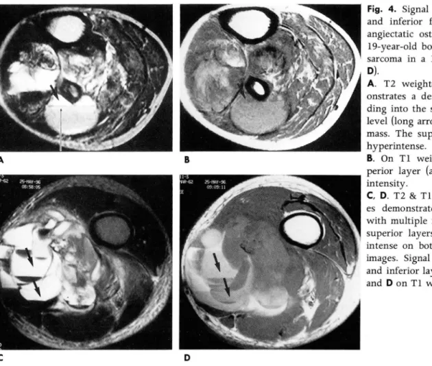
A B
C D
Fig. 4. Signal intensity of superior and inferior fluid layers in a tel- angiectatic osteogenic sarcoma in a
19-year-old boy (A, B) and synovial sarcoma in a 34-year-old woman{C, 0).
A. T2 weighted axial image dem- onstrates a destructive mass exten- ding into the soft tissue. Fluid-fluid level (Iong arrow) is seen within the mass. The superior layer (arrow) is hyperintense.
B. On Tl weighted image, the su- perior layer (arrow) has low signal intensity.
C, D. T2 & Tl weighted axial imag- es demonstrate a soft tissue mass with mu1tiple fluid-fluid levels. The superior layers (arrows) are hyper intense on both Tl & T2 weighted images. Signal intensity of superior and inferior layers are different in B and 0 on Tl weighted images.
전 일정시간 이상 움직임이 없는 자세를 유지하고 액체액체층 에 수직으로 횡단면 또는 시상면 영상을 얻어야 한다(1).
액체액체층은 여러 종양들의 전산화단층촬영 또는 자기공명 영상에서 관찰될 수 있는데 동맥류성 골냥에서 흔히 보이지만 (1 -4) 모세혈관확장성 골육종 (2, 5) , 거대세포종 (2, 6), 연골 아세포종 (2, 5, 7), 섬유성 이형성 (Fibrous dysplasia) (2), 고
정혜원 오1: 자기공명영상의 액처|액처|층
렵 골냥 (2, 5, 8) 악성섬유조직구종 (2) , 전형적 골육종 (classic osteogenic sarcoma)(2), 혈관종 (2) , 그리고 활막육종 (2) 에서 도드물지 않게 관찰되므로 액체액체층의 존재는특정 종양의 진단에 도움이 되지않는 비특이적인 소견으로 알려져 있다.
액체액체층은 종양내부의 여러 차례에 걸친 출혈에 의하여 형성되는 것으로 (1, 4) 이 연구에 포함된 질환 중 거대세포종,
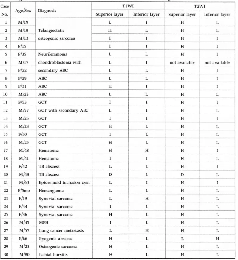
Table 1. Signal Intensity of the Fluid Within the Musculoskeletal Diseases on MR Images
Case TlWI
No. Age/Sex Diagnosis
Superior layer Inferior layer
l M/19 L I
2 M/l8 Telangiectatic H L
3 M/l3 osteogenic sarcoma I I
4 F/l5 I I
5 F/35 Neurilemmoma L L
6 M/l7 chondroblastoma with L I
7 F/22 secondary ABC L L
8 F/29 ABC L L
9 F/31 ABC H I
10 M/23 ABC L L
11 F/53 GCT I I
12 Mβ7 GCT with secondary ABC L I
13 M/26 GCT I I
14 M/28 GCT H L
15 F/30 GCT I L
16 M/25 GCT H L
17 M/48 Hematoma H H
18 M/41 Hematoma I I
19 F/42 TB abscess L L
20 M/48 TB abscess D L
21 Mι3 Epidermoid inclusion cyst L I
22 F/5mo Hemangioma L L
23 F/l9 Synovial sarcoma L H
24 F/34 Synovial sarcoma I L
25 F/46 Synovial sarcoma H L
26 M/45 MFH I L
27 Mβ7 Lung cancer metastasis L H
28 F/66 Pyogenic abscess H L
29 M/23 Osteogenic sarcoma H L
30 M/80 Ischial bursitis H L
H: hyperintense than the signal of muscle, 1: iso siganl intensity with the muscle L: low signal intensity than that of muscle
- 148 -
T2WI
Superior layer Inferior layer
H L
H L
H I
H I
H I
not available not available
H I
H I
H I
H L
H I
H L
H I
H L
H L
H L
H I
H L
H I
D L
H I
H L
H L
H L
H L
H L
H L
L H
H L
H L
대한방시선의학호|지 1998; 38: 145-150
모세혈관확장성 골육종, 동맥류성 골낭 각 1예와 활막육종 3예 에서는 병리 육안조직에서 출혈을 확인 할 수 있었다. 내부물 출혈시기에 따라 자기공명영상에서 다른 신호강도를 나타내게 되므로(9, 10) 종양 내부에서 각기 다른 신호강도로 보인다. 그 러나 화농성 골수염에서 골수강 (medullary cavi ty)내에 지방 -액층 (fat -fluid level)이 보이는 경우도 있으므로 (ll) 출혈 외에 괴사 또는 염증에 의한 침전물에 의해서도 액체액체층이 형성될 수 있으리라 생각되고 실제로 저자들이 경험한 바에서 도 종양이나 혈종 외에 결핵성 농양에서도 액체액체층이 관찰 되었다. 양성 질환의 경우 T1 강조영상에서 상층액이 다양한 신호강도를 보이지만 이와는 달리 악성종양의 경우 상층부가 모두 높은 신호강도를 보였다는 보고가 있으나 (2) 저자들의 경 우에는 양성 빛 악성 질환 모두가 다양한 신호강도를 보여 다른 결과로 나타났으며 T2 강조영상에서도 양성 또는 악성 여부나 질환의 종류에 관계없이 상층은고신호강도,하층은저 또는동 등신호강도로 보이는 경우가 많았다 (90%). 액체액체층이 병변 내부출혈에 의해 관찰되는경우와괴사또는염증에 의해 생긴 경우 침전물의 구성성분에 따라 자기공명영상상의 신호강도가 다르지 않을까기대하였으나각각의 질환에서 신호강도에 의한 차이점은 찾을 수 없었다.
액체액체층의 개수에 있어서는 유표피 봉업 낭종을 제외한 진성 골종양 또는 연부조직 종양은 모두 (23/23) 여러 개의 액 체액체층을 포함하고 있고 연부조직 내의 혈종과 결핵성 농양 전 예 그리고유표피 봉업 낭종에서 단하나의 액체액체층이 관 찰되었다. 이제까지의 보고에서도 종양의 경우는 고립골냥에서 하나의 액체액체층이 보였던한예 (8) 를제외하고는모든종양 에서 다방성 낭포 내에서 여러 개의 액체액체층이 관찰되었다
(1-7).
결론적으로 액체액체층은 다양한 질환에서 관찰되는 비특이 적인 소견으로 액체액체층의 존재유무, 개수 또는 내부 신호강 도에 의해 칠환간의 감별진단을하는것은어렵다.그러나자기 공명영상에서 액체액체층이 관찰된 다양한 질환 중 거대세포
종, 모세혈관확장성 골육종, 동맥류성 골냥과 활막육종이 높은 빈도를 차지하였다. 그리고 종양이외의 질환에서도 액체액체층 이 관찰될 수 있고 이 경우 연부조직 내의 명변으로 하나의 액 체액체층을 동반한 단방성 낭포로 나타나는 경우가 많았다. 따 라서 이러한 소견들은 자기공명영상상에서 액체액체층이 관찰 될 경우 그 영상 분석에 있어 큰 도움을 줄 것으로 사료된다.
송} τ1 드프 등4
1. Beltran JB, Simon DC, Levy M, Herman L, Weis L, Mueller CF Aneurysmal bone cysts: MR imaging at 1.5T. Radi%gy 1986; 158: 689-690
2. Tsai JC, Dalinka MK, Fallon MD, Zlatkin MB, Kressek HY.
Fluid-fluid level: a nonspecific findings in tumors of bone and soft tissue. Radi%
‘
gy 1990; 175:779-7823. Hudson TM. Fluid levels in aneurysmal bone cysts: a CT fea- ture. AJR 1984; 141: 1001-1004
4. Hertzanu Y, Mendelsohn DB, Gottschalk FG. Aneurysmal bone cyst of calcaneus. Radi%gy 1984; 151: 51-52
5. Burr BA, Resnick D, Syklawer R, Haghighi P. Fluid-fluid levels in a unicameral bone cyst: CT and MR findings. J Comput Tomogr 1993; 17: 134-136
6. Kaplan PA, Murphey M, Greenway G, et al. Fluid-fluid levels in giant cell tumors of bone: report of two cases. J Comput Tomogr 1987; 11: 151-155
7. Kahmann R, Gold RH, Echkardt JJ, Mirra JM. Case report 337.
Ske/eta/ Radio/ 1985; 14: 301-304
8 김성문, 강홍식 고럽골냥‘자기공명영상 소견 대한방사선의학 회지 1994;31: 1173-1177
9. Gomori JM, Grossman RI. Mechanisms responsible for the ap- pearance and evolution of intracranial hemorrhage. RadioGrap- hics 1988; 8: 427-440
10. Osborn AG. lntracrania/ hemorrhage. In Osborn AG. Diagnostic Neuroradi%gy. 1st ed. St. Louis:Mosby, 1994: 154-197 11. Rafii M, Firooznia H, Golimbu C, McCauley DI. Hematogeneous
Osteomyelitis with fat-fluid level shown by CT. Radi%gy 1984
; 153: 493-494
정혀|원 오1: 자기공명영상의 액처|맥체충
J Korean Radiol Soc 1998; 38: 145-150
Fluid-fluid Level on
MR
Image: Significance in Musculoskeletal Diseases1Hye Won Chung, M.D.l. 2, Kyung Won Lee, M.D., Chi Sung Song, M.D. 2 Sang Wook Han, M.D., Heung Sik Kang, M.D.
1 Department Of Radiology, Seoul National University College Of Medicine 2Department Of Radiology, Seoul City Boramae Hospital
Purpose: To evaluate the frequency, number and signal intensity of fluid-fluid levels of musculoskeletal diseases on MR images, and to determine the usefulness of this information for the differentiation of musculoskeletal diseases.
Materials and Methods: We retrospectively reviewed 30 cases in which fluid-fluid level was seen on MR images; they were diagnosed histopathologically(24/30), clinically(S/30) or clinicopath이
ogically(1/30). To differntiate these diseases, we determined their frequency, the number of fluid-fluid levels and the signal intensity of fluid.
Results: MR images revealed fluid-fluid levels in the following diseases: giant cell tumor(6), tel- angiectatic osteosarcoma( 4), aneurysmal bone cyst(3), synovial sarcoma(3), chondroblastoma(2), soft tissue tuberculous abscess(2), hematoma(2), hemangioma(l), neurilemmoma(l), metastasis(l), malig- nant fibrous histiocytoma(l), bursitis(l), pyogenic abscess(l), and epidermoid inclusion cyst(l). Fourteen benign tumors and ten malignant, three abscesses, and two hematomas were included.
Unlike true tumors, hematomas, tuberculous abscesses and the epidermoid inclusion cyst showed only one fluid-fluid level in a unilocular cyst. On Tl-weighted images, the signal intensities of fluid varied, but on T2-weighted images, superior layers were in most cases more hyperintense than in- ferior layers.
Conclusion : Because fluid-fluid layers are a nonspecific finding, it is difficu1t to specifically diag- nose each disease according to the number of fluid-fluid levels or signal intensity of fluid. In spite of the nonspecificity of fluid-fluid levels, they were frequent1y seen in cases of giant cell tumor, telangiectatic osteosarcoma, aneurysmal bone cyst, and synovial sarcoma. Nontumorous diseases such as abscesses and hematomas also demonstrated this finding.
Index words : Bone neoplasms, MR Soft tissues, neoplasms Bones, abscess
Bones, hemorrhage
Address reprint requests to ‘ Heung Sik Kang, M.D., Department of Radiology, Seoul National University College of Medicine,
# 28 Yongon-Dong, Chongno-Gu, Seoul 110-744, Korea. Tel. 82-2-760-2584 Fax.82-2-743-6385
%