logic organisms, which might play a role as an etiological factor in late implant complications.
Aplastic anemia is a rare hematological disease character- ized by pancytopenia and bone marrow hypoplasia, where normal hematopoietic tissue is replaced by fatty tissue. The incidence rate of aplastic anemia is two per one million per- sons per year, as reported by Young3. Though the etiology of aplastic anemia is typically unknown, it can be inherited, idiopathic, or acquired. Acquired aplastic anemia can be caused by intake of certain drugs and chemicals4,5, radiation therapy6, viral infection7,8, or pregnancy. There is also con- vincing evidence that aplastic anemia can be an autoimmune disease. This characterization indicates that the immune sys- tem attacks the bone marrow, resulting in insufficient blood cell production. Patients that suffer from aplastic anemia complain of general fatigue, shortness of breath, and pale ap- pearance due to low red blood cell (RBC) count. The severity of those symptoms depends on the severity of the aplastic anemia. Prolonged pancytopenia that results from neutrope- nia contributes to susceptibility to infectious diseases. In ad- dition, thrombocytopenia increases the risk of severe bleed- ing at various sites and is associated with oral manifestations such as petechia, ecchymosis, hematoma, gingival bleeding, and bleeding after tooth extraction9. In patients with severe
I. Introduction
Patients with hematologic disorders are at a greater risk for prolonged bleeding; therefore, it is important to reduce op- portunities for bleeding during surgical implant procedures.
Prolonged bleeding can interfere with the initial process of osseointegration, and recurrent bleeding from a wound site leads to persistence of iron in the tissue. Excessive tissue iron is associated with fibrosis, poor wound healing, and excessive angiogenesis1. If bone apposition does not occur, fibrous-scar tissue is formed between the implant surface and the sur- rounding bone2, which results in the absence of an anchoring function of the endosseous implant. Additionally, a lack of white blood cells (WBCs) increases the amount of oral patho-
Seunggon Jung
Department of Oral and Maxillofacial Surgery, Dental Science Research Institute, School of Dentistry, Chonnam National University, 33 Yongbong-ro, Buk-gu, Gwangju 61186, Korea
TEL: +82-62-220-5436 FAX: +82-62-220-5437 E-mail: [email protected]
ORCID: http://orcid.org/0000-0003-2008-3205
This is an open-access article distributed under the terms of the Creative Commons Attribution Non-Commercial License (http://creativecommons.org/licenses/by-nc/4.0/), which permits unrestricted non-commercial use, distribution, and reproduction in any medium, provided the original work is properly cited.
CC
Aplastic anemia and dental implant rehabilitation: a clinical trial
Jun-Hwa Kim, Uttom Kumar Shet, Byeong-Guk Kim, Myung-In Kim, Min-Suk Kook, Hee-Kyun Oh, Sun-Youl Ryu, Hong-Ju Park, Seunggon Jung
Department of Oral and Maxillofacial Surgery, School of Dentistry, Chonnam National University, Gwangju, Korea
Abstract(J Korean Assoc Oral Maxillofac Surg 2015;41:265-269)
The purpose of this study was to investigate implant-supported restoration as a technique for restoring missing teeth in patients with aplastic anemia.
Recurrent bleeding from wound sites leads to persistent release of iron in the tissue. Excessive iron in tissue is related to clinical findings, including fibrosis, poor wound healing, and high level of angiogenesis, which are possible etiological factors of reduced osseointegration. A 44-year-old female patient with aplastic anemia was treated with multiple endosseous implants throughout the mandible and in the posterior region of the maxilla. After 14 implants were placed, radiological and clinical parameters were assessed during the follow-up period. Marginal bone did not change significantly dur- ing the follow-up period. The fine trabecular bone in intimate contact and enclosing the implant fixture was sufficient for successful osseointegration.
None of the 14 implants were associated with compilations during the seven-year experimental period. This study suggests that dental implant proce- dures are a safe and reliable treatment option for restoration of missing dentition in patients with aplastic anemia.
Key words: Aplastic anemia, Dental implants, Rehabilitation
[paper submitted 2015. 5. 4 / revised 1st 2015. 7. 30, 2nd 2015. 9. 16 / accepted 2015. 9. 25]
Copyright Ⓒ 2015 The Korean Association of Oral and Maxillofacial Surgeons. All rights reserved.
performed without any special consideration.
After five months of healing, the full mucoperiosteal flap was elevated with a midcrestal incision and two vertical re- leasing incisions under intravenous sedation with midazolam (0.05 mg/kg) and the local anesthesia lidocaine plus with epinephrine 1:100,000. Ten Osstem US II (Osstem Implant Co., Busan, Korea) implants were installed in the spaces left by teeth #32, #33, #34, #36, #37, #42, #43, #44, #46, and
#47 (Fig. 1. A), and then provisional implants were placed in the sockets of teeth #31, #35, #41, #45.(Fig. 1. D) Primary closure of the flap involved periosteal releasing incisions, and 3-0 Vicryl (Ethicon Inc., Cornelia, GA, USA) was used as suture material.
Three months after implant installation, a second surgery was performed with an apically repositioned flap in order to produce keratinized gingiva, which helps maintain a healthier gingival environment.(Fig. 1. B) Four months postopera- tively, the provisional prostheses were replaced with the final prostheses.(Fig. 1. C)
We obtained panoramic radiographs using the KODAK 8000C System (Carestream Health Inc., Atlanta, GA, USA) and evaluated all panoramic and periapical radiographs with PiView Star (INFINITT Healthcare Co., Ltd., Seoul, Korea).
Marginal bone change was estimated from the border be- tween the implant platform and implant abutment to the low- ermost part of the absorbed marginal bone around the implant fixture, based on radiographs performed immediately after the operation. The magnification ratio on the radiographs was corrected by comparing the actual length of an implant fix- ture with that on the radiographic image.
After two months of loading on the final mandible prosthe- ses, bilateral sinus elevation was performed with ramal bone graft. The oval-shaped bony windows were removed from the lateral walls of the sinuses, and sinus membranes were care- fully elevated, followed by bone grafting. Particulated ramal bone graft material was used in combination with Bio-Oss (Geistlich Pharma AG, Wolhusen, Switzerland), and four Os- stem US II implants were simultaneously placed at sites #16,
#17, #26, and #27.(Fig. 1. E) Fibrin glue was injected into the bone graft site to enhance the bone healing process. All of the procedures were performed under intravenous sedation and local anesthesia with a vasoconstrictor, and the medications were the same as listed above.
Seven months after implant installation in the sinus eleva- tion site, prosthetic loading was applied, and peri-implant bone change was evaluated in panoramic and periapical radiographs. After one year and three months of prosthetic aplastic anemia, infection and bleeding can be life threaten-
ing, and such patients have demonstrated a high mortality rate. Although patients with moderate aplastic anemia might not require treatment, frequent and regular blood cell count- ing is important. Recent therapeutic advances, such as immu- no-suppressive therapy with bone marrow transplantation, use of growth factor alone or in conjunction with immune- suppressive therapy after bone marrow transplantation, blood component transfusion, and improved infection control, have increased survival rates10,11 of such patients.
This study evaluated the following parameters; 1) the amount of peri-implant bone resorption, 2) level of osseointe- gration as measured by the amount of fine trabecular bone in close contact with the fixture, 3) probing depth, and 4) peri- abutment gingival connective tissue contour. The purpose of this study was to demonstrate the effectiveness of implant- supported restoration in aplastic anemic patients by evaluat- ing radiographic and clinical features.
II. Case Report
A 44-year-old woman presented to the Department of Oral and Maxillofacial Surgery of Chonnam National University Hospital (Gwangju, Korea) to evaluate missing teeth in the maxilla and the mandible and possible implant-supported res- toration. The patient had been diagnosed with severe aplastic anemia at the Department of Hemato-Oncology of Chonnam National University Hospital in July 1995 and had received immunosuppressive therapy. She has been under regular follow-up and had taken 1 mg of folin (folic acid) and 50 mg of pyridoxine (vitamin B6) from May 2004 to June 2005.
A general assessment was performed based on panoramic radiograph and cone-beam computed tomography, and we planned to install 14 endosseous implants. Ten implants were inserted in the mandible in the first stage, and four implants were inserted in the maxilla in the second stage, with eleva- tion of both sinuses and autogenous bone grafts. During the preoperative evaluation, the Department of Hemato-Oncol- ogy confirmed that the patient would be able to maintain a greater than 1,000 absolute neutrophil count and have lower chances of postoperative bleeding if she was able to main- tain a greater than 80,000 platelet count during the surgery.
A complete blood cell count investigation revealed that the patient had an RBC of 3.51 million, a WBC count of 3,000, and a count of 84,000 platelets per milliliter of blood. These results indicated that both extraction of teeth #17, #16, #26, and #27 with poor prognosis and implant surgery could be
postoperative complications were noted.
On follow-up, the marginal bone had not changed signifi- cantly one year and three months after prosthetic loading. In periapical radiographs, fine trabecular bone was observed around each fixture, which indicates that osseointegration was successful for all of the implants. We measured the prob- ing depth, which is the distance from the gingival margin to the periodontal pocket, on the mesiobuccal and distobuccal sides of each tooth. The average of the measured values for each tooth was calculated, and a mean probing depth of 1.66 mm was obtained using the Williams Colorvue ProbeKit loading on the lower jaw, marginal bone change was evalu-
ated around the implants using panoramic and periapical radiographs (Fig. 1. F), which revealed peri-implant bone resorption of 1.37 mm on #16, 1.73 mm on #17, 1.30 mm on
#26, 1.11 mm on #27, 1.32 mm on #32, 1.70 mm on #33, 1.89 mm on #34, 1.0 mm on #36, 0.86 mm on #37, 0.50 mm on
#42, 0.72 mm on #43, 1.0 mm on #44, 2.52 mm on #46, and 2.12 mm on #47.
Tranexamic acid at a concentration of 500 mg/5 mL was available for perioperative bleeding. However, no severe bleeding occurred intraoperatively or postoperatively, and no
A B
C D
E F
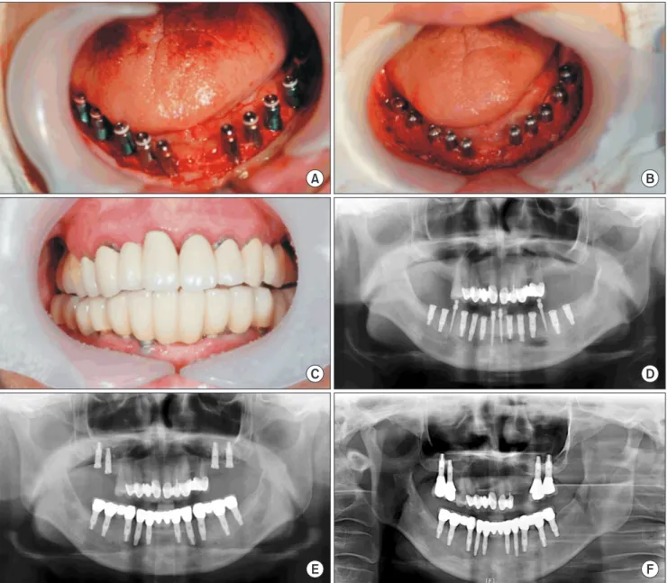
Fig. 1. Photographs and radiographs of a 44-year-old female with aplastic anemia who underwent implant-supported restoration. A. Ten implants were placed in the mandible with a surgically-guided stent. B. Healing abutment connection and apically repositioned flap during the second surgery. C. Frontal view 20 months after prosthetic loading. D. Panoramic view after implant installation. Panoramic view after implant placement surgery with a few provisional implants. E. Panoramic view; final prosthesis in the mandible. Four implants were placed on each side of the posterior maxilla with sinus elevation. F. Panoramic view; seven years after implant placement surgery.
Jun-Hwa Kim et al: Aplastic anemia and dental implant rehabilitation: a clinical trial. J Korean Assoc Oral Maxillofac Surg 2015
bone resorption, observed while loading dental prostheses, other implants were well-maintained, fulfilling the success criteria cited by Albrektsson et al.14. The first two implants were categorized into group II (satisfactory survival), and the others into group I (success, optimum health), according to the health scale developed by the International Congress of Oral Implantologists Consensus Conference for Implant Success in Pisa in 200715. Fine trabecular bone structure was observed around all of the implant fixtures, which created sufficient anchorage necessary for long-term implant main- tenance. Peri-implant resorption did not change significantly during the follow-up period. Stable, rigid, fixated implants have been reported with pocket depths ranging from 2 to 6 mm15; in the presented case, the mean probing depth was 1.66 mm after 21.4 months of prosthetic loading. There were no complications during the seven-year follow-up period.
Thus, based on these results, implant surgery can be per- formed safely even for patients with hematologic disorders by applying three principles of implant installation; Induction of initial hemostasis, maintenance of hemostasis during the healing period, and immobilization of the implant. These can be achieved through appropriate systemic treatment and pre- cise management of local soft tissue.
Conflict of Interest
No potential conflict of interest relevant to this article was reported.
ORCID
Jun-Hwa Kim, http://orcid.org/0000-0002-6870-1028 Uttom Kumar Shet, http://orcid.org/0000-0002-0137-072X Byeong-Guk Kim, http://orcid.org/0000-0001-6972-8276 Myung-In Kim, http://orcid.org/0000-0002-1253-3846 Min-Suk Kook, http://orcid.org/0000-0002-8053-8534 Hee-Kyun Oh, http://orcid.org/0000-0003-0391-7095 Sun-Youl Ryu, http://orcid.org/0000-0002-9718-6096 Hong-Ju Park, http://orcid.org/0000-0001-9132-6433 Seunggon Jung, http://orcid.org/0000-0003-2008-3205
References
1. Hoffman M, Harger A, Lenkowski A, Hedner U, Roberts HR, Monroe DM. Cutaneous wound healing is impaired in hemophilia B. Blood 2006;108:3053-60.
2. Esposito M, Thomsen P, Ericson LE, Lekholm U. Histopathologic observations on early oral implant failures. Int J Oral Maxillofac
(Hu-Friedy, Chicago, IL, USA).
All procedures, from surgery to restoration, were unevent- ful over a follow-up period of seven years.(Fig. 1. F) No implants showed mobility, and the patient did not complain of pain or discomfort during the seven-year follow-up period.
In addition, there was no evidence of peri-implant radiolu- cency seven years after implant installation. Finally, only two implants (#46, #47) showed marginal bone resorption greater than 2 mm after seven years of follow-up.
III. Discussion
Implant-supported restoration has become common prac- tice in dentistry and is associated with long-term successful outcomes. This could increase the popularity of implant- supported restoration among dentists and the general public even though its effect in medically compromised patients, especially those with bleeding disorders, has not been thor- oughly demonstrated.
Special considerations for these medically compromised patients include bleeding control during operative and post- operative periods in order to avoid life-threatening situations, achieving successful osseointegration, and long-term implant maintenance. Restoration of initial hemostasis can modulate some of the parameters of wound healing. An extended pe- riod of adequate hemostatic function is necessary to accom- modate the normal healing process, probably because the risk of hemorrhage is increased by vascular remodeling and an- giogenesis during the healing process12. Furthermore, patients with aplastic anemia are susceptible to peri-implant infection as aplastic anemia is characterized by marked hypoplastic bone marrow and pancytopenia (anemia, leucopenia, and thrombocytopenia).
We were not able to collect data regarding specific blood management strategies for successful implant placement in cases of aplastic anemia, so we followed the general guide- lines. We prepared a blood transfusion to maintain platelet count during surgery, since a platelet count less than 25,000/
mL indicates a high risk of spontaneous oral bleeding9. Tranexamic acid was also prepared for prolonged retention of intact weak blood clots and to prevent bleeding in plasmin- rich areas such as the oral cavity. Tranexamic acid can sig- nificantly reduce blood loss after oral surgery in patients with hemophilia and can be used topically or systemically13.
Radiographic examinations with implant mobility tests are the most reliable for assessing osseointegration. Although two of the present implants showed moderate peri-implant
10. Storb R, Thomas ED, Buckner CD, Clift RA, Fefer A, Fernando LP, et al. Allogeneic marrow grafting for treatment of aplastic ane- mia: a follow-up on long-term survivors. Blood 1976;48:485-90.
11. Camitta BM, Storb R, Thomas ED. Aplastic anemia (second of two parts): pathogenesis, diagnosis, treatment, and prognosis. N Engl J Med 1982;306:712-8.
12. McDonald A, Hoffman M, Hedner U, Roberts HR, Monroe DM.
Restoring hemostatic thrombin generation at the time of cutaneous wounding does not normalize healing in hemophilia B. J Thromb Haemost 2007;5:1577-83.
13. Castellanos-Cosano L, Núñez-Vázquez RJ, Segura-Egea JJ, Torres- Lagares D, Corcuera-Flores JR, Machuca-Portillo G. Protocol for oral implant rehabilitation in a hemophilic HIV-positive patient with type C hepatitis. Implant Dent 2014;23:622-5.
14. Albrektsson T, Zarb G, Worthington P, Eriksson AR. The long-term efficacy of currently used dental implants: a review and proposed criteria of success. Int J Oral Maxillofac Implants 1986;1:11-25.
15. Misch CE, Perel ML, Wang HL, Sammartino G, Galindo-Moreno P, Trisi P, et al. Implant success, survival, and failure: the Interna- tional Congress of Oral Implantologists (ICOI) Pisa Consensus Conference. Implant Dent 2008;17:5-15.
Implants 1999;14:798-810.
3. Young NS. Acquired aplastic anemia. Ann Intern Med 2002;136:
534-46.
4. Vincent PC. Drug-induced aplastic anaemia and agranulocytosis.
Incidence and mechanisms. Drugs 1986;31:52-63.
5. Betticher DC, Wolfisberg HP, Krapf R. Aplastic anemia in carbam- azepine therapy. Schweiz Med Wochenschr 1991;121:583-8.
6. Muir KR, Chilvers CE, Harriss C, Coulson L, Grainge M, Dar- byshire P, et al. The role of occupational and environmental expo- sures in the aetiology of acquired severe aplastic anaemia: a case control investigation. Br J Haematol 2003;123:906-14.
7. Honkaniemi E, Gustafsson B, Fischler B, Nemeth A, Frost BM, Papadogiannakis N, et al. Acquired aplastic anaemia in seven chil- dren with severe hepatitis with or without liver failure. Acta Paedi- atr 2007;96:1660-4.
8. Safadi R, Or R, Ilan Y, Naparstek E, Nagler A, Klein A, et al. Lack of known hepatitis virus in hepatitis-associated aplastic anemia and outcome after bone marrow transplantation. Bone Marrow Trans- plant 2001;27:183-90.
9. Sepúlveda E, Brethauer U, Rojas J, Le Fort P. Oral manifestations of aplastic anemia in children. J Am Dent Assoc 2006;137:474-8.