The Prevalence of Lumbar Spine Facet Joint Osteoarthritis and Its Association with Low Back
Pain in Selected Korean Populations
Sangbong Ko, MD, Alexander R. Vaccaro, MD*, Sangwook Lee, MD, Jaekun Lee, MD, Hojin Chang, MD
Department of Orthopaedic Surgery, Catholic University of Daegu School of Medicine, Daegu, Korea,
*Department of Orthopaedic Surgery, Thomas Jefferson University, Philadelphia, PA, USA
Received March 12, 2014; Accepted May 15, 2014 Correspondence to: Sangbong Ko, MD
Department of Orthopaedic Surgery, Catholic University of Daegu School of Medicine, 33 Duryugongwon-ro 17-gil, Nam-gu, Daegu 705-718, Korea Tel: +82-53-650-4283, Fax: +82-53-626-4272
E-mail: [email protected]
The lumbar facet joint was first suggested as the source of low back pain (LBP) and lower extremity pain in 1911.1) Since then, facetogenic back pain has become widely ac- cepted by many authors; however, the entity of facetogenic back pain is still controversial.2-9) Although the role of lumbar spine facet joint osteoarthritis (LSFJOA) has been supported by relief of LBP following intra-articular or periarticular injection,2,6) the relationship between LSFJOA and clinically significant LBP continues to be the subject
Background: This study was to evaluate the association of lumbar spine facet joint osteoarthritis (LSFJOA) identified by multi- detector computed tomography (MDCT) with age and low back pain (LBP) in an adult community-based population in Korea.
Methods: A sample of 472 participants (age range, 20 to 84 years) who underwent MDCT imaging for abdominal or urological lesions, not for chief complaints of LBP, were included in this study. LSFJOA based on MDCT findings was characterized using four grades of osteoarthritis of the facet joints. The prevalence of LSFJOA according to age group (below 40 years, 40–49 years, 50–59 years, 60–69 years, and above 70 years), gender, and spinal level was analyzed using chi-square tests and the association between LBP and LSFJOA adjusting for age, gender, and spine level was analyzed using multiple binary logistic regression test.
Results: Eighty-three study subjects (17.58%) had LSFJOA (grade ≥ 2). The prevalence of LSFJOA was not associated with gender (p = 0.092). The prevalence of LSFJOA increased with age (p = 0.015). The highest prevalence of LSFJOA was observed at L4–5 in men (p = 0.001) and at L5–S1 in women (p = 0.003), and at L5–S1 in the overall population (p = 0.000). LSFJOA was not associated with LBP in men (p = 0.093) but was associated with LBP in women (p = 0.003), especially at L3–4 (p = 0.018) and L5–S1 (p = 0.026).
Conclusions: The prevalence of LSFJOA based on the computed tomography imaging was 17.58% in the adult community Ko- rean population. The prevalence of LSFJOA increased with age, and the highest prevalence was noted at L5–S1. LSFJOA was not associated with LBP at any spinal level and age except at L3–4 and L5–S1 in women.
Keywords: Lumbar spine, Facet joint arthritis, Low back pain, Community-based Korean population
of controversy.
The prevalence of lumbar spine facet joint origi- nated pain based on single diagnostic blocks have been reported to range from 7.7% to 75% among patients re- porting back pain.10,11) In the United States, the prevalence of lumbar facet joint pain is 15% on the basis of local an- esthetic diagnostic blocks in a population of injured US workers.6) Similar studies report a prevalence of 40% to 45% in a pain management practice,8,9) and 40% in an Aus- tralian rheumatology practice.12)
Many published clinical studies report no correla- tion between the clinical LBP and degenerative spinal changes observed on many radiological modalities such as radiographs, magnetic resonance imaging, computed tomography (CT) and even radionuclide bone scan-
Copyright © 2014 by The Korean Orthopaedic Association
This is an Open Access article distributed under the terms of the Creative Commons Attribution Non-Commercial License (http://creativecommons.org/licenses/by-nc/3.0) which permits unrestricted non-commercial use, distribution, and reproduction in any medium, provided the original work is properly cited.
Clinics in Orthopedic Surgery • pISSN 2005-291X eISSN 2005-4408
ning.6,8,9,12,13) CT is the preferred method of imaging LSF- JOA,11) providing cross-sectional images of the opposing joint surfaces in the axial plane4) at relatively low cost with precise osseous details.
The efficacy of intra-articular or periarticular injec- tion therapy on LBP potentially associated with LSFJOA has not been clearly estabilished.11) Lewinnek and Warf- ield2) reported 96% correlation but Schwarzer et al.13) re- ported no associations. LSFJOA is a multifactorial process, and it has been suggested that the presence of interverte- bral disc degeneration leads to a greater load and motion at the facet joint, resulting in degenerative changes similar to those seen in other synovial joints.14,15) LSFJOA is inti- mately tied to degeneration of the intervertebral discs.
Few studies on the prevalence of LSFJOA have been published. Eubanks et al.16) found that LSFJOA is a univer- sal finding and characteristic osteoarthritis findings begin to appear early in more than one half of the adults younger than 30 years. The most common LSFJOA level appears to be L4–5. However, the prevalence of lumbar spine facet joint pain resulting from radiographically proven degen- erative changes has not been known in adult community- based Korean populations and the association between the facet joint pain and radiologically observed facet joint degenerative changes has not been studied and remains controversial.11)
The aim of the present study was (1) to evaluate the prevalence of LSFJOA by age, gender, and spinal level, (2) to evaluate the association of LSFJOA with LBP, and (3) to evaluate which levels of LSFJOA are most associated with LBP in an adult community-based Korean population.
METHODS
Sample
This retrospective study included patients who had un- dergone CT between January 1, 2010, and December 31, 2010, and met the following criteria: they were over the age of 20 years, they gave us their informed consent and they underwent CT examinations to assess abdominal or urological lesions for reasons unrelated to LBP. The CT scans ordered by the Departments of General Surgery and Urology were included. To prevent a result bias, we ex- cluded patients in whom a chief complaint of LBP was the primary indication for the CT examination ordered by the Departments of Orthopaedic Surgery, Neurologic Surgery, Rehabilitation and Pain Clinic. A total of 472 participants who were aged from 20 to 84 years were consecutively en- rolled.
LBP Evaluation
All the participants who had undergone multidetector CT scanning were asked to complete two questionnaires, which were administrated by senior trained nurses who were not involved in this study. The question on LBP that was translated into Korean in the questionnaire was “Have you had LBP that needed medication almost every day for at least 1 month in the last 12 months?” This was modified from Nordic Low Back Pain Questionnaire.17) The indi- vidual’s answers of “yes” or “no” to the above question was used in the present study as the LBP outcome. The ques- tion has been widely used by several authors18,19) for work- related compensation.
Scanning Parameters
CT was performed on one of three 16-multidetector com- puted tomography (MDCT) machines or a dual source 64- MDCT system (Lightspeed Ultra, GE Healthcare, Milwau- kee, WI, USA). The axial slice thickness varied from 0.75 to 2.5 mm because the images were obtained from patients with different indications and by different protocols.
LSFJOA Evaluation
All the CT scans were analyzed in a blinded fashion by two orthopedic surgeons independently. The images were reviewed on a secure-access picture-archiving commu- nication system (Philips Sectra, Linköping, Sweden). All the CT images that were initially reviewed were the axial images at the intervertebral disc level. Lumbar facet joints were graded on both sides at levels of L1–2, L2–3, L3–4, L4–5, and L5–S1. Four grades of facet joint osteoarthritis were defined using criteria similar to those suggested by Pathria et al.20) and Weishaupt et al.21) (Table 1). LSFJOA was defined as at least one joint affected by facet joint dis- ease between the spinal levels L1 and S1 (grade ≥ 2).
Statistical Analysis
Before the analysis, the study population was dichoto- mized on the basis of the presence of LSFJOA (≥ grade 2) on any side at any level. We analyzed LSFJOA in 5 differ- ent age groups (< 40, 40–49, 50–59, 60–69, and ≥ 70 years) and according to gender. The prevalence of LSFJOA in males and females each was compared according to the age group and according to the involved spinal level using chi-square (χ2) test for trend or multiple binary logistic re- gression analysis. Multiple binary logistic regression analy- sis was used to show the association between LBP and LSFJOA after adjusting for age, gender and spinal level. All the statistical analyses were performed with IBM SPSS ver.
19.0 (IBM Co., Armonk, NY, USA). A p-value of less than
0.05 was considered to be statistically significant.
RESULTS
Epidemiologic Characteristics
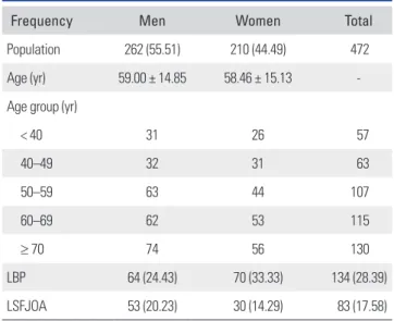
The epidemiologic characteristics of the 472 participants are listed in Table 2.
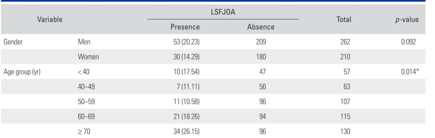
Prevalence of LSFJOA According to Gender and Age The prevalence of LSFJOA according to gender and age is listed in Table 3. Fifty-three have LSFJOA but 209 have no LSFJOA in men (20.23%) and thirty have LSFJOA but 180 have no LSFJOA in women (14.29%). There is no statisti- cally significant difference between men and women on the prevalence of LSFJOA (p = 0.092). The increasing age demonstrated a higher prevalence of facet joint osteoar- thritis with statistical significance (p = 0.015).
Prevalence of LSFJOA According to Spinal Level
In men, the difference in the prevalence of LSFJOA ac- cording to the spinal level was statistically significant (p = 0.001) and the highest prevalence of LSFJOA was found at L4–5. In women, the different in the prevalence of LS- FJOA across spinal levels was statistically significant (p = 0.003) and the highest prevalence of LSFJOA was found at L5–S1. In all population, the difference in the prevalence of LSFJOA according to the spinal level was statistically significant (p = 0.000) and the L5–S1 level exhibited high- est prevalence. The gender difference at each level was not statistically significant (L1–2, p = 0.377; L2–3, p = 0.741;
L3–4, p = 0.567; L5–S1, p = 0.893) except at L4–5 (p = 0.002). Men demonstrated a higher prevalence of LSFJOA compared to women at L4–5 level (Table 4).
LSFJOA and LBP
The prevalence of LSFJOA according to spinal level in
individuals with or without LBP is listed in Table 5. The prevalence of LSFJOA was not associated with LBP in men (p = 0.093), whereas the prevalence of LSFJOA was associ- ated with LBP in women (p = 0.003). The prevalence of LSFJOA according to age in individuals with or without LBP is shown in Table 6. The prevalence of LSFJOA was not statistically significantly associated with LBP (all, p >
0.05). LSFJOA at L3–4 and L5–S1 was related to LBP in women at a statistically significant level (p = 0.018 and p = 0.026, respectively). No significant difference in the preva- lence of LSFJOA was identified between individuals with and without LBP in the study population as a whole or in the subgroup analysis based on age and gender except for the above-mentioned cases.
Table 1. Criteria for Grading Osteoarthritis of the Facet Joint
Grade Criteria
0 Normal facet joint space (2–4 mm width)
1 Narrowing of the facet joint space (< 2 mm) and/or small osteophyte and/or mild hypertrophy of the articular process
2 Narrowing of the facet joint space and/or moderate osteophyte and/or moderate hypertrophy of the articular process and/or mild subarticular bone erosions
3 Narrowing of the facet joint space and/or large osteophytes and/or severe hypertrophy of the articular process and/or severe subarticular bone erosions and/or subchondral cyst and/or vacuum phenomenon in the joints
Adapted from Ashton et al.26) with permission from John Wiley & Sons and from Yang and King27) with permission from Wolters Kluwer Health.
Table 2. Epidemiologic Descriptive Statistics of the Study Population (n = 472)
Frequency Men Women Total
Population 262 (55.51) 210 (44.49) 472
Age (yr) 59.00 ± 14.85 58.46 ± 15.13 -
Age group (yr)
< 40 31 26 57
40–49 32 31 63
50–59 63 44 107
60–69 62 53 115
≥ 70 74 56 130
LBP 64 (24.43) 70 (33.33) 134 (28.39)
LSFJOA 53 (20.23) 30 (14.29) 83 (17.58)
Values are presented as number (%) or mean ± standard deviation.
LBP: low back pain, LSFJOA: lumbar spine facet joint osteoarthritis (≥ grade 2) at one joint or more between spinal levels L1 and S1.
Table 3. Prevalence of LSFJOA According to Gender and Age
Variable LSFJOA
Total p-value
Presence Absence
Gender Men 53 (20.23) 209 262 0.092
Women 30 (14.29) 180 210
Age group (yr) < 40 10 (17.54) 47 57 0.014*
40–49 7 (11.11) 56 63
50–59 11 (10.58) 96 107
60–69 21 (18.26) 94 115
≥ 70 34 (26.15) 96 130
Values are presented as number (%).
LSFJOA: lumbar spine facet joint osteoarthritis (≥ grade 2) at one joint or more between spinal levels L1 and S1.
*p < 0.05.
Table 4. Prevalence of LSFJOA According to Spinal Level
Spinal level Men (n = 262) Women (n = 210) Total (n = 472) Chi-square test (p-value)
L1–2 10 (3.82) 5 (2.38) 15 (3.18) 0.781 (0.377)
L2–3 12 (4.58) 11 (5.24) 23 (4.87) 0.109 (0.741)
L3–4 10 (3.82) 6 (2.86) 16 (3.39) 0.328 (0.567)
L4–5 29 (11.07) 7 (3.33) 36 (7.63) 9.900 (0.002)*
L5–S1 24 (9.16) 20 (9.52) 44 (9.32) 0.018 (0.893)
Chi-square test (p-value) 19.878 (0.001)* 16.141 (0.003)* 24.099 (> 0.001)* -
Values are presented as number (%) unless otherwise indicated.
LSFJOA: lumbar spine facet joint osteoarthritis (≥ grade 2) at one joint or more between spinal levels L1 and S1.
*p < 0.05.
Table 5. The Prevalence of LSFJOA According to Spinal Level in Individuals with or without LBP Spinal level
Men Women Total
With LBP Without
LBP Chi-square
test (p-value)† With LBP Without
LBP Chi-square
test (p-value)‡ With LBP Without
LBP Chi-square
test (p-value)§
L1–2 3 7 0.072 (0.789) 2 3 0.018 (0.894) 5 10 0.035 (0.852)
L2–3 2 10 0.606 (0.436) 6 5 1.506 (0.220) 8 15 0.149 (0.699)
L3–4 4 6 1.001 (0.317) 5 1 5.644 (0.018)* 9 7 4.868 (0.027)*
L4–5 13 16 5.748 (0.017)* 2 5 0.228 (0.633) 15 21 2.012 (0.156)
L5–S1 6 18 0.024 (0.876) 12 8 4.947 (0.026)* 18 26 2.158 (0.142)
Whole spine 28 57 2.830 (0.093) 27 22 8.829 (0.003)* 55 79 8.554 (0.003)*
LSFJOA: lumbar spine facet joint osteoarthritis (≥ grade 2) at one joint or more between spinal levels L1 and S1, LBP: low back pain.
*p < 0.05. †Chi-square test (LBP vs. non-LBP by spinal level in men). ‡Chi-square test (LBP vs. non-LBP by spinal level in women). §Chi-square test (LBP vs. non-LBP by spinal level).
Multiple Logistic Regression Test
Regarding the multiple logistic regression analysis, LBP was a dependent variable and LSFJOA at each spinal level and gender were included as independent variables. LBP was not associated with spinal level and age group but was more common in women than men (p = 0.002) (Table 7).
DISCUSSION
This is the first study to describe the prevalence of LSFJOA identified by MDCT in an adult community-based Korean
population. The prevalence of LSFJOA in this study was lower than that in the study by Kalichman et al.11) This study also evaluated the association between LSFJOA and LBP in the adult community-based Korean population.
Several authors have reported that LSFJOA is more com- mon in the most caudal motion segments.22-24) Fujiwara et al.25) found that the median grade of LSFJOA at L4–5 was significantly higher than that at L3–4, while no significant differences were found between L3–4, L5–S1, and between L4–5 and L5–S1. Kalichman and Hunter23) stated that the possible reason for the high prevalence and severity of LS- Table 6. The Prevalence of LSFJOA According to Age in Individuals with or without LBP
Age group (yr)
Men Women Total
With
LBP Without
LBP Chi-square
test (p-value)* With
LBP Without
LBP Chi-square
test (p-value)† With
LBP Without
LBP Chi-square
test (p-value)‡
< 40 3 4 0.078 (0.780) 1 2 0.010 (0.919) 4 6 0.398 (0.528)
40–49 0 1 0.238 (0.625) 2 4 0.220 (0.639) 2 5 0.184 (0.668)
50–59 4 5 0.583 (0.445) 2 0 3.328 (0.068) 6 5 1.939 (0.164)
60–69 5 10 0.585 (0.444) 4 2 1.764 (0.184) 9 12 1.118 (0.290)
≥ 70 6 15 0.519 (0.471) 6 7 0.181 (0.671) 12 22 0.443 (0.506)
All ages 18 35 1.781 (0.182) 15 15 2.478 (0.115) 33 50 3.304 (0.069)
LSFJOA: lumbar spine facet joint osteoarthritis (≥ grade 2) at one joint or more between spinal levels L1 and S1, LBP: low back pain.
*Chi-square test (LBP vs. non-LBP according to age in men). †Chi-square test (LBP vs. non-LBP according to age in women). ‡Chi-square test (LBP vs. non-LBP according to age).
Table 7. Results of the Multiple Logistic Regression Analysis with LBP (Yes vs. No) Used as a Dependent Variable
Parameter Odds ratio estimates
p-value Point estimate 95% Wald confidence interval
FJ OA L1L2 (yes vs. no) 1.300 0.486–3.474 0.601
FJ OA L2L3 (yes vs. no) 1.363 0.560–3.321 0.495
FJ OA L3L4 (yes vs. no) 0.472 0.186–1.194 0.113
FJ OA L4L5 (yes vs. no) 0.784 0.336–1.831 0.574
FJ OA L5S1 (yes vs. no) 0.963 0.414–2.243 0.931
Gender (women vs. men) 3.837 1.637–8.994 0.002*
Age group (yr) 40–49 (vs. < 40) 3.413 0.517–22.520 0.202
50–59 (vs. < 40) 0.669 0.124–3.609 0.640
60–69 (vs. < 40) 0.958 0.227–4.038 0.953
≥ 70 (vs. < 40) 2.256 0.553–9.203 0.257
FJ OA: facet joint osteoarthritis (≥ grade 2).
*p < 0.05.
FJOA at the L4–5 spinal level may be its position as a tran- sition between the more mobile lumbar segments and the relatively stiff L5–S1 segment. Unlike previous studies, the highest prevalence of LSFJOA was found at the L4–5 spi- nal level in men and at L5–S1 in women. Approximately 15%–40% of chronic LBP is attributed to LSFJOA.10) The histologic basis for facet joint pain has been scientifically established, but the precise clinical etiology remains un- determined.10) Hyperextension increases the load on the lumbar facet joint and stretches the capsule. This mechani- cal deformation may stimulate nociceptors in the joint capsule causing pain.26,27)
The observation that the age is associated with the incidence of LSFJOA is not surprising.11)
Lewin22) stated that facet joints showed only minor cartilage changes before the age of 45 years and that the osteoarthritis advanced with age. Kalichman and Hunter23) also reported that the prevalence of LSFJOA increases with age. However, some authors28,29) have reported the presence of LSFJOA in younger patients. The prevalence of LSFJOA in our study was more common in subjects less than 40 years of age than 40–49 years. The cause of this observation is unclear.
Gender has not been associated with the prevalence of LSFJOA in other studies.23,24) Fujiwara et al.25) found that motion segments in women showed significantly greater motion in lateral bending, flexion, and extension, but not in axial rotation, than in men in a cadaveric study. In our study, the prevalence of LSFJOA was not different between men and women.
In terms of specific spinal level, the present study did reveal statistically significant differences in the preva-
lence of LSFJOA between men and women at L4–5 spi- nal level where men demonstrated a significantly higher prevalence of LSFJOA than women. The result is same as the study by Eubanks et al.16) but is in contrast to the study of Kalichman et al.11)
The relationship of radiographic LSFJOA to the clinical syndrome of LBP is inconsistent.3) The cardinal role of facet joint abnormalities in patients with LBP is still debated.5-7) Schwarzer et al.13) even questioned the clini- cal importance of facet joint osteoarthritis. They were not able to demonstrate a significant correlation between the degree of osteoarthritis observed on CT and the pain score during the facet block. In our study, the prevalence of LSF- JOA was not associated with LBP.
One of the limitations of this study is that it was a cross-sectional investigation without any longitudinal fol- low-up. In addition, we did not adjust for the bias such as occupation, which should be addressed in further analysis.
The prevalence of LSFJOA based on CT imaging was 17.58% (20.23% in men and 14.29% in women), was not associated with gender, increased with age, and was the highest at the L5–S1 spinal level in an adult commu- nity-based Korean population. At the L4–5 spinal level, LSFJOA was more common in men than in women. No significant association was observed between LSFJOA and LBP at any spinal level and age except at L3–4 and L5–S1 levels where LSFJOA was related to LBP in women.
CONFLICT OF INTEREST
No potential conflict of interest relevant to this article was reported.
REFERENCES
1. Goldthwait JE. The lumbo-sacral articulation: an explana- tion of many cases of “lumbago”, “sciatica” and paraplegia.
Boston Med Surg J. 1911;164:365-72.
2. Lewinnek GE, Warfield CA. Facet joint degeneration as a cause of low back pain. Clin Orthop Relat Res. 1986;(213):
216-22.
3. Helbig T, Lee CK. The lumbar facet syndrome. Spine (Phila Pa 1976). 1988;13(1):61-4.
4. Carrera GF, Haughton VM, Syvertsen A, Williams AL.
Computed tomography of the lumbar facet joints. Radiol- ogy. 1980;134(1):145-8.
5. Badgley CE. The articular facets in relation to low-back pain and sciatic radiation. J Bone Joint Surg Am. 1941;23(2):481-
96.
6. Nachemson AL. Newest knowledge of low back pain: a criti- cal look. Clin Orthop Relat Res. 1992;(279):8-20.
7. Schwarzer AC, Aprill CN, Derby R, Fortin J, Kine G, Bog- duk N. Clinical features of patients with pain stemming from the lumbar zygapophysial joints. Is the lumbar facet syndrome a clinical entity? Spine (Phila Pa 1976). 1994;
19(10):1132-7.
8. Manchikanti L, Pampati V, Fellows B, Bakhit CE. Prevalence of lumbar facet joint pain in chronic low back pain. Pain Physician. 1999;2(3):59-64.
9. Manchikanti L, Pampati V, Fellows B, Bakhit CE. The diag- nostic validity and therapeutic value of lumbar facet joint
nerve blocks with or without adjuvant agents. Curr Rev Pain. 2000;4(5):337-44.
10. Dreyer SJ, Dreyfuss PH. Low back pain and the zygapophy- sial (facet) joints. Arch Phys Med Rehabil. 1996;77(3):290- 300.
11. Kalichman L, Li L, Kim DH, et al. Facet joint osteoarthritis and low back pain in the community-based population.
Spine (Phila Pa 1976). 2008;33(23):2560-5.
12. Schwarzer AC, Wang SC, Bogduk N, McNaught PJ, Laurent R. Prevalence and clinical features of lumbar zygapophysial joint pain: a study in an Australian population with chronic low back pain. Ann Rheum Dis. 1995;54(2):100-6.
13. Schwarzer AC, Wang SC, O'Driscoll D, Harrington T, Bog- duk N, Laurent R. The ability of computed tomography to identify a painful zygapophysial joint in patients with chron- ic low back pain. Spine (Phila Pa 1976). 1995;20(8):907-12.
14. Goode AP, Carey TS, Jordan JM. Low back pain and lumbar spine osteoarthritis: how are they related? Curr Rheumatol Rep. 2013;15(2):305.
15. Varlotta GP, Lefkowitz TR, Schweitzer M, et al. The lumbar facet joint: a review of current knowledge: part 1: anatomy, biomechanics, and grading. Skeletal Radiol. 2011;40(1):13- 23.
16. Eubanks JD, Lee MJ, Cassinelli E, Ahn NU. Prevalence of lumbar facet arthrosis and its relationship to age, sex, and race: an anatomic study of cadaveric specimens. Spine (Phila Pa 1976). 2007;32(19):2058-62.
17. Kuorinka I, Jonsson B, Kilbom A, et al. Standardised Nordic questionnaires for the analysis of musculoskeletal symp- toms. Appl Ergon. 1987;18(3):233-7.
18. Dovrat E, Katz-Leurer M. Cold exposure and low back pain in store workers in Israel. Am J Ind Med. 2007;50(8):626-31.
19. Ghaffari M, Alipour A, Jensen I, Farshad AA, Vingard E.
Low back pain among Iranian industrial workers. Occup
Med (Lond). 2006;56(7):455-60.
20. Pathria M, Sartoris DJ, Resnick D. Osteoarthritis of the facet joints: accuracy of oblique radiographic assessment. Radiol- ogy. 1987;164(1):227-30.
21. Weishaupt D, Zanetti M, Boos N, Hodler J. MR imaging and CT in osteoarthritis of the lumbar facet joints. Skeletal Radiol. 1999;28(4):215-9.
22. Lewin T. Osteoarthritis in lumbar synovial joints: a mor- phologic study. Acta Orthop Scand Suppl. 1964:Suppl 73:1- 112.
23. Kalichman L, Hunter DJ. Lumbar facet joint osteoarthritis: a review. Semin Arthritis Rheum. 2007;37(2):69-80.
24. Fujiwara A, Tamai K, Yamato M, et al. The relationship be- tween facet joint osteoarthritis and disc degeneration of the lumbar spine: an MRI study. Eur Spine J. 1999;8(5):396-401.
25. Fujiwara A, Lim TH, An HS, et al. The effect of disc de- generation and facet joint osteoarthritis on the segmental flexibility of the lumbar spine. Spine (Phila Pa 1976). 2000;
25(23):3036-44.
26. Ashton IK, Ashton BA, Gibson SJ, Polak JM, Jaffray DC, Eisenstein SM. Morphological basis for back pain: the dem- onstration of nerve fibers and neuropeptides in the lumbar facet joint capsule but not in ligamentum flavum. J Orthop Res. 1992;10(1):72-8.
27. Yang KH, King AI. Mechanism of facet load transmission as a hypothesis for low-back pain. Spine (Phila Pa 1976). 1984;
9(6):557-65.
28. Tischer T, Aktas T, Milz S, Putz RV. Detailed pathological changes of human lumbar facet joints L1-L5 in elderly indi- viduals. Eur Spine J. 2006;15(3):308-15.
29. Gries NC, Berlemann U, Moore RJ, Vernon-Roberts B. Early histologic changes in lower lumbar discs and facet joints and their correlation. Eur Spine J. 2000;9(1):23-9.