49
Immune Network
급성 골수성 백혈병 환자에서 G-CSF를 포함한 고용량 화학요법 후 자가 말초혈 조혈모세포이식의 효과
1고려대학교 의과대학 내과학교실, 2전남대학교 의과대학 소아과학교실
김병수
1․국 훈
2․황태주
2․최철원
1․김준석
1Autologous Peripheral Blood Stem Cell Transplantation Using G-CSF Combined Conditioning in AML Patients
Byung Soo Kim
1, Hoon Kook
2, Tai Ju Hwang
2, Chul Won Choi
1and Jun Suk Kim
11Department of Internal Medicine, Korea University Medical Center, Seoul, Korea,
2Department of Pediatrics, Chonnam National University Hospital, Gwangju, Korea
ABSTRACT
Background: The possibility that G-CSF recruits leukemic cells from the G0 to S phase, which may lead to a greater susceptibility to cytotoxic drugs, such as ara-C, has been presented in Harada’s study. Methods: In this study, we referred to the protocol of Harada et al 1 to try G-CSF combined marrow-ablative chemotherapy and autologous PBSCT, for the treatment of AML patients in CR1 status. Between January 1997 and March 1998, six AML patients (3: children, 3: adults) in CR1 status were autografted and followed up to 3 years. Results: The major regimen related toxicity was composed of mucositis and diarrhea without death. The time of ANC recovery to 500/L and 1,000/L was 11~48 and 16~81 days, respectively. The mean time of platelet recovery to 20,000/L and 50,000/L was 21 ~233 and 35~370 days, respectively. The platelet recovery time to 50,000/L was markedly prolonged for more than 100 days in four patients (66.7%). Moreover, four patients (66.7%) experienced a relapse of leukemia after transplantation, with a mean interval of 147.5 days after PBSCT. Two patients were in CR status for 53 and 51 months after PBSCT, respectively. Conclusion: The G-CSF combined marrow-ablative chemotherapy and autologous PBSCT resulted in a markedly delayed platelet recovery and no advantages for decreasing the relapse rate of AML.
But, further studies will be warranted. (Immune Network 2002;2(1):49-52) Key Words: G-CSF combined myeloablative chemotherapy, autologous PBSCT
책임저자:국 훈, 전남대학교병원 소아과
501-191, 광주광역시 동구 학 1동 5번지 Tel: 062-220-6645, Fax: 062-222-6103 E-mail: [email protected]
서 론
G-CSF가 백혈병 세포의 활성도를 촉진시킬 수 있다는 실험적 사실이 증명됨으로써(1), 화학제와 G-CSF를 병 용 투여하면 활성도가 증가된 백혈병세포의 비율이 높 아짐에 비례하여 화학제의 항암효과가 증강되리라는 가 설이 가능해졌다. 이에, 조혈모세포이식의 전처치로 시 행되는 고용량 화학요법에서도 G-CSF를 병용 투여하여 G0phase의 백혈병 세포를 세포주기로 끌어들여 Ara- C 와 같은 화학제의 항백혈병 효과를 극대화시키는 이른
바 ‘Priming' 효과를 유도시키려는 시도가 일부에서 진 행되었다(2,3,6). Harada 등(2)은 16명의 급성 골수성 백 혈병(acute myelogenous leukemia, AML) 환자를 대상으 로 G-CSF가 포함된 골수박멸 전처치 요법(myeloablative conditioning regimen) 후 자가 말초혈 조혈모세포이식 (peripheral blood stem cell transplantation, PBSCT)을 시도 한 결과 5년간 무병 생존율이 74.5%라는 매우 양호한 결 과를 보고한 바 있었다. 이에 저자 등은 제 1 관해 상태 의 소아 및 성인 AML 환자들에서 G-CSF 병합 고용량 화학요법 후 자가 PBSCT의 효과를 규명하고자 본 연구 를 시행하였다. 그러나 저자 등의 연구에서는 당초 기대 와는 달리 이식 후 혈소판 회복의 상당한 지연과 높은 초기 재발률이 관찰되었으며 이를 발표한 바가 있었다 (3). 본 고에서는 저자 등이 G-CSF 병합 고용량 화학요법
50
Byung Soo Kim, et al.Table II. The G-CSF combined myeloablative high dose chemotherapy regimen
Day* G-CSF Chemotherapy
-12, 11 5μg/Kg 6 hr IV
-10 5μg/Kg 6 hr IV Ara-C 100 mg/m2 24h CI;
-9 5μg/Kg 6 hr IV Ara-C 100 mg/m2 24h CI; Bu 4 mg/Kg PO -8 5μg/Kg 6 hr IV Ara-C 100 mg/m2 24h CI; Bu 4 mg/Kg PO -7 10μg/Kg 6 hr IV Ara-C 100 mg/m2 24h CI; Bu 4 mg/Kg PO -6 10μg/Kg 6 hr IV Ara-C 100 mg/m2 24h CI; Bu 4 mg/Kg PO
-5 20μg/Kg 6 hr IV Ara-C 100 mg/m2 24h CI; VP-16 15~20 mg/Kg IV over 8h -4 20μg/Kg 6 hr IV Ara-C 3.0 g/m2 over 3 h bid; VP-16 15~20 mg/Kg IV over 8h
-3 Ara-C 3.0 g/m2 over 3 h bid;
-2 -1 PBSCT day
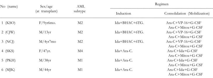
*Day before peripheral blood stem cell transplantation (PBSCT), Ara-C; Cytosine arabinoside, Bu; busulfan, VP-16; etoposide Table I. Main characteristics of the patients before transplantation
Regimen
No (name) Sex/age AML
(at transplant) subtype
Induction Consolidation (Mobilization)
1 (KSO) F/9yr6mo. M2 Ida+BHAC+6TG. Ara-C+VP-16+G-CSF
Ara-C+Mitox+G-CSF
2 (CJW) M/13yr M2 Ida+BHAC+6TG. Ara-C+VP-16+G-CSF
Ara-C+Mitox+G-CSF
3 (NCJ) M/4yr7mo M2 Ida+BHAC+6TG. Ara-C+VP-16+G-CSF
Ara-C+Mitox+G-CSF
4 (SKS) F/47yr. M4 Ida+Ara-C. Ara-C+Ida+G-CSF
Ara-C+Mitox+G-CSF
5 (PKH) M/38yr M1 Ida+Ara-C. Ara-C+Ida+G-CSF
Ara-C+Mitox+G-CSF
6 (MJK) M/44yr M1 Ida+Ara-C. Ara-C+Ida+G-CSF
Ara-C+Mitox+G-CSF
Ida; Idarubicin, BHAC; N4-behenoyl-1-β-D-arabinosylcytosine, 6-TG; 6-Thioguanine, Ara-C; Cytosine arabinoside, VP-16; Etoposide, Mitox; Mitoxantrone
후 자가 PBSCT를 시행한 환자들의 장기 추적 관찰을 포 함한 결과를 보고하고자 한다.
대상 및 방법
고려대 의료원 혈액종양 내과 및 전남대학교병원 소아 과에 내원하여 AML (M0 및 M3은 제외)로 진단받고 관 해유도 화학요법으로 일차 완전관해(First Complete Remission, CR1)를 이룬 후 주요 장기기능이 정상인 상 태에서 1997년 1월부터 1998년 3월까지 G-CSF가 포함된 고용량 화학요법 및 자가 말초혈 조혈모세포이식을 받 은 환자 6명을 대상으로 하였으며 그 결과를 3년 이상 추적 관찰하였다. 본 환자들의 자세한 내역 및 관해유도
와 공고 화학요법은 Table I과 같다. 말초혈에서의 조혈 모세포 채집은 총 단핵구수가 4×10⁸/Kg 혹은 CD34+
세포수가 5×10⁶/Kg 이상을 목표로 하여 공고요법 기간 (G-CSF 사용) 동안에 시행하였다. 그리고, 2차 공고 및 가동화 요법이 끝난 후 고용량 화학요법 및 자가 PBSCT 를 시행하기 전에 골수조직검사를 하여 관해상태가 지 속되는가를 확인하였다. 저자 등은 자가 PBSCT 시 Harada 등의 전처치 요법을 그대로 원용하였다(Table II).
Harada 등의 연구와 차별이 되는 사항은 Harada 등은 이 식 후 G-CSF를 사용하지 않았는데 반하여 저자 등은 자 가 PBSCT 후 조혈기능의 촉진을 위하여 G-CSF (5 g/Kg) 를 이식 후 7일째부터 투여하였다는 점에 있다. 저자 등
Autologous Peripheral Blood Stem Cell Transplantation Using G-CSF Combined Conditioning
51
Table III. The details of infused cell doses, duration of hematologic recovery, and outcome of transplantation
Infused cell dose Time to recovery (days)
Outcome No.
of transplantation MNC CD34+Cell ANC (500/μL) ANC (1000/μL) Platelet (20000/μL) Platelet (50000/μL)
1 8.9 5.7 15 26 31 123 CR (+1615 days)
2 17.5 20.1 17 17 21 35 Recur (+123 days)
3 8.2 2.2 48 81 233 370 CR (+1553 days)
4 7.0 9.3 11 16 34 73 Recur (+106 days)
5 7.3 9.2 16 21 86 - Recur (+121 days)
6 6.5 8.8 11 16 34 103 Recur (+241 days)
MNC; Mononuclear cell count (×108/Kg), CD34+cell; CD34 positive cell (×106/Kg), ANC; Absolute neutrophil count, CR; Complete remission
은 본 환자들에서 이식 후 합병증 발생, 조혈기능의 회 복, 재발률, 그리고 생존기간 등을 파악하여 그 결과를 분석하여 보았다.
결 과
고용량 화학요법과 관련된 주요 독성으로 점막염 (grade II~III, 6~18일, 중앙값: 10일)과 설사(grade II~
III, 7~16일, 중앙값: 9일)가 관찰되었으나, 이와 관련된 사망은 없었다. 이식된 세포 수 및 이식 후 말초혈액내 과립구와 혈소판의 회복양상은 Table III과 같다. 절대과 립구수(absolute neutrophil count, 이하 ANC)가 500/μL, 1,000/μL 이상으로, 혈소판수가 20,000/μL, 50,000/μL 이상으로 회복되기까지 각각 11~48일, 16~81일과 21~
233일, 35~370일이 소요되었다. 그런데 특이하게도 이 식된 조혈모세포수가 충분하였음에도 불구하고 4명 (66.7%)에서 혈소판수가 50,000/μL 이상으로 회복되는 데 100일 이상이나 걸리는 지연회복의 양상을 보였다.
또한, 4명의 환자에서 이식 후 106~241일 사이에 백혈 병의 재발을 경험하였다. 나머지 2명은 현재 이식 후 53, 51개월째로 정상 말초혈액검사 소견과 함께 관해상태를 유지하고 있다.
고 찰
본 연구에서는 당초 기대와는 달리 혈소판의 지연 회 복과 더불어 높은 초기 재발률이 관찰되었다. 이식 후 조혈기능회복의 지연은 보통 이식된 조혈모세포수의 부 족에 기인한다. 그런데 본 연구에서는 단지 1명에서 이 식된 CD34+ 세포 수가 2.2×10⁶/Kg으로 약간 낮은 외에 는 모두 충분한 세포들이 이식되었다. 그런데, 최근에 화 학요법과 G-CSF의 투여를 병용하면 조혈기능의 회복이 저하된다는 연구결과를 Tjan-Heijnen 등(4)이 제시하였 다. 그들은 소세포성 폐암 환자에서 적정량의 화학요법 을 시행 시 G-CSF를 병용한 경우가 그렇지 않은 때에 비하여 유의하게 화학요법 후 말초혈액내 과립구 및 혈
소판의 회복이 지연됨을 관찰하였다. 그러나, 이 보고는 관례적인 비골수제거성(non-myeloablative) 적정용량 화 학요법을 대상으로 한 연구이었기 때문에 그 결과를 고 용량 화학요법 및 자가 조혈모세포이식에서의 G-CSF의
‘Priming' 효과에 적용하기에는 문제가 있을 수 있기 때 문에 이의 규명을 위한 연구가 계속 필요하리라고 생각 된다. 그리고 저자 등이 본 연구에서 발생한 높은 초기 재발률의 원인을 추적한 결과 적은 CD34+ 세포 수 (2.2&5.7×10⁶/Kg)를 이식받은 환자들에서는 재발이 되 지를 않았고 많은 CD34+ 세포 수(8.8-20.1×10⁶/Kg)를 이식받은 환자들에서는 재발이 되었다는 사실을 발견할 수 있었다. 결국 본 사실은 채집된 조혈모세포 수가 많은 때에는 그 속에 백혈병 세포의 오염이 많았을 가능성을 암시한다고 여겨진다. 이에 관한 최근의 연구로 Pecora (5)는 말초혈에서 많은 양의 조혈모세포를 채집하기 위 하여 대부분에서 혈액 분반술의 횟수가 증가하고 이는 말초혈액내 암세포 오명의 가능성을 높인다고 하면서 특히 채집된 CD34+ 세포 수가 5×10⁶/Kg 이상인 경우 에는 암세포 오염이 증가한다고 주장한 바 있었다. 그리 고, 본 연구에서는 조혈모세포의 채집을 1차 공고요법부 터 시작하였는데 이 또한 채집된 조혈모세포 속에 백혈 병 세포의 오염이 많을 가능성을 시사할 수 있다. 이에 대한 지적으로 Miyamoto 등(6)은 화학요법을 반복할수 록 그에 따라 조혈모세포를 말초혈액에서 채집 시 백혈 병 세포의 오염 가능성이 낮아진다고 밝힌 바 있었다.
그러나, 본 연구에서는 백혈병 세포의 오염을 객관적으 로 증명하지는 못하였기 때문에 이에 관한 추가 연구가 계속 필요하리라고 생각된다.
본 연구 초기결과의 발표 후, Harada 등(7)은 50명의 AML 환자들을 대상으로 G-CSF 병합 고용량 화학요법 후 자가 PBSCT를 시도한 결과를 제시하였다. 그들은 8% (4/50)의 100일 이내 초기 치료 관련 사망률 및 일부 (자세한 내용은 제시되지 않음)의 혈소판의 지연회복을 관찰하였으나 534일의 중앙 추적기간 중 62%의 관해유
52
Byung Soo Kim, et al.지를 보였다고 하면서 본 치료가 AML의 높은 재발률은 보이지 않는다고 주장하였다. 그러나 Harada 등의 연구 (7)에는 CR1에서 이식받은 AML 환자의 상당수(52%
(22/42): M2와 M3)가 본 연구에서는 제외한 M3을 포함 하고 있었으므로 정확한 비교를 하기에는 문제가 있다 고 생각된다.
결국 저자 등은 G-CSF 병합 고용량 화학요법 후 자가 PBSCT를 시도한 결과 이식 후 혈소판의 회복이 상당히 지연됨을 경험하였고 이를 극복하기 위하여 다량의 조 혈모세포를 말초혈액에서 채집하면서 높은 초기 재발률 을 관찰하였기 때문에 잠정적으로 본 연구를 중단한 상 태이다. 그러나, 본 치료 후 생존한 2명에서는 이식 후 3년 이상 경과한 시점(53, 51개월)까지 백혈병의 재발 없 이 정상적인 조혈기능을 유지하고 있기 때문에 좀 더 개 선된 G-CSF 병합 고용량 화학요법 후 자가 PBSCT 치료 계획을 저자 등은 구상하고 있다.