STRENGTH OF GLASS FIBER REINFORCED PMMA RESIN AND SURFACE ROUGHNESS CHANGE AFTER ABRASION TEST
Sang-Il Lee, D.D.S., M.S.D., Chang-Whe Kim, D.D.S., M.S.D., Ph.D.,
Young-Jun Lim, D.D.S., M.S.D., Ph.D., Myung-Joo Kim, B.Sc., D.D.S., M.S., Ph.D., Suk-Dae Yun, D.D.S., M.S.D., Ph.D.
Department of Prosthodontics, Graduate School, Seoul National University
Statement of the problem.The fracture of acrylic resin dentures remains an unsolved problem. Therefore, many investigations have been performed and various approaches to strength- ening acrylic resin, for example, the reinforcement of heat-cured acrylic resin using glass fibers, have been suggested over the years. But problems such as poor workability, rough surface, poor adhesion of glass fiber resin complex are not solved yet.
Purpose. The aim of the present study was to investigate the effect of short glass fibers on the transverse strength of heat-polymerized denture base acrylic resin and roughness of resin complex after abrasion test.
Material and methods. To avoid fiber bunching and achieve even fiber distribution, glass fiber bundles were mixed with acrylic resin powder in conventional mixer with a non-cutting blade, to produce the glass fiber(10㎛ diameter, 3㎜ length, silane treated) resin composite. Glass fibers were incorporated at 0%, 3%, 6% and 9% by weight. Transverse strength were measured.
After abrasion test, surface roughness was evaluated and scanning electron microscope view was taken for clinical application.
Results.
1. 6% and 9% incorporation of 3mm glass fibers in the acrylic resin enhanced the transverse strength of the test specimens(p<0.05).
2. Before abrasion test, incorporation of 0%, 3%, 9% glass fiber in the resin showed no dirrerence in roughness statisticaly(p>0.05).
3. After abrasion test, incorporation of 0%, 3%, 6% glass fiber in the resin showed same sur- face roughness value statistically(p>0.05).
4. In SEM, surface roughness increased as the percentage of the fibers increased.
5. In the areas where glass fiber bunchings are formated, a remarkably high roughness was noticed.
Conclusion. 6% and 9% addition of silane-treated short glass fibers into denture base acrylic resin increased transverse strength significantly. Before and after abrasion test, incor- poration of 0%, 3%, 6% glass fiber in the resin showed same surface roughness value statistically.
Key Words
Glass fiber, PMMA resin, Surface roughness, Transverse strength
J Korean Acad Prosthodont : Volume 45, Number 3, 2007
F
or multiple teeth missing patients, removable partial denture, complete denture, or implant prosthesis are made, mostly using heat-poly- merized PMMA resin.Although mechanical failure of acrylic den- tures during services still occurs, many advantages such as excellent appearance, ease of manipula- tion, minimum expense, accuracy of fit and ease in repair ensure its continued use.
Breakage may result from impact fracture or from fatigue fracture often seen in complete maxil- lary dentures, where continual flexing of the base during use leads to crack development.1,2
Much research have been undertaken with a view toward reinforcing acrylic resins to enhance their physical and mechanical properties.
Much investigation have bee done such as reinforcement by metal wire or metal plate, glass fiber reinforcement, kevlar fiber reinforcement, car- bon fiber reinforcement, polyethylene fiber rein- forcement, rubber material and so on.
It has been reported that carbon fibers reduce the fatigue and improve tensile strength, transverse deflection, and the elastic modulus of PMMA resins.
However, the black color of carbon fibers caus- es esthetic problems.3-7
Inclusion of metal fillers improved the ther- mal conductivity of PMMA and enhanced its strength, but at the expense of poor esthetics of the complete denture.8
Aramid fibers have been shown to significant- ly increase the impact strength of PMMA resin and enhance the fracture resistance of acrylic resin den- ture base material. However, the yellow color of the aramid fibers might limit their use to cer- tain intraoral applications.9-11
In recent years, there has been considerable interest in the reinforcement of denture bases with polyethylene fibers.12-14Unlike carbon, met-
al, and aramid, polyethylene fibers are almost invis- ible in pink resin denture bases. Polyethylene fibers are used in three forms, namely continuous parallel, woven, and short. As has been reported by some investigators, these different forms enhance selected mechanical properties of the PMMA resin. The use of polyethylene fibers in three forms, namely continuous parallel, woven, and short, was reported by some investigators to enhance select mechanical properties of PMMA resin. Other investigators have reported that no significant increase was found in the overall strength of polyethylene fiber-reinforced acrylic resin.15-16
Much investigation using glass fiber have been made on account of esthetic properties.
Many investigators have examined different forms of glass fibers in an attempt to improve the mechanical properties of dental polymers.17-20In some studies, glass fibers have been used in a woven or short form, whereas others have used unidirectional continuous roving.
Investigations about ideal length of glass fiber for strength reinforcement have bee done.
After incorporation of glass fiber, impact strength increased significantly and transverse strength increased reasonably.
Optimal adhesion between the fibers and the polymers matrix can be obtained by mixing with silane-coupling compounds.21 Various kind of silane material has been used showing differ- ent binding effects.
Glass fiber is already used in fixed partial den- ture, but in case of removable partial denture or complete denture usage is not common.
Even distribution of glass fiber is limiting fac- tor for glass fiber resin complex and heavy inclu- sion of glass fiber causing viscosity brought about incomplete resin packing. Glass fiber can irri- tate skin and make the surface rough.
Especially, surface roughness can irritate mucous
membrane, cause discoloration, and induce plaque deposition.
In this study, various content of glass fiber were inserted into resin and transverse strength was tested. After abrasion test, surface roughness was evaluated and scanning electron microscope view was taken for clinical application.
MATERIAL AND METHODS
The acrylic resin used in this study was heat- polymerized PMMA (Vertex RS, Vertex Dental B.V., Zeist, Netherlands). 4 groups of PMMA specimens(10 specimens per group) were pre- pared(Tabel I).
3mm short E-glass fibers (Chopped strand, Hankuk fiber Co., Milyang, Korea) were used for the study.
The bundle form of the glass fibers had a diam- eter of 10μm , which consist of about 100 single glass fibers (Fig. 1).
A specially designed stainless steel mold was fab- ricated to produce 4 specimens at a time.
Mixing method and powder-liquid-ratio.
The desired mass of fibers were first mixed thoroughly with a predetermined volume of polymethylmethacrylate powder, then the required mass of liquid (methylmethacrylate) was added to the mix and stirred so that the fibers were randomly oriented to give isotropic properties to the composite using a mixing device(CGS-2800 mixer, Cheon-woo machinery, Seon-bo preci- sions Co, Seoul, Korea).
The mixing device divided the fiber bundles and produced even mixture.
To avoid damaging the fibers, the sharp blade was covered with resin shield.
Acrylic resins containing 3%, 6% and 9% of short glass fibers (by weight) were prepared by mixing thoroughly.
The PMMA polymer/monomer ratio was 30ml/12cc for all samples.
This higher than normal liquid to powder ratio was used to ensure better impregnation of the glass fiber.
The polymer composite and the monomer were mixed, and allowed to stand for 10 to 15 minutes.
The unpolymerized acrylic resin dough was then packed and pressed slowly and incrementally to a pressure of 250 bar in a mold to produce four specimens at a time. Two trial closures were made in the mold to remove excess material and each mold was left for 30 minutes in the clamp before placing the mold in hot water. The resin
Table I. Classification of test specimens according to type of reinforcement
Group Glass fiber inclusion Number of specimens
1 0% 10
2 3% 10
3 6% 10
4 9% 10
Fig. 1.Chopped glass fiber bundles.
composite was polymerized in boiling water for 30 minutes according to the manufacturer’s instructions. Molds were cooled slowly and dipped in cold water after 30 minutes.
4 groups of PMMA specimens (10 specimens per group) were prepared.
Control group had no glass fiber and group 2,3,4 had glass fiber increasely.
The specimen dimensions were 60mm in length, 10mm in width, and 3.3mm in thickness, in accordance with ISO specification.22
The specimens were removed from the molds, and all faces and edges were wet-ground until smooth and flat on metallographic grinding paper of 500 FEPA and 1200 FEPA (grain size of approximately 30μm and 14μm).
All specimens were tested for transverse strength using a three-point bending apparatus in a uni- versal testing machine (Instron model 4466, Instron, Massachusetts, USA) at a crosshead speed of 5mm/min. The distance between the sup- port centers was 50mm. Specimens were loaded at their centers until fracture occurred
The thickness of each specimen was measured with a fine digital micrometer (Digimatic outside micrometer, Mitutoyo, Kawasaki, Japan) at three different sites, and the mean calculated.
The maximum load required to fracture the specimens in each treatment group was record- ed, along with the maximum deformation at the point of load application.
The fracture load of each specimen was converted to transverse strength by calculation using the fol- lowing formula:
S = 3PL/2bd2
S = the transverse strength P = the maximum load applied L = the span between the two supports B = the width of the sample
D = the thickness of the sample
The modulus of elasticity E, of the tested spec- imens in each group was calculated using the fol- lowing formulae:
E = PL3/4δbd3(δ= deformation)
Abrasion test and roughness estimation of specimens.
After strength test, one specimen from each group was selected and trimmed to 4 specimens from each group was tested by surface roughness tester (Form Talysurf plus, Taylor Hopson Ltd., Leicester, England) before and after abrasion test 10 times.
Estimated length was 2.5mm and filtration was ---. Ra and Rq was estimated.
After abrasion test, roughness test direction was perpendicular to abrasion trough for maxi- mum roughness value.
Abrasion test was done in the reciprocating machine(The 858 Mini Bionix II Test System, MTS System Co., Minnesota, USA), where depth of tooth brush was 2mm and reciprocating move- ment width was 11mm with 1 herts (Fig. 2) .
Fig. 2.Three-point bending test, Abrasion test.
Oral B 60 Tooth brush(Oral B, Gillette, Massachusetts, USA) was used.
Reciprocating movement simulating 3 year use was done 30,000 times in saline solution.
SEM
Scanning electron microscope(JSM 840A, Jeol Ltd., Tokyo, Japan) photographs were taken at a mag- nification of ⅹ100 for specimens.
Statistics
One-way analysis of variance (ANOVA) was used to compare the 4 types of specimens for transverse strength and surface roughness.
Furthermore, Duncan’s multiple range tests was used to determine any difference between groups.
RESULTS
1. Transverse strength
The Table presents a summary of the calcu- lated means and standard deviations of the trans-
verse strength for each group examined (Table II).
The acrylic resin specimens without glass fibers (controls) exhibited a mean transverse strength of 82.7 MPa. Fiber reinforced specimens showed higher transverse strength than the un-reinforced resin.
6% and 9% incorporation of 3mm glass fibers in the acrylic resin enhanced the mean transverse strength of the test specimens to 93.0 Mpa and 96.8 Mpa respectively.
Transverse strength of the test groups treated with 3% fiber were similar to those of untreated (0%) groups, and the standard deviations observed in the results from specimens containing fiber were much greater than those of the controls (Table III).
2. Surface roughness
Glass fiber free PMMA resin had glossy surface and there was little difference after abrasion test.
Glass fiber included resin showed no statistical roughness difference after abrasion test but glass fiber clusters can be recognized.
Table II. Transverse strength - Mean and standard deviation
Glass fiber inclusion Average N S.D.
Glass fiber 0% 82.7 10 4.7
Glass fiber 3% 85.6 10 9.0
Glass fiber 6% 93.0 10 11.1
Glass fiber 9% 96.8 10 8.8
Total 89.5 40 10.1
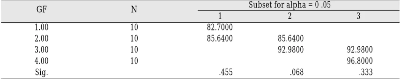
Table III. Duncan’s multiple comparison tests for transverse strength
GF N Subset for alpha = 0 .05
1 2 3
1.00 10 82.7000
2.00 10 85.6400 85.6400
3.00 10 92.9800 92.9800
4.00 10 96.8000
Sig. .455 .068 .333
Means for groups in homogeneous subsets are displayed.
Before abrasion test, surface of glass fiber resin complex was glossy in case of proper proportion of glass fiber inclusion.
There was no extrusion of glass fiber out of resin surface, and generally tight contact between glass fiber and resin was observed in the SEM.
In some cases, gaps between glass fiber and resin
can be found, and holea by exfoliation of glass fiber were found.
After abrasion, roughness increased a llittle by naked eye. Luster and surface roughness val- ue also sustained statistically.
But in case of 9% inclusion of glass fiber, rough- ness value increased statistically.
Table V. Duncan’s multiple comparison tests for Ra. before abrasion
BA N Subset for alpha = 0 .05
1 2
Glass fiber 0% 10 .0850
Glass fiber 3% 10 .1270 .1270
Glass fiber 6% 10 .1670 .1670
Glass fiber 9% 10 .2270
Sig. .209 .127
Means for groups in homogeneous subsets are displayed.
Fig. 3.Surface roughness (Ra) before abrasion test.(Ra) Fig. 4.Surface roughness (Ra) after abrasion test.(Ra)
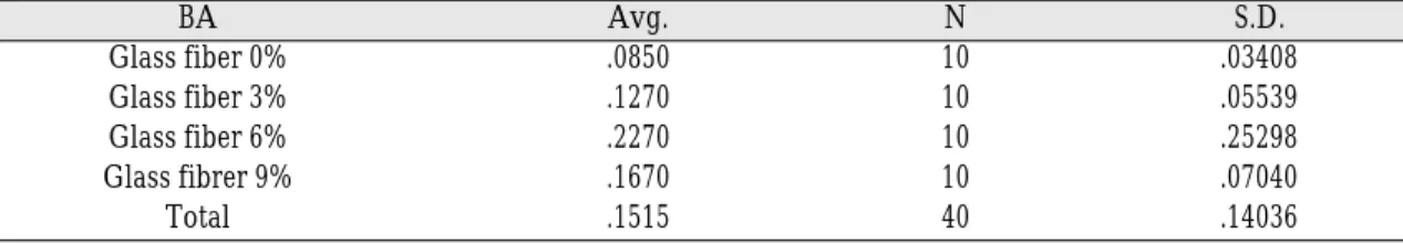
Table IV. Roughness(Ra.) - before abrasion test
BA Avg. N S.D.
Glass fiber 0% .0850 10 .03408
Glass fiber 3% .1270 10 .05539
Glass fiber 6% .2270 10 .25298
Glass fibrer 9% .1670 10 .07040
Total .1515 40 .14036
3. SEM
In SEM, intimate contact between fibers and resin
matrix was found and there was some void.
Glass fibers were generally distributed evenly in the resin matrix with little bunching.
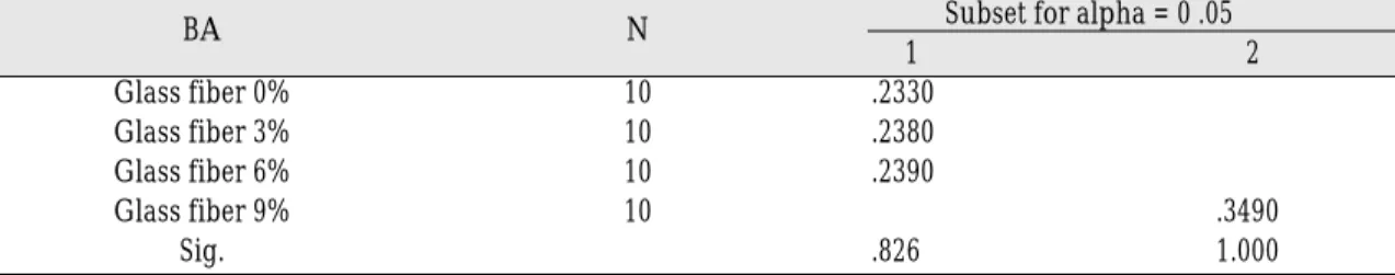
Table VII. Duncan’s multiple comparison tests for Ra. after Abrasion
BA N Subset for alpha = 0 .05
1 2
Glass fiber 0% 10 .2330
Glass fiber 3% 10 .2380
Glass fiber 6% 10 .2390
Glass fiber 9% 10 .3490
Sig. .826 1.000
Means for groups in homogeneous subsets are displayed.
Fig. 5. SEM of group 1 specimen before abrasion test. Fig. 6. SEM of group 2 specimen before abrasion test.
Table VI. Roughness(Ra.) after abrasion test
AA Avg. N S.D.
Glass fiber 0% .2390 10 .02132
Glass fiber 3% .2330 10 .02751
Glass fiber 6% .2380 10 .03259
Glass fiber 9% .3490 10 .10344
Total .2648 40 .07366
SEM before abrasion test (0%, 3%, 6%, 9%) (Fig. 5, 6, 7, 8)
Fig. 7.SEM of group 3 specimen before abrasion test. Fig. 8. SEM of group 4 specimen before abrasion test.
Fig. 9.SEM of group 1 specimen after abrasion test. Fig. 10. SEM of group 2 specimen after abrasion test.
Fig. 11. SEM of group 3 specimen after abrasion test. Fig. 12. SEM of group 4 specimen after abrasion test.
SEM after abrasion test (0%, 3%, 6%, 9%) (Fig. 9, 10, 11, 12)
DISCUSSION
The fracture of acrylic resin dentures remains an unsolved problem. Many investigations have been performed and various approaches ways of strengthening the acrylic resin have been exam- ined, for example, the reinforcement of heat- polymerized PMMA resin using glass fibers, have been suggested in the past.
However, few applications in a clinical setting have been reported due to the surface treatment problem and the complexities of the procedures involved.
Although the use of glass fiber increased trans- verse strength, it is impossible to lay glass fiber in the resin matrix exactly. On the other hand, increased strength was observed when short glass fiber was included exactly.
In this experiment, simple mixing of short glass fiber and resin powder can be performed easily.
The short fiber lengths were convenient for inclu- sion into the acrylic resin dough.
When glass fiber was used, the transverse strength increased continuously with fiber con- centration.
The relatively large standard deviations encoun- tered with results from the fiber containing spec- imens demonstrated a possible drawback of the technique. A similar problem has been encountered by other workers, upon mechanically testing denture resin containing randomly orientated carbon fibers. They emphasized that reinforcement is optimized when fibers are laid in a strategic fash- ion, running parallel to the surface of the denture base. In this way, their contribution to rein- forcement is maximized, whereas fibers at right angles to the surface produce no beneficial effect.
However, they concluded that the technical dif- ficulties of ensuring that fibers were aligned might outweigh the potential advantage, by complicating the technique to such an extent
that it became impractical. This study has shown that a significant effect is produced by glass fibers randomly orientated in specimens.
Presumably, some fibers are orientated to produce beneficial effects and others little or no benefit. The ease and simplicity of their inclusion would make this technique more acceptable for wide- spread use, avoiding the necessity of interrupting the packing procedures, and time-consuming placement of orientated fibers or woven fiber mats.
Further work is clearly needed to investigate the nature of the reinforcement afforded by short glass fiber. The effects of excess monomer on dimensional stability and biocompatibility are of particular importance.
Complete chemical bonding of glass fibers with resins may reduce the roughness dramati- cally.
Using SEM, an especially huge interface gap was observed.
Without using silane, glass fibers lead to a reduction of strength on the contrary. So, it can be assumed that the silane induces a chemical bond- ing.
The viscosity of resin doughs with more than 9%
glass fibers has been remarkably reduced, which made its treatment difficult. The roughness of the resin surfaces was also very high.
Therefore, the oral mucous membrane irritation is expected without resin coatings.
Mostly, the rough surface causes discoloration and plaque deposition.
Discoloration is hardly expected, as the acrylic resins containing 3% and 6% chopped glass fibers show little change in roughness. Concdrning this, more practical investigations are needed.
Oral mucous membrane irritation can be exclud- ed, as long as glass fibers don’t stick out of resins.
However, there is a possibility that glass fibers
come out of resins and cause oral mucous mem- brane irritation.
The glass fibers are fractured as soon as they come out of the resins. They can be stuck into skins or suck in during breathing.
The higher the content of glass fibers was, the lower the gloss of the resins was.
Most of all, the formation of glass fiber bunch- ings affected the esthetics negatively.
Other than asbestos, the glass fiber has no car- cinogenic substance and can be used safely.
However, there is a possibility that fine chopped glass fibers are suck into the lung. More investi- gations must be performed.
The abrasion test was performed simulating practical use of dentures over several years.
CONCLUSION
The purpose of this study is to evaluate the difference in surface roughness after abrasion of glass fibers reinforced PMMA resin.
0%, 3%, 6%, and 9% glass fibers were inserted and transverse strength was estimated.
Abrasion test of glass fiber reinforced resin was done in the saline solution, and roughness was estimated before and after abrasion.
Quantitative analysis was done by 2-dimen- sional surface roughness tester and qualitative analysis was done by electric microscope.
The results are
1. The more glass fiber was inserted, strength increased statistically.
2. Viscosity decreased as quantity of glass fiber increased.
3. Roughness increased a little after abrasion test.
4. Before abrasion test, incorporation of 0%, 3%, 9% glass fiber in the resin showed no dirrerence in roughness statistically.
5. After abrasion test, incorporation of 0%, 3%, 6%
glass fiber in the resin showed same surface roughness value statistically.
6. In SEM, surface roughness increased as the per- centage of the fibers increased.
7. In the areas where glass fiber bunchings are for- mated, a remarkably high roughness was noticed.
REFERENCES
1. Kelly E. Fatigue failure in denture base polymers.
J Prosthet Dent 1969;21:257-66.
2. Hargreaves AS. The prevalence of fractured den- tures. A survey. Br Dent J 1969;126:451-55.
3. Yazdanie N, Mahood M. Carbon fiber acrylic resin composite: An investigation of transverse strength. J Prosthet Dent 1985;54:543-47.
4. DeBoer J, Vermilyea SG, Brady RE. The effect of car- bon fiber orientation on the fatigue resistance and bending properties of two denture resins. J Prosthet Dent 1984;51:119-21.
5. Kilfoil BM, Hesby RA, Pelleu GB. The tensile strength of a composite resin reinforced with car- bon fibers. J Prosthet Dent 1983;50:40-3.
6. Malquarti G, Berruet RG, Bois D. Prosthetic use of carbon fiber-reinforced epoxy resin for esthetic crowns and fixed partial dentures. J Prosthet Dent 1990;63:251-7.
7. Sehajpal SB, Sood VK. Effect of metal fillers of some physical properties of acrylic resins. J Prosthet Dent 1989;61:746-51.
8. Berrong JM, Weed RM, Young JM. Fracture re- sistance of Kevlar-reinforced poly(methyl methacry- late)resin : A Preliminary Study. Int J Prosthodont 1990;3:391-5.
9. Mullarky RH. Aramid fiber reinforcement of acrylic appliances. J Clin Orthod 1985;19:655-8.
10. Pourdeyhimi B, Robinson HH, Schwartz P, Wagner HD. Fracture toughness of kevlar 29/poly(methyl- methacrylate) composite materials for surgical implantations. Ann Biomed Eng 1986;14:277-94.
11. Chow TW, Cheng YY, Ladizesky NH. Polyethylene fibre reinforced poly(methylmethacrylate) -wa- ter sorption and dimensional changes during im- mersion. J Dent 1993;21:367-72.
12. Braden M, Ladizesky NH. Denture base poly(methyl methacrylate) reinforced with ultra-high modulus polyethylene fibres. Br Dent J 1998;164:109-13.
13. Ladizesky NH, Chow TW. Reinforcement of com- plete denture bases with continuous high perfor- mance polyethylene fibers. J Prosthet Dent 1992;68:934-9.
14. Ladizesky NH, Cheng YY, Chow TW, Ward IM.
Acrylic resin reinforced with chopped high per- formance polyethylene fiber-properties and den- ture construction. Dent Mater 1993;9:128-35 15. Gutteridge DL. Reinforcement of poly(methyl
methacrylate) with ultra-high-modulus polyethylene fibre. J Dent 1992;20:50-4.
16. Williamson DL, Boyer DB, Aquilino SA, Leary JM. Effect of polyethylene fiber reinforcement on the strength of denture base resins polymerized by microwave energy. J Prosthet Dent 1994;72:635-8.
17. Solnit GS. The effect of methylmethacrylate rein- forcement with silane-treated and untreated glass fibers. J Prosthet Dent 1991;66:310-4.
18. Vallitu PK. Acrylic resin-fiber composite-part 2: The effect of polymerization shrinkage of polymethyl methacrylate applied to fiber roving on trans- verse strength. J Prosthet Dent 1994;71:613-7.
19. Gutteridge DL. The effect of including ultra-high-
modulus polyethylene fibre on the impact strength of acrylic resin. Br Dent J 1998;164:177-80.
20. Goldberg AJ, Burstone CJ. The use of continu- ous fiber reinforcement in dentistry. Dent Mater 1992;8:197-202.
21. Clark HA, Pluedemann EP. Bonding of silane coupling agents in glass-reinforced plastics. Modern plastics 1963;June:133-8.
22. International standard ISO 1567, ed 3. 1999(E) 02-15.
Reprint request to:
CHANG-WHEKIM, D.D.S., M.S.D., PH.D.
DEPARTMENT OFPROSTHODONTICS,COLLEGE OFDENTISTRY, SEOULNATIONALUNIVERSITY
28, YONKON-DONG,CHONGRO-GU, SEOUL,110-749, KOREA [email protected]