KJTCVS
The Korean Journal of Thoracic and Cardiovascular SurgeryClinical Research
Surgical Outcomes of Kommerell Diverticulum
Young Kern Kwon, M.D., Sung Jun Park, M.D., Suk Jung Choo, M.D., Ph.D., Tae Jin Yun, M.D., Ph.D., Jae Won Lee, M.D., Ph.D., Joon Bum Kim, M.D., Ph.D.
Department of Thoracic and Cardiovascular Surgery, Asan Medical Center, University of Ulsan College of Medicine, Seoul, Korea
ARTICLE INFO Received June 9, 2020 Revised September 8, 2020 Accepted September 19, 2020 Corresponding author Joon Bum Kim Tel 82-2-3010-5416 Fax 82-2-3010-6966
E-mail [email protected] ORCID
https://orcid.org/0000-0001-5801-2395
Background: We aimed to assess the clinical outcomes of patients who underwent sur- gical repair of Kommerell diverticulum (KD) with individualized surgical methods.
Methods: A retrospective analysis was performed of adult patients (aged ≥17 years) who underwent surgery to treat KD between June 2008 and October 2019.
Results: Nine patients (median age, 45 years; range, 19–67 years; 7 men) underwent sur- gical repair. The indications for surgical therapy were acute aortic dissection in 2 patients, the presence of compressive symptoms due to dilated KD in 4 patients, and aneurysm growth in 3 patients. Various surgical techniques were used: (1) resection of the divertic- ulum stump and revascularization of the aberrant subclavian artery (n=3), (2) one-stage total-arch replacement including the diverticulum segment (n=3), and (3) hybrid repair (n=3). Early mortality occurred in 1 case of hybrid repair. Transient paraparesis occurred in a patient who underwent total arch repair as part of complicated acute aortic dissection.
During follow-up (median duration, 30 months; range, 7–130 months), no late death or associated aortic complications were documented. All survivors were free from symptoms and had no abnormal findings on follow-up computed tomography.
Conclusion: With a customized surgical approach and appropriate consideration of pa- tient-specific anatomy and associated comorbidities, KD can be repaired with favorable outcomes.
Keywords: Aorta, Anomaly, Kommerell, Surgical operation, Outcomes
Copyright©The Korean Society for Thoracic and Cardiovascular Surgery. 2020. All right reserved.
This is an Open Access article distributed under the terms of the Creative Commons Attribution Non-Commercial License (http://creativecommons.org/licenses/
Introduction
Kommerell diverticulum (KD) is a rare congenital anom- aly of the aortic arch resulting from the failed involution of the fourth primitive dorsal arch. It is a segmental aortic aneurysmal dilatation associated with aberrant subclavian arteries (ASAs) and can occur on either the left or the right side. The natural history of this disease is still not well un- derstood due to its rarity, and issues surrounding the sur- gical treatment of KD remain controversial. Compressive symptoms, such as dysphagia lusoria, stridor, or compres- sive chest pain, may require therapeutic interventions when the surrounding structures (the esophagus or tra- chea) are mechanically compressed by the enlarged diver- ticulum. In contrast, most patients with KD are asymp- tomatic and are diagnosed incidentally. In such patients, a specific threshold for therapeutic intervention based on aneurysm size has not yet been established because of the
unclear natural history of this disease. The levels of risk of catastrophic aortic events associated with KD, including rupture or dissection, vary widely across reports, from 4%
to 19% and from 11% to 53%, respectively [1,2]. Although some authors have advocated generous prophylactic inter- ventions for this condition, the technically demanding sur- gical procedures attributable to the complex anatomy in- volved have complicated the issue [3].
While various techniques have been proposed for the treatment of KD, including open repair and endovascular or hybrid approaches, the majority of reports are limited to case series with small sample sizes, and a knowledge gap still exists regarding the optimal management of this rare disease. Although we were also limited by a small number of cases, we sought to share our experiences with the surgi- cal treatment of KD over the past 12 years.
https://doi.org/10.5090/kjtcs.20.071 pISSN: 2233-601X eISSN: 2093-6516
Korean J Thorac Cardiovasc Surg. 2020;53(6):346-352
Young Kern Kwon, et al. Surgical Outcomes of Kommerell Diverticulum
KJTCVS
Methods
Study subjects
We retrospectively reviewed the records of adult patients at least 17 years old who underwent open surgical repair of KD or a hybrid operation between June 2008 and October 2019 at Asan Medical Center, Seoul, Korea. A total of 9 pa- tients were identified in the institutional cardiac surgical database. The data collected included details regarding each patient’s preoperative symptoms, comorbidities, oper- ative profiles, and follow-up. This study was approved by the institutional ethics committee and review board of Asan Medical Center (approval no., 2020-0026). The re- quirement for informed consent was waived due to the ret- rospective nature of the study.
Imaging data
Preoperative computed tomography (CT) scans, includ- ing aortic CT angiography, were performed for all patients.
By reanalyzing the CT scans, the orientation of the aorta, the origin of the ASA, the associated aortic anomaly, and the size of the KD were identified. Two measurements of KD size were made: the maximal distance from the tip of the aneurysm to the opposite aortic wall and the diameter of the diverticular orifice (Fig. 1). Postoperative CT scans were also performed for all patients during follow-up to as-
sess the aortic anatomy after surgery.
Surgical profile
The indications for surgical repair included compressive symptoms associated with KD, aortic rupture, acute or chronic aortic dissection, and distal arterial obstruction by the diverticulum. Patients without symptoms or cata- strophic aortic events were not treated surgically.
Four surgeons performed the surgical management of KD during the study period. The surgical approaches and techniques were determined at the operating surgeon’s dis- cretion in consideration of the patient’s anatomy and asso- ciated comorbidities. Concomitant procedures were per- formed when needed.
Results
Baseline patient characteristics and preoperative findings
The median age of the 9 patients at operation was 45 years (range, 19–67 years), and 7 (77.8%) were men. Preop- erative CT scans revealed a right aortic arch (RAA) with a left ASA in 7 patients (77.8%) and a mean maximal aneu- rysmal diameter of 6.1±2.3 cm.
The indications for surgical treatment were complicated acute aortic dissection in 2 patients (patients 4 and 8), the presence of compressive symptoms exerted by the dilated KD in 4 patients (patients 1, 2, 7, and 3), and aneurysm growth in the remaining patients (patients 5, 6, and 9).
Three patients presented typical compressive symptoms requiring surgical treatment. In patient 3, KD repair was performed as a concomitant procedure during the surgical excision of a left atrial myxoma, since the patient exhibited dysphagia attributed to KD.
Perioperative profiles depending on surgical approach
The surgical approaches and techniques were deter- mined at the operating surgeon’s discretion in consider- ation of the patient’s anatomy and associated comorbidi- ties. Diverticular stump resection and revascularization of the ASA were chosen for young patients with relatively small diverticula. For patients who required concomitant ascending aorta replacement, such as those with aortic dis- section, sternotomy was preferred, and one-stage total- arch replacement was performed. However, a hybrid opera-
Fig. 1. Two size measurements of the Kommerell diverticulum: (1) the maximal distance from the tip of the aneurysm to the opposite aortic wall (yellow dashed arrow) and (2) the maximal diameter of the diverticular orifice (blue arrow).
https://doi.org/10.5090/kjtcs.20.071
KJTCVS
tion combined with endovascular vascular repair was an alternative for those deemed to be otherwise at high risk associated with extensive surgery.
Resection of diverticulum stump with aberrant subclavian artery revascularization
Resection of the diverticulum stump and revasculariza- tion of the ASA were performed in 3 patients (33.3%). Graft interposition of the proximal ascending aorta and su- pra-aortic vessels was not performed in these patients. The
mean age of these 3 patients was 25.0±7.1 years, and the mean maximal diameter of the ascending aorta was 2.6±0.2 cm. The mean diameter of the diverticulum was 3.9±0.3 cm in these patients. All 3 patients had RAAs asso- ciated with left ASAs. The distal left ASAs were transferred to the left common carotid arteries, after which simple stump closure of the diverticulum was performed (Fig. 2).
The approach for the stump closure was made through a median full sternotomy in 2 patients (patients 1 and 3), while patient 2 underwent this procedure through a left posterolateral thoracotomy (Table 1). Cardiopulmonary bypass (CPB) was used in 2 patients (patients 1 and 3).
One-stage total-arch replacement including the diverticulum segment
Proximal segments of the aorta were replaced in 3 pa- tients. Graft interposition was performed on the ascending aorta, aortic arch, and proximal descending thoracic aorta including the diverticulum segment under CPB via median full sternotomy. Arch vessels were reimplanted to graft branches of a 4-branch graft. The mean age of these pa- tients was 50.0±16.1 years, and the mean maximal divertic- ular size was 7.1±0.6 cm. The indications for surgery were acute aortic dissection (in patient 4), chronic aortic dissec- tion (in patient 5), and symptomatic aortic aneurysm in- volving the aortic arch (in patient 6). The mean pump time was 310.0±113.8 minutes, and all 3 patients underwent sur- gery under moderate hypothermia (mean lowest body tem- perature, 23.4°C±3.3°C). Patient 4 presented with acute type B aortic dissection complicated by left upper-limb malperfusion. Detailed information regarding the anatom- ic profile and surgical procedure was described previously [4]. The intimal tear was located at the mid-arch involving
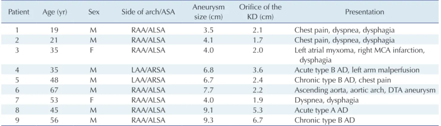
Table 1. Patient characteristics
Patient Age (yr) Sex Side of arch/ASA Aneurysm size (cm)
Orifice of the
KD (cm) Presentation
1 19 M RAA/ALSA 3.5 2.1 Chest pain, dyspnea, dysphagia
2 21 M RAA/ALSA 4.1 1.7 Chest pain, dyspnea, dysphagia
3 35 F RAA/ALSA 4.0 2.0 Left atrial myxoma, right MCA infarction,
dysphagia
4 35 M LAA/ARSA 6.8 3.6 Acute type B AD, left arm malperfusion
5 48 M LAA/ARSA 6.7 2.4 Chronic type B AD, chest pain
6 67 M RAA/ALSA 7.7 2.2 Ascending aorta, aortic arch, DTA aneurysm
7 53 F RAA/ALSA 4.0 1.9 Dyspnea, dysphagia
8 45 M RAA/ALSA 9.1 5.3 Acute type A AD
9 56 M RAA/ALSA 9.3 6.7 Chronic type B AD
ASA, aberrant subclavian artery; KD, Kommerell diverticulum; M, male; F, female; RAA, right aortic arch; LAA, left aortic arch; ALSA, aberrant left subclavian artery; ARSA, aberrant right subclavian artery; MCA, middle cerebral artery; AD, aortic dissection; DTA, descending thoracic aorta.
Fig. 2. Resection of the diverticulum stump and revascularization of the aberrant subclavian artery. (A) All 3 patients had a right aortic arch with an aberrant left subclavian artery. The aortic arch vessels branched in the following order: left carotid artery, right carotid artery, right subclavian artery, and aberrant left subclavi- an artery. (B) The aberrant left subclavian artery was divided and anastomosed to the left common carotid artery with the end-to- side technique. The proximal stump of the left subclavian artery was repaired with an oversewing stitch. LCCA, left common carot- id artery; RCCA, right common carotid artery; RSCA, right subcla- vian artery; ALSA, aberrant left subclavian artery.
A B
Young Kern Kwon, et al. Surgical Outcomes of Kommerell Diverticulum
KJTCVS
dissection flaps spanning from the opening of the right common carotid artery to the renal arteries and including the KD and the left ASA. Emergency total-arch replace- ment was performed through median full sternotomy in this patient.
Hybrid repair of Kommerell diverticulum
Hybrid repair of KD was performed in the other 3 pa- tients. Three distinct hybrid approaches were used in the respective patients: (1) graft interposition of the total aortic arch just before the diverticular segment of the descending thoracic aorta, followed by retrograde thoracic endovascu- lar aorta repair (TEVAR) (in patient 9), (2) type 2 zone 0 debranching and elephant trunk insertion followed by ret- rograde TEVAR (in patient 8) (Fig. 3), and (3) zone 2 deb- ranching through a neck incision and subsequent retro- grade TEVAR (in patient 7) (Fig. 4). The mean age of the 3 patients was 51.3±4.6 years, and the size of the diverticu- lum ranged from 4.0 to 9.3 cm. Hybrid procedures were performed due to compressive symptoms attributable to a huge diverticulum (in patients 7 and 9) and acute type A aortic dissection in patient 8. Two patients underwent sur-
A B
C D E
Fig. 3. Hybrid repair of Kommerell diverticulum. Endovascular repair is a potential alternative in patients with aortic diverticulum compli- cated by aortic dissection. (A, B) A computed tomography scan of patient 8 shows a type A aortic dissection with an acute angle of aortic arch curvature. (C) Illustrates aortic disease. (D) Ascending aor- ta replacement with the elephant trunk technique, along with zone 0 debranching surgery, was per- formed to create a proximal land- ing zone. (E) The procedure was followed by thoracic endovascular aortic repair for the exclusion of aortic dissection in the descending thoracic aorta. LCCA, left common carotid artery; RCCA, right com- mon carotid artery; RSCA, right subclavian artery; ALSA, aberrant left subclavian artery.
A B
*
Fig. 4. Minimally invasive hybrid surgery to treat Kommerell diver- ticulum with bypass of the common carotid artery to the subclavi- an artery and endovascular repair. (A) Bypass of the left common carotid artery to the aberrant subclavian artery was performed with a ring-reinforced polytetrafluoroethylene graft through a cervical approach. The aneurysm of the Kommerell diverticulum was covered with a stent graft. This technique made it possible to repair the Kommerell diverticulum with a minimal incision and without cardiopulmonary bypass. (B) A computed tomography scan of patient 7 shows a ringed polytetrafluoroethylene graft (ar- row) connecting the left common carotid artery and left subcla- vian artery and stent graft (star) in the aorta. LCCA, left common carotid artery; RCCA, right common carotid artery; RSCA, right subclavian artery; ALSA, aberrant left subclavian artery; LVA, left vertebral artery.
https://doi.org/10.5090/kjtcs.20.071
KJTCVS
gery under CPB (patients 8 and 9), while the procedure for patient 7 did not require CPB.
Clinical outcomes
One in-hospital death (14.2%) occurred. This death oc- curred after a hybrid procedure in patient 9, who under- went surgery due to an extensive aneurysm compressing the aorta. A type 3 endoleak had led to persistent growth of the diverticulum and the aneurysmal sac after the endo- vascular repair. The patient could not be weaned from the ventilator and eventually died of sepsis attributed to a tra- cheoesophageal fistula, a complication that arose within 2 weeks of the hybrid procedure.
One patient (patient 4) experienced temporary parapare- sis but recovered without sequelae. This patient also expe- rienced mediastinitis. Several rounds of debridement and irrigation followed by omental and pectoralis muscle flap interposition were performed. On follow-up, he showed a progressively enlarging thoracoabdominal aorta (maximal diameter, 5.1 cm) due to remnant distal aortic dissection and consequently underwent thoracoabdominal aorta re- placement a year after the index procedure.
No late mortality occurred, and all patients were free from relevant symptoms of KD during the follow-up peri- od (median duration, 30 months; range, 7–130 months).
Postoperative serial CT scans performed for the other 7 patients (with the exception of patients 4 and 9) showed no abnormal findings related to the repaired aorta (median time of CT scan, 6 months; range, 0–115 months) (Table 2).
Discussion
KD is a congenital aortic anomaly that arises due to failed involution at the fourth primitive dorsal arch [1].
The prevalence of right ASA from the left aortic arch and left ASA from the right aortic arch has been reported to be 0.7%–2.0% and 0.04%–0.4%, respectively. Approximately 20%–60% of cases of ASAs are associated with KD. Only a small portion of patients with KD present symptoms. Most of these aortic conditions are identified incidentally on CT scans performed for reasons unrelated to KD. Due to its rare clinical presentation, the natural history of this dis- ease is still not well known. The reported incidence of cat- astrophic aortic events associated with KD, such as aortic rupture and aortic dissection, has varied widely across re- Table 2. Operative details and complications
Patient Procedure Urgency Approach(es) CPB time
(min)
ACC time (min)
Lowest
BT (°C) Outcome
1 Resection of diverticulum stump and revascularization of ASA
Elective Sternotomy 93 NA 33.6 F/U without symptoms
over 100 months 2 Resection of diverticulum stump
and revascularization of ASA
Elective Thoracotomy NA NA NA F/U without symptoms
over 105 months 3 Resection of diverticulum stump
and revascularization of ASA;
cardiac myxoma excision
Elective Sternotomy 128 30 22.4 F/U without symptoms
over 106 months 4 Asc. aorta, total-arch and
proximal DTA replacement
Emergency Sternotomy 431 15 20.1 Mid-DTA replacement due
to progressive remnant distal aortic dissection 5 Asc. aorta, total-arch and
proximal DTA replacement
Elective Sternotomy 205 132 26.8 F/U without symptoms
over 7 months 6 Asc. aorta, total-arch and
proximal DTA replacement
Elective Sternotomy 294 197 23.4 F/U without symptoms
over 8 months 7 Hybrid operation (LSCA to LCCA
bypass, retrograde TEVAR)
Elective Cervical, femoral
NA NA NA F/U without symptoms
over 125 months 8 Hybrid operation (asc. aorta
replacement, debranching surgery, retrograde TEVAR)
Urgent Sternotomy, femoral
217 33 17.5 F/U without symptoms
over 130 months 9 2-Stage hybrid operation (asc.
aorta, total-arch replacement, second-stage retrograde TEVAR)
Elective Sternotomy, femoral
357 234 23.3 In-hospital death
CPB, cardiopulmonary bypass; ACC, aortic cross-clamping; BT, body temperature; ASA, aberrant subclavian artery; NA, not available; F/U, follow- up; asc. aorta, ascending aorta; DTA, descending thoracic aorta; LSCA, left subclavian artery; LCCA, left common carotid artery; TEVAR, thoracic endovascular aorta repair.
Young Kern Kwon, et al. Surgical Outcomes of Kommerell Diverticulum
KJTCVS
ports by different researchers. An early study by Austin and Wolfe [5] reported a 19% rate of aortic dissection in 32 KD patients, and Cina et al. [6] reported a 53% rate of aor- tic dissection or rupture among 33 patients with KD. Tana- ka et al. [1] reviewed 212 cases of KD and reported an 11%
frequency of aortic dissection and a 4% frequency of diver- ticular rupture.
Compressive symptoms of the esophagus or trachea, in- cluding dysphagia, dyspnea, chest pain, and cough, are definite indications for the surgical treatment of KD. For asymptomatic patients, however, a generally accepted, spe- cific threshold for prophylactic surgery based on diverticu- lar size or baseline aortic dimensions has not yet been es- tablished. Several groups have suggested criteria based on diverticulum size, but these size criteria and the associated measurement methods have been inconsistent. Cina et al.
[6] advocated the aggressive surgical treatment for KD larger than 3 cm in diameter at the level of the orifice, and Ota et al. [7] suggested surgical management for symptom- atic aneurysms larger than 5 cm. Considering the incre- mental complexities of surgery as the diverticulum grows, Vinnakota et al. [2] and Kouchoukos and Masetti [3] also advocated early surgery to eliminate the risk of aortic dis- section or aneurysm rupture. In contrast, asymptomatic ASA and KD are generally thought to be benign anomalies of the aorta. According to a study by Erben et al. [8], the natural course of aneurysmal growth seems to be relatively minimal, at approximately 1.4–2.4 mm per year.
Given this knowledge gap regarding the natural history of KD and the divergent points of view on the surgical treatment of KD, close outpatient follow-up has been con- ducted for asymptomatic patients at our center. Elective surgery is planned for patients with: (1) newly developed symptoms, (2) evidence of aneurysm growth, or (3) newly discovered aortic rupture/dissection in serial CT angiogra- phy. Various surgical techniques have been introduced for the repair of KD. We attempted to employ these techniques in a customized fashion, with consideration of the under- lying anatomy and associated comorbidities of each pa- tient.
Resection of the diverticular stump and subclavian-to- carotid bypass are standard procedures for patients with relatively small aneurysms. Various approaches, including left or right thoracotomy, median sternotomy, and a com- bination of thoracotomy and sternotomy, have been report- ed with favorable clinical outcomes [9-11]. Median full sternotomy provides exposure for the treatment of con- comitant heart disease, as in the case of patient 3. When the patient’s anatomy allows, ASA revascularization and
repair of the diverticulum through a left thoracotomy can be performed without using CPB (as in the case of patient 2).
When aortic dissection involves the aortic arch, graft in- terposition of the aorta encompassing the diverticular seg- ment may be more suitable. Recent studies have shown sat- isfactory surgical outcomes in patients who underwent one-stage total-arch replacement including the diverticular segment with a branched vascular graft [12]. We conducted one-stage graft interposition using a 4-branched graft for 3 patients in whom aortic dissection involved the aortic arch.
In patients with huge diverticula or a greatly dilated aor- ta, surgical procedures can be more demanding, especially if the patient has multiple comorbidities. Endovascular re- pair can be an alternative to surgical repair, minimizing postoperative complications and maximizing postoperative recovery. The pressure of the aneurysmal sac decreases af- ter successful sealing of the aneurysm, relieving the com- pressive symptoms of KD. For patients with steep aortic arch angles and limited proximal landing zones, partial aorta graft interposition with a debranching procedure provides easier access and an adequate landing zone for a stent graft.
However, KD repair using TEVAR could be unsuitable for patients with compressive symptoms. The literature in- cludes a case in which compressive symptoms did not im- prove after endovascular repair. Reoperations due to en- doleak have also been reported [2]. In our study, patient 9 had a type 3 endoleak from the ASA after exclusion via an endovascular graft. Even after subclavian artery occlusion using a device, the remnant aneurysmal sac compressed the surrounding organs. This eventually induced pneumo- nia and pulmonary abscess, which caused the death of the patient. Previous studies have shown medial attenuation of resected diverticular segments on histologic analysis [10,13]. This could explain the vulnerability of the aorta to endoleak after endovascular repair.
A right aortic arch with an ASA frequently creates a vas- cular ring when it is accompanied by ligamentum arterio- sus. However, not all KD patients have a vascular ring that encircles the trachea and esophagus. Cases of recurrent compressive symptoms have been reported in patients with KD who previously underwent ligamentum division sur- gery [13,14]. This suggests that KD alone usually compress- es the surrounding organs independently of ligamentum arteriosus, and decompression of the enlarged aneurysm can release the compressive symptoms of KD in the vast majority of cases. In cases with segmental aortic resection via sternotomy in our series, the total arch and proximal
https://doi.org/10.5090/kjtcs.20.071
KJTCVS
descending aorta were replaced, a procedure that inevitably involves the removal/release of the ligamentum arteriosus.
For other cases involving KD resection or a hybrid ap- proach, the ligamentum arteriosus was left unidentified to avoid recurrent laryngeal nerve injury; however, we did not find any residual structure compressing adjacent organs in either symptom-based or imaging-based (CT) evaluations in all 8 successful cases.
One limitation of this study is that it was a retrospective review of a limited number of cases. As KD is a congenital aortic anomaly, each patient has distinctive anatomy; ac- cordingly, 4 different surgeons applied modified and indi- vidualized surgical techniques for each patient. A cohesive strategy to treat this aortic disease should be established on the basis of further research.
In conclusion, among 9 patients, 1 case of early mortality (11.1%) occurred after surgery. Because KD is associated with vulnerability to severe aortic disease, a certain degree of mortality and morbidity after surgical repair is expect- ed. However, with modified and individualized surgical techniques designed in consideration of each patient’s anatomy and comorbidities, KD can be treated with mini- mal morbidity and mortality and a reasonable rate of symptom resolution.
Conflict of interest
No potential conflict of interest relevant to this article was reported.
ORCID
Young Kern Kwon: https://orcid.org/0000-0003-4795-340X Sung Jun Park: https://orcid.org/0000-0002-0244-062X Suk Jung Choo: https://orcid.org/0000-0003-4291-302X Tae Jin Yun: https://orcid.org/0000-0002-0336-1720 Jae Won Lee: https://orcid.org/0000-0003-0751-2458 Joon Bum Kim: https://orcid.org/0000-0001-5801-2395
References
1. Tanaka A, Milner R, Ota T. Kommerell’s diverticulum in the current
era: a comprehensive review. Gen Thorac Cardiovasc Surg 2015;63:
245-59.
2. Vinnakota A, Idrees JJ, Rosinski BF, et al. Outcomes of repair of Kommerell diverticulum. Ann Thorac Surg 2019;108:1745-50.
3. Kouchoukos NT, Masetti P. Aberrant subclavian artery and Kom- merell aneurysm: surgical treatment with a standard approach. J Tho- rac Cardiovasc Surg 2007;133:888-92.
4. Kim JB, Yang DH, Kang JW. Right aortic arch and an aberrant left subclavian artery arising from a Kommerell diverticulum complicat- ed by acute aortic dissection. J Thorac Cardiovasc Surg 2012;144:
978-9.
5. Austin EH, Wolfe WG. Aneurysm of aberrant subclavian artery with a review of the literature. J Vasc Surg 1985;2:571-7.
6. Cina CS, Althani H, Pasenau J, Abouzahr L. Kommerell’s diverticu- lum and right-sided aortic arch: a cohort study and review of the lit- erature. J Vasc Surg 2004;39:131-9.
7. Ota T, Okada K, Takanashi S, Yamamoto S, Okita Y. Surgical treat- ment for Kommerell’s diverticulum. J Thorac Cardiovasc Surg 2006;
131:574-8.
8. Erben Y, Brownstein AJ, Velasquez CA, et al. Natural history and management of Kommerell’s diverticulum in a single tertiary referral center. J Vasc Surg 2020;71:2004-11.
9. Backer CL, Mavroudis C, Rigsby CK, Holinger LD. Trends in vas- cular ring surgery. J Thorac Cardiovasc Surg 2005;129:1339-47.
10. Kim KM, Cambria RP, Isselbacher EM, et al. Contemporary surgical approaches and outcomes in adults with Kommerell diverticulum.
Ann Thorac Surg 2014;98:1347-54.
11. Uchino G, Yunoki K, Hattori S, et al. Outcomes of anterolateral tho- racotomy with or without partial sternotomy for Kommerell divertic- ulum. Ann Thorac Surg 2017;103:1922-6.
12. Chang Y, Guo HW, Yu CT, Sun XG, Chang Q, Qian XY. Surgical treatment for Kommerell’s diverticulum associated with aortic dis- section involving aortic arch. J Card Surg 2019;34:1273-8.
13. Luciano D, Mitchell J, Fraisse A, Lepidi H, Kreitmann B, Ovaert C.
Kommerell diverticulum should be removed in children with vascu- lar ring and aberrant left subclavian artery. Ann Thorac Surg 2015;
100:2293-7.
14. Backer CL, Hillman N, Mavroudis C, Holinger LD. Resection of Kommerell’s diverticulum and left subclavian artery transfer for re- current symptoms after vascular ring division. Eur J Cardiothorac Surg 2002;22:64-9.