• 검색 결과가 없습니다.

Pseudo-Aneurysm in Internal Maxillary Artery Caused by Radiofrequency Ablation: Literature Review with a Case Report JOMP

N/A
N/A
Protected

Academic year: 2021

Share "Pseudo-Aneurysm in Internal Maxillary Artery Caused by Radiofrequency Ablation: Literature Review with a Case Report JOMP"

Copied!
4
0
0

로드 중.... (전체 텍스트 보기)

전체 글

(1)

www.journalomp.org

pISSN 2288-9272 eISSN 2383-8493 J Oral Med Pain 2020;45(2):44-47 https://doi.org/10.14476/jomp.2020.45.2.44

Pseudo-Aneurysm in Internal Maxillary Artery Caused by Radiofrequency Ablation: Literature Review with a Case Report

Hyun-Woo Yang

1

, Ji-Hyun Oh

2

, Ok-Hyung Nam

3

, Chunui Lee

1

1

Department of Oral and Maxillofacial Surgery, Wonju College of Medicine, Yonsei University, Wonju, Korea

2

Department of Oral and Maxillofacial Surgery, School of Dentistry, Gangneung-Wonju National University, Gangneung, Korea

3

Department of Pediatric Dentistry, School of Dentistry, Kyunghee University, Seoul, Korea

Received May 13, 2020 Revised May 26, 2020 Accepted May 27, 2020

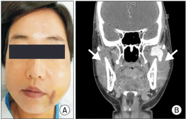
The case of pseudo-aneurysm of internal maxillary artery (IMA) in oral and maxillofacial region is known to be very rare. The etiology of this case was regarded as IMA injury by radiofrequency ablation (RFA) and such incidence was not reported previously. One case of false aneurysm in the IMA was referred from local dental clinic to our department. Left facial swelling was observed with severe trismus immediately after radiofrequency proce- dure for masseteric nerve block in local dental clinic. Despite of medication and surgical intervention, the swelling did not subside and there was massive bleeding and pulsation on one of the follow ups. The traumatic vascular disorder was suspected and finally diagnosed with angiography and treated by embolization procedure. RFA targeting masseteric nerve or trigeminal ganglion may cause traumatic injury to adjacent anatomic structures such as IMA, resulting in pseudo-aneurysm. Clinicians must be aware of potential damages of RFA.

Angiography enables the solid diagnosis for pseudo-aneurysm, and selective embolization can be optimum treatment method.

Key Words:

Key Words: Embolization; Pseudoaneurysm; Radiofrequency ablation

Correspondence to:

Chunui Lee

Department of Oral and Maxillofacial Surgery, Wonju College of Medicine, Yonsei University, 20 Ilsan-ro, Wonju 26426, Korea Tel: +82-33-741-1451

Fax: + 82-33-741-1442 E-mail: [email protected]

https://orcid.org/0000-0002-6846-5176 This work was supported by the Yonsei University Wonju Campus Future-Leading Research Initiative of 2020 (2020-52- 0063).

Case Report

JOMP

Journal of Oral Medicine and Pain

Copyright

2020 Korean Academy of Orofacial Pain and Oral Medicine. All rights reserved.

CC

This is an open-access article distributed under the terms of the Creative Commons Attribution Non-Commercial License (http://creativecommons.org/licenses/by-nc/4.0/), which permits unrestricted non-commercial use, distribution, and reproduction in any medium, provided the original work is properly cited.

INTRODUCTION

An arterial aneurysm is defined as a focal dilation of a blood vessel with respect to the original artery [1]. A pseu- do-aneurysm or traumatic arterio-venous fistula can be formed as a result of arterial wall dehiscense caused by blunt trauma and penetrating trauma. In the past, most of the pseudo-aneurysms were reported as a result of knife wound, gunshot wound, or adjacent bone fractures [2- 4]. However, nowadays some reports reveal pseudo-aneu- rysm caused by therapeutic radiofrequency ablation (RFA).

Recently some reports have introduced pseudo-aneurysms of various etiologies in oral and maxillofacial region, but only one of them was caused by RFA [5]. We experi- enced previously unreported case of the patient with inter- nal maxillary artery (IMA) pseudo-aneurysm presumably

occurred by RFA on masseteric nerve. We hereby report a case on the etiology, symptoms and treatment.

CASE REPORT

A 44-year-old woman visited Department of Emergency

with left facial swelling and spontaneous pain. The medical

history revealed that she underwent masseteric nerve block

therapy due to chronic temporo-mandibular disorder 10

days before the visit. The patient explained that facial swell-

ing appeared immediately after the treatment and pain fol-

lowed consecutively. Clinically we could confirm the facial

swelling with its tenderness, spontaneous pain, and severe

trismus (Fig. 1A). On radiological finding, low density radi-

opaque lesion with contrast extravasation in left masticator

space and peripheral contrast enhancing low density lesion

(2)

45 Hyun-Woo Yang et al. Pseudo-Aneurysm of IMA Caused by RFA

www.journalomp.org

at right masticator space was noted especially lateral to both coronoid notch (Fig. 1B). The white blood cell count, neutrophil count, C-reactive proteins were within normal limits, leading to rule out infection. However, concern- ing suspected hematoma formation and possible second- ary infection, incision and drainage with silastic tube inser- tion on left masticatory space was done on the day of visit.

Despite few weeks of observation with dressing, the patient showed little pain relief and no significant progression in facial swelling and tenderness. Two days after the first vis- it, excessive bleeding occurred on irrigation through silas- tic drain and palpable pulsation was confirmed. Suspecting vascular pathology, pressure hemostasis on pulsating le- sion was done immediately and on the same day angiogra- phy on left carotid artery was performed. The angiography

revealed a large vascular sac on the medial portion of IMA of the left side; the lesion was diagnosed traumatic pseudo- aneurysm (Fig. 2A, B). On the same day, the patient was referred to Department of Neurosurgery, and embolization was performed under simultaneous angiography. Under the guidance of 5.5F femoral sheath (Radifocus Introducer II, Terumo, Tokyo, Japan) and 5F guiding catheter (IR Co Ltd., Tokyo, Japan) to left external carotid artery, the IMA was embolized with multiple coils and histoacryl glue (Fig. 2C).

After the embolization, no post-operative hemorrhage oc- curred, and within 2 weeks of progress observation, the left facial swelling and related symptoms disappeared (Fig. 3)

The study protocol and access to patient records were approved by the Institutional Review Board of Wonju Severance Christial Hospital (CR319303), Wonju, Korea.

Fig. 3.

Fig. 3. Two weeks after embolization procedure. The left facial swelling and relating symptoms were disappeared.

Fig. 2.

Fig. 2. Internal maxillary artery (IMA) of the left side. (A) Left IMA angiography, embolization. (B) The angiography revealing a large vascular sac on the medial portion of left IMA, presumably traumatic pseudo aneurysm. (C) Under the guidance of 5.5F femoral sheath and 5F guiding catheter to left external carotid artery, the IMA was embolized with multiple coils and histoacryl glue.

A B C

Fig. 1.

Fig. 1. Clinical and radiographic findings. (A) The patient on the first visit after the masseteric nerve block at local clinic. Facial swelling with its tenderness, spontaneous pain, and severe trismus has been observed. (B) About 32×30×40 mm sized low density radio opaque lesion at left masticatory space with contrast extravasation.

Peripheral contrast enhancing low density lesion at right (arrows).

A B

(3)

46 J Oral Med Pain Vol. 45 No. 2, June 2020

www.journalomp.org

Written informed consent was obtained from the patient for publication of this case report and accompanying images.

DISCUSSION

According to the patient’s past history and referral to the local clinician, the etiology of the pseudo-aneurysm on this case was suspected as iatrogenic trauma caused by radiofre- quency device (Thermocon; Sometech, Seoul, Korea) while performing masseteric nerve block. There have been reports of pseudo-aneurysms after RFA in number of large arter- ies [4,5]; however, pseudo-aneurism in IMA due to RFA has never been reported.

RFA is well known as minimally invasive surgical method applied to selective indications of hepatic, pulmonary, ver- tebral tumor and also can be utilized in chronic pain disor- ders by thermo-coagulating ganglions and peripheral nerve, resulting in nerve block. Hematologists use RF as an alter- native method of endovenous obliteration of saphenous vein reflux and it is also applied to the surgical therapy of sleep apnea in fields of otolaryngology and oral surgery [6-9].

RFA involves percutaneous insertion of an electrode into a lesion under thorough guidance of radiologic, ultrasonic devices. Then a high frequency, alternating current (350-500 kHz) is delivered through the lesion causing agitation of the tissue ionic molecules, which in turn produces frictional heat. This energy heats the tissue temperature up to 100°C.

Unlike electrocautery or CO

2

laser, which directly delivers high energy to destroy targeting tissues, the heat generated from RFA device is created by the tissues’ resistance to ra- diofrequency current. Such mechanism of RFA emits rela- tively uniform zone of radiant and conductive heat which is about 5-10 mm radius from the electrode [10].

Radiofrequency energy emitted through the electrode generates heat leading to coagulative necrosis. Necrotic tis- sues are then metabolized resulting in tissue contraction or obliteration. Percutaneous RFA has been used as a less in- vasive alternative to surgical resection of primary of met- astatic lesions who have failed or are not candidates for surgery.

RFA, as mentioned above, is a useful therapy which can be applied in many fields, however, its usage may often

lead to serious complications including pseudo-aneurysms.

Improper location of electrode around trigeminal nerve branches may result in paresthesia of skin segments, facial muscle weaknesses, diplopia, and anesthesia dolorosa [11].

Tamai et al. [12] reported the intrahepatic pseudo-aneurysm after RFA therapy of hepatocellular carcinoma. Michael et al. [5] reported pseudo-aneurysm of lingual artery af- ter tongue base reduction using RFA due to patient’s sleep apnea. Schmerber et al. [13] reported the case of internal carotid artery false aneurysm due to gasserian ganglion thermo-coagulation under the diagnosis of trigeminal neu- ralgia. Many reports on complications due to radiofreqeun- cy ablation especially that of pseudo-aneurysm formation made authors to presume that electrode insertion, or heat energy of RFA may harm surrounding structures of targeted operation site.

Sakurai et al. [14] reported the case of pulmonary artery pseudoaneurysm after RFA of pulmonary epitheloid he- mangioendothelioma and suggested its etiology as the pen- etration trauma and vessel wall damage by the heat energy.

Performers of RFA suggest that the likelihood of potential thermal injury induced by RFA is low because of blood flow. However, we speculated that the friable arterial wall cannot always withstand the thermal energy depending on the proximity of the artery.

In our case, it is presumable that local clinician focused only on detecting the masseteric nerve without thorough consideration about surrounding structures. Considering the contrast computed tomography of the patient, the targeted site of the RFA is the coronoid notch area which is adja- cent to the IMA. The left IMA of the patient may have been damaged by improper location of the electrode, vessel wall damage due to RFA energy, or both.

RFA must be performed with thorough knowledge of an- atomic structures adjacent to the targeted site and real-time monitoring of the operation with C-arm or ultrasonography is highly recommended in order to prevent complications. It must also be done by a skilled operator who has sufficient understanding of the targeted anatomy. Moreover, the pa- tient must be warned about possible damages of anatomic structures nearby and consequent complications including pseudo-aneurysm.

Several therapeutic options are introduced for the

(4)

47 Hyun-Woo Yang et al. Pseudo-Aneurysm of IMA Caused by RFA

www.journalomp.org

treatment of IMA pseudo-aneurysm, such as external ca- rotid artery ligation and IMA ligation under surgery [3,15].

However, of many treatment options, selective embolization with angiography is less invasive and more sophisticated than traditional surgical procedures. It is possible to gain control of the damaged vessel in places difficult to reach surgically, and it reduces the risk of bleeding and revascu- larization of the pseudo-aneurysm by collateral circulation [16].

In conclusion, we report previously unreported case of pseudo-aneurysm of IMA iatrogenically caused by RFA.

The patient described in this report presented excessive fa- cial swelling immediately after RFA therapy. Under prop- er diagnosis with angiography the patient’s left IMA was successfully embolized. Pseudo-aneurysm may cause life- threatening event followed by massive bleeding. Therefore, when performing RFA, thorough understanding of ana- tomic structures adjacent to the targeting lesion is required.

And the therapy must be performed by experienced spe- cialist under radiologic or ultrasonic guidance particularly when complications are expected.

CONFLICT OF INTEREST

No potential conflict of interest relevant to this article was reported.

ORCID

Hyunwoo Yang

https://orcid.org/0000-0003-2673-9623 Ji-Hyun Oh

https://orcid.org/0000-0002-6050-7175 Ok-Hyung Nam

https://orcid.org/0000-0002-6386-803X Chunui Lee

https://orcid.org/0000-0002-6846-5176

REFERENCES

1. Aggarwal S, Qamar A, Sharma V, Sharma A. Abdominal aortic aneurysm: a comprehensive review. Exp Clin Cardiol 2011;16:11- 15.

2. Clark S, Wood N. Traumatic aneurysms of the internal maxillary artery and their treatment by embolisation: a report of two cases.

Br J Oral Maxillofac Surg 1990;28:302-305.

3. Banks P, Redpath TH. Closed carotid artery hemorrhage as a com- plication of minor gunshot wounds of the face and jaws. J Oral Surg 1972;30:176-183.

4. Lee JH, Hong SM, Lee EJ, et al. False aneurysm in internal maxil- lary artery. J Kor Oral Maxillofac Surg 2003;29:56-59.

5. Herzog M, Schmidt A, Metz T, et al. Pseudoaneurysm of the lingual artery after temperature-controlled radiofrequency tongue base reduction: a severe complication. Laryngoscope 2006;116:665-667.

6. Merchant RF, Pichot O. Long-term outcomes of endovenous ra- diofrequency obliteration of saphenous reflux as a treatment for superficial venous insufficiency. J Vasc Surg 2005;42:502-509.

7. Sweet WH, Wepsic JG. Controlled thermocoagulation of trigemi- nal ganglion and rootlets for differential destruction of pain fi- bers. 1. Trigeminal neuralgia. J Neurosurg 1974;40:143-156.

8. Dupuy DE, Hong R, Oliver B, Goldberg SN. Radiofrequency abla- tion of spinal tumors: temperature distribution in the spinal ca- nal. AJR Am J Roentgenol 2000;175:1263-1266.

9. Blumen MB, Dahan S, Fleury B, Hausser-Hauw C, Chabolle F.

Radiofrequency ablation for the treatment of mild to moderate obstructive sleep apnea. Laryngoscope 2002;112:2086-2092.

10. Hong K, Georgiades C. Radiofrequency ablation: mechanism of action and devices. J Vasc Interv Radiol 2010;21(8 Suppl):S179- S186.

11. Raj PP, Lou L, Erdine S, Staats PS, Waldman SD. Radiographic imaging for regional anesthesia and pain management. New York: Churchill Livingstone; 2003.

12. Tamai F, Furuse J, Maru Y, Yoshino M. Intrahepatic pseudoaneu- rysm: a complication following radio-frequency ablation therapy for hepatocellular carcinoma. Eur J Radiol 2002;44:40-43.

13. Schmerber S, Vasdev A, Chahine K, Tournaire R, Bing F. Internal carotid false aneurysm after thermocoagulation of the gasserian ganglion. Otol Neurotol 2008;29:673-675.

14. Sakurai J, Mimura H, Gobara H, Hiraki T, Kanazawa S. Pulmo- nary artery pseudoaneurysm related to radiofrequency ablation of lung tumor. Cardiovasc Intervent Radiol 2010;33:413-416.

15. Allen GW. Ligation of the internal maxillary artery for epistaxis.

Laryngoscope 1970;80:915-923.

16. Gerbino G, Roccia F, Grosso M, Regge D. Pseudoaneurysm of the

internal maxillary artery and Frey’s syndrome after blunt facial

trauma. J Oral Maxillofac Surg 1997;55:1485-1490.

수치

Fig. 3. Two weeks after embolization procedure. The left facial  swelling and relating symptoms were disappeared.

참조

관련 문서