Ann Hepatobiliary Pancreat Surg 2017;21:84-87
https://doi.org/10.14701/ahbps.2017.21.2.84
Case Report
Laparoscopic cholecystectomy in a case of situs inversus totalis:
a review of technical challenges and adaptations
Azhar Alam, and Abhijit Santra
Department of Surgery, B.R. Singh Hospital and Centre for Medical Education and Research, Eastern Railways, Kolkata, West Bengal, India
Situs inversus totalis is a rare congenital condition, characterized by the transposition of the thoracic and abdominal viscera, resulting in a mirror image of normal anatomy. Even though situs inversus does not predispose to gall stones, a laparoscopic cholecystectomy, in a case of situs inversus, can prove to be a technically challenging procedure, espe- cially for the right-handed surgeon. In this case report, we present an unusual case of cholelithiasis in a patient with situs inversus totalis. A laparoscopic cholecystectomy, which is considered the gold standard procedure for sympto- matic gallstones, was performed. The technical challenges that were anticipated due to anatomical anomalies were managed by various preoperative and intraoperative modifications. Through this present case report, we concluded that a laparoscopic cholecystectomy is a feasible and safe procedure in patients with situs inversus totalis and can be precisely performed by a right-handed surgeon, with necessary adaptations. (Ann Hepatobiliary Pancreat Surg 2017;21:84-87)
Key Words: Laparoscopic cholecystectomy; Situs inversus totalis; Adaptations
Received: December 24, 2016; Revised: January 27, 2017; Accepted: February 2, 2017 Corresponding author: Azhar Alam
Department of Surgery, B.R. Singh Hospital and Centre for Medical Education and Research, Eastern Railways, 18 C, Smith Lane, 4th Floor, Kolkata 700013, West Bengal, India
Tel: +918017306836, E-mail: [email protected]
Copyright Ⓒ 2017 by The Korean Association of Hepato-Biliary-Pancreatic Surgery
This is an Open Access article distributed under the terms of the Creative Commons Attribution Non-Commercial License (http://creativecommons.org/
licenses/by-nc/4.0) which permits unrestricted non-commercial use, distribution, and reproduction in any medium, provided the original work is properly cited.
Annals of Hepato-Biliary-Pancreatic Surgery ∙ pISSN: 2508-5778ㆍeISSN: 2508-5859
INTRODUCTION
‘Situs inversus totalis’ is a rare congenital anomaly characterized by the transposition of the thoracic and ab- dominal viscera through the sagittal plane, resulting in a mirror image of normal anatomical structures. The disease is, generally, an autosomal recessive genetic condition, though it may be X-linked, and has also been found in identical twins with an estimated incidence of 1 per 5000-20,000 live births.1-4
Anatomically, the condition is associated with the liver and gallbladder being situated on the left side of the abdo- men, the stomach and spleen on the right side of the abdo- men, and the heart located on the right side of the thorax.
It may also be associated with several other abnormalities including bronchiectasis, sinusitis and deficient tra- cheo-bronchial cilia, known as Kartagener’s syndrome. The presence of symptomatic cholelithiasis, in a case with situs inversus totalis, can be a diagnostic dilemma with an atyp-
ical, left, hypochondrium pain. The management of this con- dition also poses its own share of technical challenges for the right-handed surgeon. The mirror image of the anatomy leads to difficulties dissecting the ‘Calot’s Triangle’ and hence appropriate adaptations are a necessity, both in the preoperative setting, as well as intraoperatively.5 There have been about 40 reports of open cholecystectomies and around 20 reports of laparoscopic cholecystectomies in patients with situs inversus, according to the published literature.6,7
Herein, we report a case of a laparoscopic chol- ecystectomy performed on a patient with situs inversus to- talis, discussing the technical aspects and the necessary pre- and intraoperative adaptations.
CASE
A 20-year-old female presented with dyspepsia and pain in her left, upper abdomen for the past five days.
The pain was insidious in onset, originated in the left hy-
Azhar Alam and Abhijit Santra. Laparoscopic cholecystectomy in situs inversus totalis 85
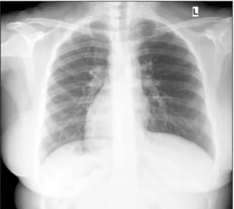
Fig. 1. Chest X-ray (PA view) showing dextrocardia.
Fig. 3. Section of computed tomography scan showing re- versed positions of the viscera.
Fig. 2. Coronal section of computed tomography scan showing the liver on the left and the stomach and the heart on the right.
pochondrium with radiation to the left shoulder, was con- stant and dull aching in nature, aggravated with fatty foods, and was relieved with analgesics and antacids.
There was no history of fever, jaundice, vomiting, urinary complaints, altered bowel habits or weight loss. The men- strual history of the patient was regular. She had experi- enced a similar episode around three months prior, which was relieved by conservative medical therapy. She did not give any history of any medical or surgical comorbidities and had no significant drug history.
The general examination of the patient was essentially normal with no signs of jaundice, fever or anemia. When examining the abdomen, there was mild tenderness from deep palpation in the left hypochondrium, with normal bowel sounds. The rest of the abdominal examination was unremarkable. Examination of the cardiovascular system revealed an apex beat in the right, 5th intercostal space in the right, mid-clavicular line.
The patient’s routine blood investigation revealed a normal complete blood count, normal liver and kidney function tests, normal thyroid function test and normal glycemic status. The urine examination also did not reveal any abnormalities. The ultrasonography of the abdomen showed as follows: 1) A gallbladder, small in size, with a thickened wall and situated on the left side. The lumen was filled with multiple, small calculi with posterior acoustic shadowing; 2) The liver situated on the left side, normal in size, with a homogenous parenchymal echo pattern. The intrahepatic biliary channels were not dilated;
3) The common bile duct was normal in size and diameter without any intraluminal lesions; 4) The spleen was nor- mal in size and shape without any focal lesions and situ- ated on the right side; and 5) Features were consistent with situs inversus. The electrocardiograph showed a right axis deviation while the chest x-ray (posteroanterior view) showed dextrocardia, the left hemidiaphragm slightly raised compared to the right side and a fundic gas shad- ow, of the stomach, on the right side, all findings con- sistent with situs inversus totalis (Fig. 1). The findings were re-confirmed with a computed tomography of the abdomen and the diagnosis was established to be a case of cholelithiasis in a patient with situs inversus totalis (Figs. 2 and 3).
86 Ann Hepatobiliary Pancreat Surg Vol. 21, No. 2, May 2017
Fig. 5. Intraoperative photograph showing the common bile duct, cystic duct, and cystic artery.
Fig. 4. Operation field photograph showing ports placed in the mirror positions.
After obtaining necessary anesthetic fitness, the patient was scheduled to undergo an elective laparoscopic cholecystectomy. The procedure began by adjusting the theatre equipment, including the carbon dioxide insuf- flator, the diathermy set and the monitor, which were placed on the left side of the patient, almost mirroring their normal positions. The patient was then positioned in the reverse Trendelenberg position after adequate anesthesia. The primary surgeon and the first assistant stood on the right side of the patient, whereas the second assistant was on the left side. A total of four ports were made - the two 10 mm ports were placed in the in- fraumbilical and subxiphoid regions, respectively and the two 5 mm ports were placed in the left hypochondrium in the left mid-clavicular line and in the left anterior axil- lary line at the level of the umbilicus (Fig. 4).
The technical challenges anticipated included creating a pneumoperitoneum from the left side, dissection of the Calot’s Triangle with the right-handed surgeon using the non-dominant hand and chances of the surgeon’s arms crossing during retraction of the Hartmann’s pouch. The pneumoperitoneum was induced using a Veress needle through the infraumbilical incision, by the surgeon on the left side of the patient, as done conventionally. An in- spection of the abdominal cavity was done and the diag- nosis of situs inversus was confirmed. The camera was maneuvered from the infraumbilical port by the first assis- tant and the fundus of the gallbladder was retracted by the second assistant, with a toothed grasper, from the 5 mm port in the left, anterior axillary line. In order to pre-
vent the primary surgeon from crossing arms while re- tracting the Hartmann’s pouch from the right side of the patient, the assistant retracted it from the left side. This not only prevented the primary surgeon from crossing arms, but also enabled him to dissect the Calot’s Triangle with his right hand using Maryland dissecting forceps in- serted through the subxiphoid/epigastric port. The cystic duct and artery were identified and dissected free from the surrounding structures. They were clipped using tita- nium clips, the applicator being introduced through the 10 mm epigastric port. The gall bladder was dissected from the gall bladder fossa using a hook diathermy and was delivered outside through the epigastric port. Adequate hemostasis was ensured and the port sites were closed with non-absorbable sutures (Fig. 5).
The estimated operating time was around 70 minutes and the postoperative period was uneventful. She was dis- charged on the second postoperative day and the sutures were removed on the seventh postoperative day, in the outpatient department. She followed up one month after surgery and was found to have recovered well. She has not developed any postoperative complications to date.
DISCUSSION
Situs inversus totalis is an uncommon condition with prevalence rates varying from 0.04 to 0.30%.8 Situs in- versus, in itself, does not predispose to the formation of gall stones.9 However, a strong index of clinical suspicion, along with imaging modalities such as ultrasonography and computed tomography scans, are needed for the accu- rate diagnosis of this condition.
Azhar Alam and Abhijit Santra. Laparoscopic cholecystectomy in situs inversus totalis 87
The first successful laparoscopic cholecystectomy in a patient with situs inversus was performed in 1991 by Campos and Sipes.10 In the following years, there have been several case reports describing this rare and techni- cally challenging procedure. Moirangthem et al.11 reported a similar case in which the primary surgeon performed the retraction, as well as the dissection, standing on the right of the patient. We believe that in such rare and technically difficult cases, it would be easier if the retraction was done by an assistant, thereby enabling the primary surgeon to perform the dissection much more meticulously. In a sim- ilar report, Arya et al.12 performed the operation using a mirror image placement of the ports and the instruments.
The primary surgeon performed the dissection, while the first assistant retracted the Hartmann’s pouch throughout the surgery. In our opinion, since the first assistant per- formed the retraction and held the camera, this could be simplified by allowing the second assistant to retract the Hartmann’s pouch, as reported in our case.
There have also been reports in the literature of this procedure being performed with modifications such as the surgeon standing in between the abducted legs of the pa- tient (Lloyd-Davis position) as described by Yaghan et al.3 The contralateral disposition of the abdominal viscera, resulting in mirror image anatomy, poses a major techni- cal challenge for the right-handed surgeon, who would now need to perform major steps of the procedure with his non-dominant hand. There was also a concern about the surgeon’s arms crossing during retraction of the Hartmann’s pouch. We adapted to these challenges by al- lowing the assistant to retract the Hartmann’s pouch, thus enabling the primary surgeon to perform the dissection of the Calot’s Triangle, as well as the clip application, using his dominant hand via the epigastric port, with adequate precision, similar to that in a conventional laparoscopic cholecystectomy. Similarly, the port placements, as well as the position of the surgical team, were also an exact mirror image of the conventional procedure. We therefore believe, that through these subtle modifications, the right-handed surgeon can perform this technically chal-
lenging procedure almost as skillfully as a left-handed surgeon, in such cases.
It may thus be concluded that a laparoscopic chol- ecystectomy in a patient with situs inversus is feasible and may be recommended as the procedure of choice in such cases. The procedure can be performed safely, as well as with precision, by a right-handed surgeon with meticulous preoperative planning and intraoperative adaptations, re- sulting in operating times comparable to conventional cases.
REFERENCES
1. Yokoyama T, Copeland NG, Jenkins NA, Montgomery CA, Elder FF, Overbeek PA. Reversal of left-right asymmetry: a situs inversus mutation. Science 1993;260:679-682.
2. Gedda L, Sciacca A, Brenci G, Villatico S, Bonanni G, Gueli N, et al. Situs viscerum specularis in monozygotic twins. Acta Genet Med Gemellol (Roma) 1984;33:81-85.
3. Yaghan RJ, Gharaibeh KI, Hammori S. Feasibility of laparo- scopic cholecystectomy in situs inversus. J Laparoendosc Adv Surg Tech A 2001;11:233-237.
4. Takei HT, Maxwell JG, Clancy TV, Tinsley EA. Laparoscopic cholecystectomy in situs inversus totalis. J Laparoendosc Surg 1992;2:171-176.
5. Hall TC, Barandiaran J, Perry EP. Laparoscopic cholecystectomy in situs inversus totalis: is it safe? Ann R Coll Surg Engl 2010;92:W30-W32.
6. Oms LM, Badia JM. Laparoscopic cholecystectomy in situs in- versus totalis: The importance of being left-handed. Surg Endosc 2003;17:1859-1861.
7. McKay D, Blake G. Laparoscopic cholecystectomy in situs in- versus totalis: a case report. BMC Surg 2005;5:5.
8. Iskandar ME, Radzio A, Krikhely M, Leitman IM. Laparoscopic cholecystectomy for a left-sided gallbladder. World J Gastroenterol 2013;19:5925-5928.
9. Crosher RF, Harnarayan P, Bremner DN. Laparoscopic chol- ecystectomy in situs inversus totalis. J R Coll Surg Edinb 1996;41:183-184.
10. Campos L, Sipes E. Laparoscopic cholecystectomy in a 39-year-old female with situs inversus. J Laparoendosc Surg 1991;1:123-125; discussion 126.
11. Moirangthem GS, Singh CA, Charaborty G, Lokendra K, Prabhu T. Laparoscopic cholecystectomy in a patient of situs inversus at regional institute of medical sciences (RIMS). J Med Soc 2014;28:60-62.
12. Arya SV, Das A, Singh S, Kalwaniya DS, Sharma A, Thukral BB. Technical difficulties and its remedies in laparoscopic chol- ecystectomy in situs inversus totalis: a rare case report. Int J Surg Case Rep 2013;4:727-730.