207
Correspondence to: Sang Woo Kim, Division of Gastroenterology, Department of Internal Medicine, Seoul St. Mary’s Hospital, The Catholic University of Korea College of Medicine, 505, Banpo- dong, Seocho-gu, Seoul 137-040, Korea. Tel: 02-2258-2059, Fax:
02-2258-2089, E-mail: viper@catholic.ac.kr
Received October 28, 2009, Accepted December 1, 2009
*These authors commonly attributed in this work.
Risk Factors for Lymph Node Metastasis in Patients with Submucosal Invasive Colorectal Carcinoma
Division of Gastroenterology, Department of Internal Medicine,
1Department of Hospital Pathology and 2Division of Colorectal Surgery,
Department of Surgery, The Catholic University of Korea College of Medicine, Seoul, Korea
Hye Young Sung, M.D.*, Won Kyung Kang, M.D.
2*, Sang Woo Kim, M.D., Kwan Woo Nam, M.D., Chan Kwon Jung, M.D.
1, Jae Hyuck Chang, M.D., Yu Kyung Cho, M.D., Jae Myung Park, M.D., In Seok Lee, M.D., Jae Im Lee, M.D.
2,
Seong Tack Oh, M.D.
2, Myung-Gyu Choi, M.D., In-Sik Chung, M.D.
Purpose: This study was designed to determine the risk factors of lymph node (LN) metastasis in patients with
submucosal invasive colorectal cancer (SICC).Methods: Between January 1998 and January 2009, we reviewed patients who had undergone radical colon
resection with LN dissection for SICC.Results: There were 36 males and 40 females (mean age, 61.1 years; range, 35∼86 years). In the univariate analysis,
the risk of LN metastasis was related to the depth (absolute and relative), lymphovascular invasion, tumor budding, and tumor differentiation (P<0.05). The relative depth by Kudo classification and lymphovascular invasion were significant predictors of LN metastasis both in univariate and multivariate analysis. In SICC with an absolute depth <1,800μm, no LN metastasis was detected. Regardless of the size of the SICC, tumors that invaded within the sm2 layer and had no lymphovascular invasion had no LN metastasis.Conclusion: In the SICC, lymphovascular invasion and depth of submucosal invasion are strong predictors of
LN metastasis. If deep invasion exceeds sm2 and positive lymphovascular invasion exists in the resected specimen, additional colectomy with LN dissection appears to be necessary. (J Korean Surg Soc 2010;78:207-212)Key Words: Submucosal invasive colorectal cancer, Lymph node metastasis, Lymphovascular invasion, Endoscopic
mucosal dissection
INTRODUCTION
Early colorectal carcinoma is defined as carcinoma in which invasion is limited to the mucosa or submucosa, regardless of the presence or absence of lymph node (LN) metastases.(1) An increase in colorectal cancer screening and progress in endoscopic techniques has resulted in an
increased frequency of detection of early stage colorectal cancer. Furthermore, advances in endoscopic instruments and techniques have increased the number of endoscopic resections for early stage colorectal cancer and provided a better quality of life for patients. Complete endoscopic resection of intramucosal carcinoma is accepted as a cura- tive therapy because there is little risk of LN metastasis.(2-4) Local therapy, such as endoscopic resection and local excision, are considered adequate therapy for early stage colorectal cancer without LN metastasis, although there is a risk of LN metastasis when cancer cells have invaded the submucosa. The incidence of LN metastasis in submucosal invasive colorectal cancer (SICC) has been reported to be approximately 7∼15%.(4-6) Accordingly, identification of
Table 1. The clinicopathologic characteristics in submucosal invasive colorectal cancer
No. of patients (n=76) Age (years) (mean±SD) (range) 61.1±11.2 (35∼86)
Gender (male:female) 36:40
Longest size (cm) 2.2±1.5
Lesion locations (%)
Cecum 2 (2.6)
Ascending colon 3 (3.9)
Transverse colon 5 (6.6)
Descending colon 1 (1.3)
Sigmoid colon 34 (44.7)
Rectum 31 (40.8)
Shapes (%)
Ip 58 (76.3)
Isp 7 (9.2)
Is 7 (9.2)
IIa+IIc 4 (5.3)
Differentiation (%)
Well 41 (53.9)
Moderate 32 (41.2)
Poor 3 (3.9)
the risk factors for LN metastasis is crucial in selecting appropriate therapeutic modalities for SICC. This study was designed to clarify the risk factors for LN metastasis in SICC and to determine the optimum criteria for endoscopic resection of early colorectal cancer.
METHODS
The study subjects included 76 patients with colorectal cancer invading the submucosa who were treated by surgical resection and lymphadenectomy at the Seoul St.
Mary’s Hospital in Seoul, Korea. The clinical data from the colorectal cancer database and clinical charts were also reviewed retrospectively. Macroscopically, tumors were class- ified as protruded (Ip, Isp, and Is) or superficial (IIa, IIb, IIc, IIa+IIc, and IIc+IIa).(1) All pathology slides were re-examined by a single pathologist. The tumor size, histo- logic type and grade, lymphovascular invasion, budding, depth of submucosal invasion, and width of tumor invasion were investigated. Histologic type and grade were classified according to the World Health Organization criteria.(7) Lymphovascular invasion was defined as the presence of cancer cells within the endothelial-lined channels. An isolated cell or a small cluster of carcinoma cells in the invasive front was defined as a ‘‘budding’’ focus, and >10 budding foci when viewed at a 200-fold magnification was considered positive for tumor budding, based on the data from Ueno et al.(8,9) The depth of submucosal invasion was measured in three ways. First, the absolute depth of submucosal invasion was defined as the vertical distance from the lower edge of the muscularis mucosa to the deepest invasive front and was measured with an optical micrometer. When the muscularis mucosa could not be identified, the vertical distance from the apex of the tumor to the deepest invasive front was measured by microscopy with an ocular lens scale. Second, Kudo’s classification was used with the relative invasion depth of the submucosal layer as follows: sm1, infiltration into the upper third of the submucosal layer; sm2, middle third; or sm3 lower third.(2) Third, Haggitt’s classification was used.(10) If the cancer invaded the stalk, the depth of the stalk invasion
was also measured as the length from the neck (Haggitt level 2) to the deepest portion of invasion. The width of the submucosal invasion was also measured at the maximum distance per section by microscopy with an ocular lens scale.
1) Statistical analysis
All statistical analyses were performed by the SPSS, version 13. The relationships between LN metastasis and age, absolute depth of submucosal invasion, and tumor size (longest) were compared using an independent sample t-test. The relationships between LN metastasis and other histopathologic factors were estimated using a χ2-test.
Multivariate binary logistic regression analysis was used to evaluate the independent risk factors for LN metastasis.
The odds ratio in the multivariate analysis was defined as the ratio of the probability that an event would occur to the probability that the event would not occur. P<0.05 was considered significant.
2) Ethical considerations
This study was approved by the Institutional Review
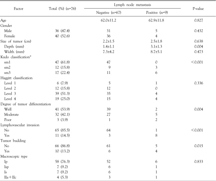
Table 2. Univariate analysis of risk factors for lymph node metastasis in patients with submucosal invasive colorectal cancer
Factor Total (%) (n=76) Lymph node metastasis
P-value Negative (n=67) Positive (n=9)
Age 62.0±11.2 62.9±11.8 0.827
Gender
Male 36 (47.4) 31 5 0.432
Female 40 (52.6) 36 4
Size of tumor (cm) 2.2±1.5 2.5±1.8 0.638
Depth (mm) 1.4±1.1 3.1±1.3 0.004
Width (mm) 7.3±4.2 8.7±5.1 0.473
Kudo classification*
sm1 47 (61.8) 47 0 <0.001
sm2 12 (15.8) 9 3
sm3 17 (22.4) 11 6
Haggitt classification
Level 1 6 (7.9) 5 1 0.336
Level 2 12 (15.8) 12 0
Level 3 39 (51.3) 35 4
Level 4 19 (25.0) 15 4
Degree of tumor differentiation
Well 41 (53.9) 39 2 0.004
Moderate 32 (42.1) 27 5
Poor 3 (3.9) 1 2
Lymphovascular invasion
No 65 (85.5) 64 1 <0.001
Yes 11 (14.5) 3 8
Tumor budding
No 66 (86.8) 61 5 0.015
Yes 10 (13.2) 6 4
Macroscopic type
Ip 58 (76.3) 52 6 0.833
Isp 7 (9.2) 6 1
Is 7 (9.2) 6 1
IIa+IIc 4 (5.3) 3 1
*sm1, sm2 and sm3 are tumor cell invasion into the upper, middle, and lower thirds of the submucosal layer, respectively.
Board of Seoul St. Mary’s Hospital. The study was carried out in accordance with the recommendations of the Decla- ration of Helsinki.
RESULTS
1) Clinical and pathologic characteristics
The clinicopathologic features of the 76 patients are summarized in Table 1. Of the 76 patients, 36 were males and 40 were females; the mean age was 61.1±11.2 years (range, 35∼86). Ten patients (13.2%) had tumors in the right side of colon, 35 (46.1%) had tumors in the left side
of colon, and 31 (40.8%) had tumors in the rectum. Nine hundred thirteen LNs (mean, 12) from 76 patients with colorectal cancer invading the submucosa were examined.
Histologically, LN metastasis was detected in 32 of the 913 LNs. The overall LN metastasis rate was 11.8% (9 of 76 patients). The mean tumor size (longest length) was 2.2±1.5 cm (range, 0.4∼8 cm). The histologic type was classed as well differentiated adenocarcinoma in 41 cases (53.9%), moderately differentiated adenocarcinoma in 32 cases (41.2%) and poorly differentiated adenocarcinoma in three cases (3.9%). Lymphovascular invasion was identified in 11 cases (14.5%). Tumor budding were noted in 10 cases
Table 3. Multivariate analysis of risk factors for lymph node meta- stasis in patients with submucosal invasive colorectal cancer
Factor Odds ratio
(95% confidence interval) P-value Lymphovascular invasion 120.0 (11.1∼1302.9) <0.001 Submucosal invasion 4.1 (1.3∼12.9) 0.017 Main differentiation 3.6 (0.7∼19.9) 0.140
Budding 4.9 (0.7∼34.6) 0.113
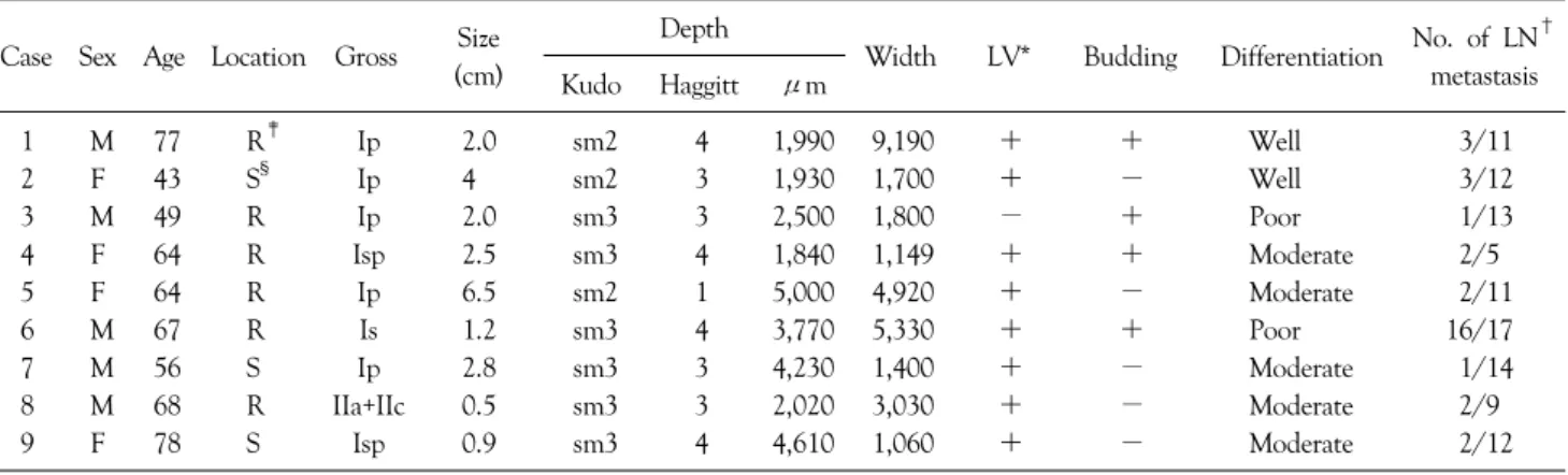
Table 4. Clinicopathologic details of submucosal invasive colorectal carcinoma in nine patients with lymph node metastasis Case Sex Age Location Gross Size
(cm)
Depth
Width LV* Budding Differentiation No. of LN† metastasis Kudo Haggitt μm
1 M 77 R‡ Ip 2.0 sm2 4 1,990 9,190 + + Well 3/11
2 F 43 S§ Ip 4 sm2 3 1,930 1,700 + − Well 3/12
3 M 49 R Ip 2.0 sm3 3 2,500 1,800 − + Poor 1/13
4 F 64 R Isp 2.5 sm3 4 1,840 1,149 + + Moderate 2/5
5 F 64 R Ip 6.5 sm2 1 5,000 4,920 + − Moderate 2/11
6 M 67 R Is 1.2 sm3 4 3,770 5,330 + + Poor 16/17
7 M 56 S Ip 2.8 sm3 3 4,230 1,400 + − Moderate 1/14
8 M 68 R IIa+IIc 0.5 sm3 3 2,020 3,030 + − Moderate 2/9
9 F 78 S Isp 0.9 sm3 4 4,610 1,060 + − Moderate 2/12
*LV = lymphovascular invasion; †LN = lymph node; ‡R = rectum; §S = sigmoid colon.
Table 5. Lymph node metastasis according to depth of invasion, tumor size, and lymphovascular invasion
Depth of invasion
LV* invasion (n=76)
LN† metastasis Absent (n=67) Present (n=9)
sm1 Absent 45 45 0
Present 2 2 0
sm2 Absent 8 8 0
Present 4 1 3
sm3 Absent 12 11 1
Present 5 0 5
*LV = lymphovascular; †LN = lymph node.
(13.2%). The mean absolute depth of submucosal invasion depth was 1.7±1.28 mm (range, 0.28∼5.0 mm). According to Kudo’s classification, the number of cases classified as sm1, sm2, and sm3 was 47 (61.8%), 12 (15.8%), and 17 (22.4%), respectively. According to Haggitt’s classification, submucosal invasion was classed as level 1 in 6 cases (7.9%), level 2 in 12 cases (15.8%), level 3 in 39 cases (51.3%) and level 4 in 19 cases (25.0%).
2) Correlation between histopathologic parameters and LN metastasis
Based on univariate analysis, the presence of LN meta- stasis was significantly associated with moderate or poor differentiation (P=0.004), lymphovascular invasion (P<
0.001), tumor budding (P=0.015), and relative depth by Kudo classification (P<0.001)(Table 2). The presence of LN metastasis was significantly associated with absolute depth of invasion (P=0.004) (Table 2). Multivariate logistic regression analyses showed that lymphovascular invasion, absolute depth of invasion, and relative depth by the Kudo
classification were a significant risk factor for LN metastasis in cases of SICC (Table 3). SICC with an absolute depth
<1,800μm had no LN metastasis (Table 4). The minimal extent of submucosal invasion in tumors with LN metastasis was 1,840μm. Cases with positive lymphovascular invasion and a depth >1,800μm accounted for 88.9% (8/9) of all LN metastasis (Table 4).
3) LN metastasis according to depth of invasion and lymphovascular invasion
To indentify the subgroup of patients who could be amenable to endoscopic mucosal resection, we classified the patients according to the relative depth of submucosal invasion and lymphovascular invasion (Table 5). Tumor size was redefined as the largest diameter and categorized as smaller or larger than 2 cm. The LN metastasis rate had no significant difference by size grouping (<2 cm vs. >2
cm [3.9% vs. 7.9%], P=0.424). Regardless of tumor size, SICC in which invasion was within the sm2 layer, and in which there was no lymphovascular invasion had no LN metastasis (Table 5).
DISCUSSION
In the treatment of early colorectal cancer, tumor cell defined in mucosal layer, it is sufficient for local treatment without adjuvant therapy because such a cancer rarely metastasizes to the LN or distant organs. However, once tumor cells have invaded through the muscularis mucosa, they are capable of metastasizing to the regional LN or even distantly to the liver or other organs. The incidence of LN metastasis in SICC has been reported to be approximately 7∼15%.(4-6) With the widespread application of endo- scopic treatment, adverse outcomes, such as tumor recu- rrence, have been observed.(11,12) Although local excision without adjuvant therapy for SICC can be safely performed in strictly selected patients, a considerably high recurrence rate, ranging from 12∼29%, has been reported.(13-17) Controversy exists with respect to the best therapeutic modality for SICC and no optimal criteria have been established for endoscopic resection of early colorectal cancer. Accordingly, identification of the risk factors for LN metastasis is crucial in selecting appropriate therapeutic modalities for SICC. In previous studies, the depth of submucosal invasion (sm3), poor histologic grade, lympho- vascular invasion, and tumor cell dissociation have been reported as adverse factors of SICC. To extend the criteria for curative endoscopic resection, the combination of a quantitative risk factor and qualitative risk factors, maybe achieve complete cure.
Based on our results, univariate analysis showed that the risk of LN metastasis was related to the depth (absolute depth and relative depth by Kudo), lymphovascular invasion, tumor budding, and tumor differentiation. Multivariate analysis showed that the depth (absolute and relative depth) of submucosal invasion and lymphovascular invasion were significant risk factors for LN metastasis. The minimal extent of submucosal invasion in tumors with LN meta-
stasis was 1,840μm (Table 4). Similarly, with our results, many previous studies have shown that the degree of submucosal invasion is an important predictor of LN metastasis.(2,5,18-20) Kobayashi et al.(19) studied SICC and reported that tumors with submucosal invasion of 1,000μm had no LN metastasis; Kitajima et al.(20) studied SICC and reached the same conclusion. These studies and our results indicate that when submucosal invasion of an endoscopically resected tumor is <1,000∼1,500 mm, com- plete cure can be achieved by endoscopic resection alone.
Measuring the vertical distance of submucosal invasion from the muscularis mucosa seems to be accurate and appropriate for endoscopic resection cases in deciding additional treatment, such as radical surgery and LN dissection. Our study clearly demonstrates that relative depth by Kudo classification (upper, sm1; middle, sm2; and lower, sm3)(2) is closely associated with the frequency of LN metastasis. Regardless of the size of the tumor in patients with SICC, invasion within the sm2 layer and no lymphovascular invasion had no LN metastasis. In the cases in which the tumor invaded beyond sm3, LN metastasis occurred without lymphovascular invasion. However, limita- tion of this classification system is relative, and it is difficult to evaluate an endoscopically-resected specimen for which the full thickness of the submucosal layer has not been removed. Accordingly, for accurate risk stratification, we should assess absolute and relative depth together. Lastly, our multivariate analysis demonstrated that the presence of lymphovascular invasion was the most significant predictor of nodal metastasis. Lymphovascular invasion occurred in 11 of 76 patients (14.5%) with SICC. Eight of nine patients (88.9%) with LN metastasis had lymphovascular invasion, whereas only 3 of 67 patients (4.5%) without LN metastasis had lymphovascular invasion. The odds ratio of LN metastasis increased 120-fold for a patient who had lymphovascular invasion compared with a patient who did not have lymphovascular invasion. Many previous publications support our finding that lymphovascular inva- sion is an important determinant in the management of patients with SICC.(6,21-24)
In summary, lymphovascular invasion and depth of
submucosal invasion are strong predictors of LN metastasis.
These factors can be used in combination to identify patients requiring additional surgery after endoscopic resec- tion. If deep invasion exceeds sm2 and positive lympho- vascular invasion exists in the resected specimen, additional colectomy with LN dissection appears to be necessary. A large scaled prospective data collection is warranted to verify our criteria for the management guidelines in patients with SICC.
REFERENCES
1) Kudo S. Endoscopic mucosal resection of flat and depressed types of early colorectal cancer. Endoscopy 1993;25:455-61.
2) Kudo S, Kashida H, Tamura T, Kogure E, Imai Y, Yamano H, et al. Colonoscopic diagnosis and management of nonpolypoid early colorectal cancer. World J Surg 2000;24:1081-90.
3) Hurlstone DP, Sanders DS, Cross SS, Adam I, Shorthouse AJ, Brown S, et al. Colonoscopic resection of lateral spreading tumours: a prospective analysis of endoscopic mucosal resection.
Gut 2004;53:1334-9.
4) Nivatvongs S. Surgical management of early colorectal cancer.
World J Surg 2000;24:1052-5.
5) Kikuchi R, Takano M, Takagi K, Fujimoto N, Nozaki R, Fujiyoshi T, et al. Management of early invasive colorectal cancer. Risk of recurrence and clinical guidelines. Dis Colon Rectum 1995;38:1286-95.
6) Nascimbeni R, Burgart LJ, Nivatvongs S, Larson DR. Risk of lymph node metastasis in T1 carcinoma of the colon and rectum. Dis Colon Rectum 2002;45:200-6.
7) Tanaka S, Yokota T, Saito D, Okamoto S, Oguro Y, Yoshida S. Clinicopathologic features of early rectal carcinoma and indications for endoscopic treatment. Dis Colon Rectum 1995;
38:959-63.
8) Ueno H, Price AB, Wilkinson KH, Jass JR, Mochizuki H, Talbot IC. A new prognostic staging system for rectal cancer.
Ann Surg 2004;240:832-9.
9) Hase K, Shatney C, Johnson D, Trollope M, Vierra M.
Prognostic value of tumor "budding" in patients with colorectal cancer. Dis Colon Rectum 1993;36:627-35.
10) Haggitt RC, Glotzbach RE, Soffer EE, Wruble LD. Prognostic factors in colorectal carcinomas arising in adenomas: impli- cations for lesions removed by endoscopic polypectomy. Gastro- enterology 1985;89:328-36.
11) Volk EE, Goldblum JR, Petras RE, Carey WD, Fazio VW.
Management and outcome of patients with invasive carcinoma arising in colorectal polyps. Gastroenterology 1995;109:1801-7.
12) Seitz U, Bohnacker S, Seewald S, Thonke F, Brand B, Braiu-
tigam T, et al. Is endoscopic polypectomy an adequate therapy for malignant colorectal adenomas? Presentation of 114 patients and review of the literature. Dis Colon Rectum 2004;47:1789- 96.
13) Garcia-Aguilar J, Mellgren A, Sirivongs P, Buie D, Madoff RD, Rothenberger DA. Local excision of rectal cancer without adjuvant therapy: a word of caution. Ann Surg 2000;231:345- 51.
14) Lamont JP, McCarty TM, Digan RD, Jacobson R, Tulanon P, Lichliter WE. Should locally excised T1 rectal cancer receive adjuvant chemoradiation? Am J Surg 2000;180:402-6.
15) Bentrem DJ, Okabe S, Wong WD, Guillem JG, Weiser MR, Temple LK, et al. T1 adenocarcinoma of the rectum: transanal excision or radical surgery? Ann Surg 2005;242:472-7.
16) Endreseth BH, Myrvold HE, Romundstad P, Hestvik UE, Bjerkeset T, Wibe A. Transanal excision vs. major surgery for T1 rectal cancer. Dis Colon Rectum 2005;48:1380-8.
17) Madbouly KM, Remzi FH, Erkek BA, Senagore AJ, Baeslach CM, Khandwala F, et al. Recurrence after transanal excision of T1 rectal cancer: should we be concerned? Dis Colon Rectum 2005;48:711-9.
18) Ueno H, Mochizuki H, Hashiguchi Y, Shimazaki H, Aida S, Hase K, et al. Risk factors for an adverse outcome in early invasive colorectal carcinoma. Gastroenterology 2004;127:385- 94.
19) Kobayashi M, Watanabe H, Maeo S. Correlation of histological atypia and cancer sprouting with vascular permeation and lymph nodal metastasis by our new histological classification of sub- mucosal invasion by colorectal carcinomas. Stomach Intest 1994;29:1151-60.
20) Kitajima K, Fujimori T, Fujii S, Takeda J, Ohkura Y, Kawamata H, et al. Correlations between lymph node metastasis and depth of submucosal invasion in submucosal invasive colorectal carcinoma: a Japanese collaborative study. J Gastroenterol 2004;
39:534-43.
21) Tominaga K, Nakanishi Y, Nimura S, Yoshimura K, Sakai Y, Shimoda T. Predictive histopathologic factors for lymph node metastasis in patients with nonpedunculated submucosal invasive colorectal carcinoma. Dis Colon Rectum 2005;48:92- 100.
22) Sakuragi M, Togashi K, Konishi F, Koinuma K, Kawamura Y, Okada M, et al. Predictive factors for lymph node metastasis in T1 stage colorectal carcinomas. Dis Colon Rectum 2003;46:
1626-32.
23) Sitzler PJ, Seow-Choen F, Ho YH, Leong AP. Lymph node involvement and tumor depth in rectal cancers: an analysis of 805 patients. Dis Colon Rectum 1997;40:1472-6.
24) Blumberg D, Paty PB, Guillem JG, Picon AI, Minsky BD, Wong WD, et al. All patients with small intramural rectal cancers are at risk for lymph node metastasis. Dis Colon Rectum 1999;42:
881-5.