Received July 15, 2015, Revised September 25, 2015, Accepted for publication October 5, 2015
Corresponding author: Weon Ju Lee, Department of Dermatology, Kyungpook National University Hospital, 130 Dongdeok-ro, Jung-gu, Daegu 41944, Korea.
Tel: 82-53-420-5838, Fax: 82-53-426-0770, E-mail: [email protected]
This is an Open Access article distributed under the terms of the Creative Commons Attribution Non-Commercial License (http://creativecommons.org/
licenses/by-nc/4.0) which permits unrestricted non-commercial use, distribution, and reproduction in any medium, provided the original work is properly cited.
Copyright © The Korean Dermatological Association and The Korean Society for Investigative Dermatology
ACKNOWLEDGMENT
This research was supported by the Basic Science Research Program through the National Research Foundation of Korea (NRF) funded by the Ministry of Science, ICT and future Planning (NRF-2015R1A2A2A11000897).
REFERENCES
1. Driskell RR, Clavel C, Rendl M, Watt FM. Hair follicle dermal papilla cells at a glance. J Cell Sci 2011;124:
1179-1182.
2. Ohyama M, Zheng Y, Paus R, Stenn KS. The mesenchymal component of hair follicle neogenesis: background, methods and molecular characterization. Exp Dermatol 2010;19:89-99.
3. Ohyama M, Veraitch O. Strategies to enhance epithelial- mesenchymal interactions for human hair follicle bioen- gineering. J Dermatol Sci 2013;70:78-87.
4. Kang BM, Kwack MH, Kim MK, Kim JC, Sung YK. Sphere formation increases the ability of cultured human dermal papilla cells to induce hair follicles from mouse epidermal cells in a reconstitution assay. J Invest Dermatol 2012;132:
237-239.
5. Strem BM, Hicok KC, Zhu M, Wulur I, Alfonso Z, Schreiber RE, et al. Multipotential differentiation of adipose tissue- derived stem cells. Keio J Med 2005;54:132-141.
6. Driskell RR, Jahoda CA, Chuong CM, Watt FM, Horsley V.
Defining dermal adipose tissue. Exp Dermatol 2014;23:
629-631.
7. Driskell RR, Lichtenberger BM, Hoste E, Kretzschmar K, Simons BD, Charalambous M, et al. Distinct fibroblast lineages determine dermal architecture in skin development and repair. Nature 2013;504:277-281.
8. Kwack MH, Sung YK, Chung EJ, Im SU, Ahn JS, Kim MK, et al. Dihydrotestosterone-inducible dickkopf 1 from balding dermal papilla cells causes apoptosis in follicular keratinocytes.
J Invest Dermatol 2008;128:262-269.
9. Zuk PA, Zhu M, Ashjian P, De Ugarte DA, Huang JI, Mizuno H, et al. Human adipose tissue is a source of multipotent stem cells. Mol Biol Cell 2002;13:4279-4295.
10. Park KC, Choi HR, Na JI, Cho HJ, Nam KM, Choi JW, et al.
Effects of murine dermal cells on the regulation of hair growth is dependent on the cell number and post-natal age of newborn mice. Ann Dermatol 2012;24:94-98.
http://dx.doi.org/10.5021/ad.2016.28.5.665
Effect of Vitamin D on the Expression of Inflammatory Biomarkers in Cultured Sebocytes Treated
with Propionibacterium acnes or Ultraviolet B Irradiation
Weon Ju Lee, Min Ji Kim, Hyo Sub Ryu
1, Mi Yeung Sohn, Yong Hyun Jang, Seok-Jong Lee, Do Won Kim
Department of Dermatology, Kyungpook National University School of Medicine, Daegu, 1Maxwell Hair Clinic, Seoul, Korea
Dear Editor:
Acne is a very common dermatologic disorder in humans.
It is a multifactorial disorder associated with follicular hy-
perkeratosis, sebaceous lipids, Propionibacterium acnes, and perifollicular inflammation. Excessive production and abnormal composition of sebaceous lipids contribute to
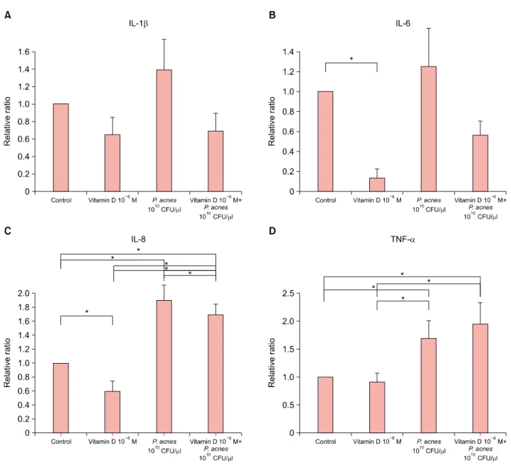
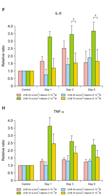
Fig. 1. Effect of vitamin D on the expression of interleukin (IL)-1β, IL-6, IL-8 and tumor necrosis factor (TNF)-α in cultured sebocytes after treatment with Propionibacterium acnes or ultraviolet B (UVB) irradiation. (A∼C) Protein expression of IL-1β, IL-6, and IL-8 (*p<0.05) in cultured sebocytes treated with 1010 CFU/μl P. acnes decreased with the treatment of 10−6 M vitamin D. (D) Protein expression of TNF-α in cultured sebocytes treated with 1010 CFU/μl P. acnes did not decrease with the treatment of 10−6 M vitamin D. (E∼H) Protein expression of IL-1β, IL-6 (*p<0.05), IL-8, and TNF-α in cultured sebocytes treated with 40 mJ/cm2 or 70 mJ/cm2 UVB showed more decreasing tendency after the addition of 10−6 M vitamin D compared with 10−8 M vitamin D. (E, F, H) Upregulation of IL-1β, IL-6, and TNF-α in cultured sebocytes by 40 mJ/cm2 UVB was inhibited 1 day after treatment with 10−6 M vitamin D compared with control.
the formation of inflammatory acne lesions1. Additionally, the upregulation of inflammatory biomarkers in sebocytes by P. acnes and ultraviolet B (UVB) irradiation can lead to inflammatory acne2. Sebocytes have been identified as bi- oactive vitamin D-responsive cells, suggesting that vitamin D analogues may be an effective therapeutic agent for acne3. This study was conducted to determine the effects of vitamin D on an increase in the expression of in- flammatory cytokines after treatment with human sebo-
cytes with P. acnes or UVB irradiation.
Primary sebocyte culture from occipital hair follicle was performed using Dulbecco’s modified Eagle’s medium (DMEM; Hyclone Laboratories, Logan, UT, USA) and Epilife (MEPI500CA; Gibco BRL, Grand Island, NY, USA).
The second passage sebocytes were obtained for the study after identification with hematoxylin and eosin (Muto Pure Chemicals Co., Tokyo, Japan) and Oil Red O (Sigma; St.
Louis, MO, USA) staining, and immunocytofluorescence with
Fig. 1. Continued.
cytokeratin 1 and 7 (Chemicon, Billerica, MA, USA). The se- bocytes were treated with 10−8 to 10−6 M 1, 25-dihydroxy- vitamin D3 (vitamin D) for 24 h as a control group. The concentrations of vitamin D were decided based on Cell Counting Kit-8 (CCK-8; Dojindo Laboratories, Kumamoto, Japan) assays. In addition, the sebocytes were treated for 5 days with 1010 CFU/μl P. acnes (ATCC1182) or a combi- nation of vitamin D (10−6 M) and P. acnes (1010 CFU/μl).
The sebocytes were also treated with vitamin D (10−8 or 10−6 M) and 40 mJ/cm2 or 70 mJ/cm2 UVB irradiation with Dermapal (Daavin, Bryan, OH, USA). The sebocytes were prepared for the evaluation of protein 5 days after treatment with vitamin D±P. acnes and 1, 3, and 5 days after treatment with vitamin D+UVB. Analysis of inter- leukin (IL)-1β, IL-6, IL-8, and tumor necrosis factor (TNF)-α protein expression was performed with ELISAs
(R&D Systems, Shanghai, China), following the manu- facturer’s advices. Briefly, samples were added to each well in triplicate. After then, 200 μl of prepared cytokine conjugate and 200 μl of premixed TMB substrate solution were mixed to each well in that order. The plates were de- veloped in the dark at room temperature for a half hour, and the reaction was stopped by mixture of 50 μl stop solution to each well. Lastly, absorbance was measured with a VersaMax Microplate Reader (Molecular Devices, Sunnyvale, CA, USA). Cultured sebocytes were also seed- ed on 60 mm dishes in quadruplicate for sebum lipid analysis. After 5 days, PBS was mixed and cells were col- lected with centrifugation (1,300g, 5 min). Lipid extraction solution (0.9% NaCl and 1% Triton X-100) was mixed to the precipitate and the mixture was homogenized with vortexing. The homogenized specimen was centrifuged
Fig. 2. Sebum production by cultured sebocytes after treatment with Propionibacterium acnes (1010 CFU/μl) was decreased by treatment with 10−6 M vitamin D. However, sebum production was increased by P. acnes (1010 CFU/μl) or 10−6 M vitamin D. There was no statistically significant difference in sebum production among the treated groups.
(13,000g, 15 min). Lipid levels were measured twice with an enzymatic method (ASAN Co., Seoul, Korea) and cor- rected for protein levels measured with the Bradford method. Data were evaluated by ANOVA. A p-value of
<0.05 was considered as statistical significance.
Upregulation of IL-1β, IL-6 and IL-8 (p<0.05) in the se- bocytes by P. acnes (1010 CFU/μl) was inhibited by vita- min D at 10−6 M (Fig. 1A∼C). Upregulation of TNF-α in the sebocytes by P. acnes (1010 CFU/μl) was not inhibited by vitamin D at 10−6 M (Fig. 1D). Upregulation of IL-1β, IL-6, IL-8 and TNF-α in the sebocytes by 40 mJ/cm2 UVB showed more decreasing tendency after treatment with 10−6 M vitamin D compared with 10−8 M vitamin D (Fig.
1E∼H). Upregulation of IL-1β, IL-6 and TNF-α in the se- bocytes by 40 mJ/cm2 UVB was inhibited 1 day after treat- ment with 10−6 M vitamin D compared with control (Fig.
1E, F, H). Upregulation of IL-1β, IL-6 (p<0.05), IL-8 and TNF-α in the sebocytes by 70 mJ/cm2 UVB showed much more decreasing tendency after treatment with 10−6 M vi- tamin D compared with 10−8 M vitamin D (Fig. 1E∼H).
Sebum production of cultured sebocytes after treatment with 10−6 M vitamin D or P. acnes (1010 CFU/μl) was increased. However, sebum production of cultured sebo- cytes after treatment with P. acnes (1010 CFU/μl) was de- creased by the addition of 10−6 M vitamin D (Fig. 2).
P. acnes plays a key role in the initiation of inflammatory acne3. Previous studies have demonstrated proliferation of the sebaceous glands and cultured sebocytes after UV irra- diation4,5. Sebocytes have shown the upregulation of in-
flammatory cytokines following treatment with UVB irra- diation6,7. Vitamin D has been reported to stimulate the proliferation of sebocytes and to inhibit their differ- entiation and lipid synthesis4. Furthermore, vitamin D de- creases the production of inflammatory biomarkers, espe- cially IL-6, IL-8, and MMP-9, from cultured sebocytes.
Krämer et al.8 also reported that vitamin D reduced the se- cretion of IL-6 and IL-8 in SZ95 sebocytes. On the basis of these, we investigated the effect of vitamin D on the in- flammatory reaction of sebocytes treated with P. acnes or UVB irradiation. In this study, as reported previously, vita- min D decreased the expression of the inflammatory cyto- kines, such as IL-1β, IL-6, IL-8, and TNF-α. In addition, vitamin D inhibited the upregulation of IL-1β, IL-6 and IL-8 in sebocytes after treatment with P. acnes. Furthermore, higher concentration (10−6 M) of vitamin D inhibited the upregulation of IL-1β, IL-6, IL-8, and TNF-α in sebocytes after treatment with 40 mJ/cm2 or 70 mJ/cm2 UVB irradi- ation compared with lower concentration (10−8 M) of vita- min D. Vitamin D decreased sebum production after treat- ment of sebocytes with P. acnes in our study. It was re- ported that treatment of slowly proliferating SZ95 sebo- cytes with vitamin D results in a statistically significant time- and dose-dependent reduction of sebum lipids8. Unlike our expectation and a previous report, the treat- ment of sebocytes with vitamin D only showed a mild in- crease in sebum production in this study. However, there was not statistically significant. Like this study, P. acnes extracts usually increase sebum production in hamster se- baceous glands both in vivo and in vitro9.
In conclusion, the treatment of sebocytes with vitamin D shows a tendency to inhibit the upregulation of in- flammatory biomarkers by P. acnes and UVB irradiation.
On the basis of these findings, the use of vitamin D for in- flammatory acne may be promising. This is supported by the report that in severe acne patients vitamin D defi- ciency significantly potentiates the inflammatory proc- ess10. In addition, the treatment of acne with vitamin D has been tried since long before. However, because of li- phophilic property and high molecular weight of vitamin D, it should be considered that vitamin D in topical agents do not easily penetrate into the deep dermis, especially se- baceous gland.
ACKNOWLEDGMENT
This research was supported by a grant from Amore-Pacific Corporation awarded in 2014.
Received December 2, 2014, Revised September 18, 2015, Accepted for publication October 6, 2015
Corresponding author: Bark-Lynn Lew, Department of Dermatology, Kyung Hee University Hospital at Gangdong, 892 Dongnam-ro, Gangdong-gu, Seoul 05278, Korea. Tel: 82-2-440-7329, Fax: 82-2-440-7336, E-mail:
This is an Open Access article distributed under the terms of the Creative Commons Attribution Non-Commercial License (http://creativecommons.
org/licenses/by-nc/4.0) which permits unrestricted non-commercial use, distribution, and reproduction in any medium, provided the original work is properly cited.
Copyright © The Korean Dermatological Association and The Korean
Society for Investigative Dermatology Fig. 1. The 1.2×1.0 cm sized flesh to reddish colored pedun- culated nodule on the right axilla.
REFERENCES
1. Zouboulis CC. Acne and sebaceous gland function. Clin Dermatol 2004;22:360-366.
2. Lee WJ, Chae SY, Ryu HS, Jang YH, Lee SJ, Kim DW.
Inflammatory cytokine expression and sebum production after exposure of cultured human sebocytes to ultraviolet a radiation and light at wavelengths of 650 nm and 830 nm.
Ann Dermatol 2015;27:163-170.
3. Shaheen B, Gonzalez M. Acne sans P. acnes. J Eur Acad Dermatol Venereol 2013;27:1-10.
4. Lesnik RH, Kligman LH, Kligman AM. Agents that cause enlargement of sebaceous glands in hairless mice. II.
Ultraviolet radiation. Arch Dermatol Res 1992;284:106-108.
5. Akitomo Y, Akamatsu H, Okano Y, Masaki H, Horio T.
Effects of UV irradiation on the sebaceous gland and sebum secretion in hamsters. J Dermatol Sci 2003;31:151-159.
6. Skiba B, Neill B, Piva TJ. Gene expression profiles of TNF-alpha, TACE, furin, IL-1beta and matrilysin in UVA- and
UVB-irradiated HaCat cells. Photodermatol Photoimmunol Photomed 2005;21:173-182.
7. Lee WJ, Park KH, Sohn MY, Lee WC, Lee SJ, Kim DW.
Ultraviolet B irradiation increases the expression of inflammatory cytokines in cultured sebocytes. J Dermatol 2013;40:993-997.
8. Krämer C, Seltmann H, Seifert M, Tilgen W, Zouboulis CC, Reichrath J. Characterization of the vitamin D endocrine system in human sebocytes in vitro. J Steroid Biochem Mol Biol 2009;113:9-16.
9. Iinuma K, Sato T, Akimoto N, Noguchi N, Sasatsu M, Nishijima S, et al. Involvement of Propionibacterium acnes in the augmentation of lipogenesis in hamster sebaceous glands in vivo and in vitro. J Invest Dermatol 2009;
129:2113-2119.
10. Siniavskiĭ IuA, Tsoĭ NO. Influence of nutritional patterns on the severity of acne in young adults. Vopr Pitan 2014;
83:41-47.
http://dx.doi.org/10.5021/ad.2016.28.5.669
Primary Cutaneous Apocrine Carcinoma
Seung-Hee Loh, Yu-Jin Oh, Bark-Lynn Lew, Woo-Young Sim
Department of Dermatology, Kyung Hee University College of Medicine, Seoul, Korea
Dear Editor:
Primary cutaneous apocrine carcinoma (PCAC), a subtype of sweat gland carcinoma, is an extremely rare malignant neoplasm1. Most of these neoplasms arise in regions of high apocrine gland density, particularly in the axilla, but