Received September 2, 2015, Revised December 9, 2015, Accepted for publication December 21, 2015
*These authors contributed equally to this work.
Corresponding author: Eun Ki Kim, Department of Biological Engineering, Inha University, 100 Inha-ro, Nam-gu, Incheon 22212, Korea. Tel:
82-32-860-7514, Fax: 82-32-872-4046, E-mail: [email protected] This is an Open Access article distributed under the terms of the Creative Commons Attribution Non-Commercial License (http://creativecommons.
org/licenses/by-nc/4.0) which permits unrestricted non-commercial use, distribution, and reproduction in any medium, provided the original work is properly cited.
Copyright © The Korean Dermatological Association and The Korean Society for Investigative Dermatology
Ann Dermatol Vol. 28, No. 5, 2016 http://dx.doi.org/10.5021/ad.2016.28.5.555
ORIGINAL ARTICLE
Kojic Acid Peptide: A New Compound with Anti-Tyrosinase Potential
Birendra Kumar Singh*, Seok Hoon Park1,*, Hyang-Bok Lee, Young-Aae Goo, Hyoung Shik Kim2, Seung Hee Cho2, Jeong Hun Lee2, Ghe Whan Ahn3, Jin Pyo Kim3, Su Myoung Kang3, Eun-Ki Kim
Department of Biological Engineering, Inha University, Incheon, 1Department of Environmental Engineering, Anyang University, Anyang,
2Anti-Ageing Research Institute of BIO-FD&C Co., Ltd., 3Natuzen Co., Ltd., Incheon, Korea
Background: Kojic acid was used for decades in the cosmetic industry as an antimelanogenic agent. However, there are two major drawbacks of Kojic acid, one is cytotoxicity and second are instability on storage. These limitations led the scientist to synthesize the active Kojic acid peptides.
Objective: In the present study, we synthesize and inves- tigate the effect of five Kojic acid peptides to overcome the limitation of Kojic acid. Methods: The peptide was analyzed and purified by high-performance liquid chromatography and matrix-assisted laser desorption ionization time of flight mass spectroscopy. Further, the tyrosinase activities of the Kojic acid and Kojic acid peptides were compared. The tox- icity was measured and the melanin content is recorded in B16F10 mouse melanoma cells.
Results: Maximum tyrosinase activity was measured by Kojic acid peptides. Therefore, Kojic acid peptides were subjected to melanin assay and cytotoxicity assay and finally the stabil- ity of the Kojic acid peptide was measured. Conclusion: It was observed that this newly synthesized Kojic acid peptide is stable and potent to inhibit the tyrosinase activity and mela- nin content of B16F10 mouse melanoma cells without ex- hibiting cell toxicity. Together, these preliminary results sug-
gest that a further exploration is being needed to establish Kojic acid peptide as antimelanogenic agent. (Ann Dermatol 28(5) 555∼561, 2016)
-Keywords-
Hypopgmentation, Kojic acid, Melanin, Peptides, Tyrosinase
INTRODUCTION
The consumption of skin care products and skin whitening agents generates billions of dollars. The world market for skin whitening is still developing and targeting all over the world1. To fulfill the demand of the cosmetic market, natu- rally occurring biologically active ingredient was ex- ploited for decades in the cosmetic industry. However, from the heap of naturally stirring biologically active con- stituents, hunt for active ingredients in the natural skin whitening agent is difficult.
The knowledge of analytical sciences, biochemistry, and other disciplines has influenced the identification of new cosmetic ingredients. Recent approaches of skin whiten- ing have provided the opportunity to choose more effec- tive cosmetic ingredients. Conventionally, plant extract, hydrolytes, essential oil, and vitamins were used as a skin whitening agent. However, because of limitations in search of clinically proven safe and effective cosmetic for- mulation, now synthetic molecules are being designed tested and prefer over the traditional ones. In recent times, peptide emerges as a more effective cosmetic agent that contains a large number of individual molecules2.
Melanin is the pigment which provides the color to skin ranges widely from black to fair. Its prime function is to protect the skin from ultraviolet exposure. The step of mel-
Fig. 1. Synthesis of activated Kojic acid by using 1,1-carbonyldiimida- zole (CDI) at room temperature.
anin synthesis in melanocytes is mediated by several en- zymes, of which tyrosinase is essential. Tyrosinase (EC 1.14.18.1) 2∼7 (monophenol or o-diphenol oxygen oxi- doreductase), also known as monophenol oxidase, mono- phenol monooxygenase, N-acetyl-6-hydroxytryptophan oxidase, phenolase, tyrosine-dopa oxidase and cresolase.
It is a copper-containing enzyme, that is widespread in na- ture and the most exploited enzymes solely due to the key role in mammalian melanogenesis. The entire process of melanin synthesis requires a combination of enzymatically catalyzed and chemical reactions for the formation of dark macromolecule i.e., melanin. There is a skin whitening agent available in the cosmetic industry as a skin whiten- ing agent which focus on modulation of the tyrosinase en- zyme in melanin synthesis pathway3. These skins whiten- ing agents like azelaic acid4,5, magnesium-L-ascorbyl-2-phos- phate, electron-rich phenols6, hydroxyanisole, corticoste- roids7, N-acetyl-4-S-cysteaminylphenol, resinoids8,9, arbutin (hydroquinone-beta-D-glucopyranoside), salicylhydroxamic acid, dioic acid, Kojic acid (KA), and hydroquinone are the most widely used in the cosmetic industry. They are recommended worldwide, but are associated with poten- tial mutagenic and adverse effects including skin irritation, contact dermatitis, low stability toward oxygen, high tox- icity and insufficient skin penetration ability and water, and exogenous ochronosis10. Because of side effect of hy- droquinone, it is under strict control of the Food and Drug Administration and prohibited in European Union10. Despite of all the efforts made so far, development of high-performance tyrosinase inhibitors is much needed and the opportunity to find the effective tyrosinase in- hibitor molecule is open.
KA [5-hydroxy-2-(hydroxymethy1)- y-pyrone], a fungal me- tabolite that has been used in many countries as skin whitening agent11. It worked through inhibition of ty- rosinase activity and thereby reducing melanin contents11. Mixed inhibitory effect on the diphenolase activity and competitive inhibitory effect on monophenolase activity were observed by KA. However, due to limitation of the cytotoxicity and instability on storage, its use in cosmetics has been limited12,13. Consequently, the researchers synthe- sized KA derivatives to enhance the properties by modify- ing the C-7 hydroxyl group14-16.
The objective of this study is to synthesize KA-peptide us-
ing solid-phase parallel peptide synthesis using Fmoc-Rink amide aminomethyl-polystyrene resin. KA was activated by carbonyldiimidazole (CDI) and coupled to the N-termi- nal of the tripeptides on the resin. After removing the res- in, KA-peptides were obtained with yield and purity. The tyrosinase activity was measured with compared to KA-PS and free KA. Further, cytotoxicity and melanin inhibition was performed to know the effects of the KA-PS on mela- nin synthesis and cell viability.
MATERIALS AND METHODS
In this study, analytical grade chemicals and reagent grade solvents were used without further purification. Dulbecco’s modified eagle’s medium (DMEM; Gibco BRL, Eggenstein, Germany), dimethyl sulfoxide (DMSO), 3-(4,5-dimethylthi- azol-2-yl)-2,5-diphenyltetrazolium bromide (MTT), 12-O-te- tradecanoylphorbol-13-acetate (TPA), were procured from Sigma Chemical Co. (St. Louis, MO, USA). Trypsin-ethy- lenediaminetetraacetic acid (EDTA) 0.5% solution (10×), phosphate buffered saline (PBS, pH 7.4) and, penicil- lin/streptomycin (10,000 U/ml) were purchased from Invitrogen Corp. (Carlsbad, CA, USA). Fmoc-l-A.A-OH (Bead- Tech, Seoul, Korea) and Fmoc-A.A-Wang resin (Merck, Frankfurt, Darmstadt, Germany) were used. Hydroxyben- zotriazole (HOBt) and diisoproylcarbodiimidazole (DIC) were obtained from GLS (Shanghai, China). KA was pur- chased from Wako (Tokyo, Japan) and 1,1-carbon- yldiimidazole (CDI) was purchased from Acros (Belgium, NJ, USA). Diisopropylethylamine, N,N’-dimethylformamide (DMF), dichloromethane (DCM), piperidine and diethyl ether were purchased from Daejung (Siheung, Korea).
1,2-ethanedithiol (EDT), thioanisole was procured from Alfa Aesar (Lancaster, UK).
Synthesis of activated Kojic acid
KA (5-hydroxy-2-(hydroxymethyl)-4#-pyran-4-one, 142.1 g, 1 mol) was dissolved in a solution of tetrahydrofurane (THF) and DMF in a proportion of 600 ml and 50 ml re- spectively and kept for stirring for 1 hour, at room tem- perature. CDI (146.0 g, 0.9 equivalent [eq.]) was dissolved in THF (200 ml) and added to the solution. The resulting yellow solid (3) were filtered, washed with THF or ether and dried in vacuum to give yellow solids (Fig. 1).
Fig. 2. Synthesis of Kojic acid peptides (KA-peptides). Reagents and conditions; a) Treat with 2% 1,8-diazabicyclo[5.4.0]undec-7-ene in N,N’-dimethylformamide (DMF) for 10 min; b) Mixed Fomc-A.A2-OH (5.0 equivalent [eq.]), hydroxybenzotriazole (HOBt) (5.0 eq.), and diisopropylcarbodiimide (5.0 eq.) with resin for 2 hours; c) repeat a) and b) procedure; d) React with KA-imidazole (3.0 eq.), HOBt (3.0 eq.) in DMF for overnight; e) React with reagent K. [trifluoroacetic acid/thioanisole/phenol/water/ethanedithiol (82.5/5/5/5/2.5(v/v)]
for 1 hour, and treat with pre-cooled diethylether.
Synthesis of Kojic acid conjugated peptides
KA conjugated peptides were synthesized by the sol- id-phase method using Fmoc chemistry. The C-terminal amino acid residue pre-loaded wang resins swelled in DCM for 1 hour. Peptide chains were elongated in the consecutive cycles of deprotection and coupling. Depro- tection was performed with 20% piperidine in the DMF for 20 minutes and 15 min respectively. HOBt/DIC chem- istry; was used for chain elongation from 5 equivalents of protected amino acid derivatives. Subsequently, Fmoc group of the final amino acid residue was removed and compound 3 (3 eq.) and HOBt (3 eq.) were added to the resin-bound peptides (Fig. 2). The mixture was in- cubated for 2 hours and the resin was treated with a cleavage solution (reagent K; trifluoroacetic acid (TFA)/thioanisole/phenol/water/EDT in the ration of 82.5/5/5/5/2.5 (v/v)) for 2 hours at room temperature. Later on, the resin was filtered. Crude peptides in the filtrate were concentrated under high vacuum, and precipitated with pre-cold ether to yield KA conjugated peptides. The crude peptide was analyzed and purified by high-perform- ance liquid chromatography (HPLC) on a Waters 600E sys- tem (Waters, Milford, MA, USA), using Phenomenex, C18, 5 μm column (300×22 mm). The solvent system which comprises of 0.1% TFA in H2O (solvent A) and 0.1% TFA in acetonitrile (solvent B) was used in a linear gradient from 4% to 14% B for 15 min with a flow rate 5 ml/min.
The elution was monitored at 230 nm. The purity of the synthesized peptide was checked on another Shim-pack col- umn, C18, 5 μm column (250×4.6 mm). The solvent sys- tem 0.1% TFA in H2O (solvent A), 0.1% TFA in acetoni- trile (solvent B) was used in a linear gradient from 5% to 65% B in 30 minutes while monitoring at 230 nm. The peptide was further characterized by matrix-assisted la- ser desorption ionization time of flight mass spectroscopy (MALDI-TOF-MS) (Voyager-DE STR; Applied Biosystems, Foster city, CA, USA), using α-cyano-4-hydroxycinnamic
acid as a matrix.
Cell lines and cell culture
Highly pigmented B16F10 mouse melanoma cell and Melan-a cell line, a highly pigmented, immortal, non-tu- morigenic mouse melanocyte cell line was used for the present study. The cells were generously provided by Prof.
Dorothy C. Bennett (St. George’s Hospital Medical School, London, UK). Culture procedures were carried out as described previously17. In brief, B16F10 murine mela- noma cells were cultured in DMEM supplemented with 10% (V/V) fetal bovine serum, 1% (V/V) penicillin /strepto- mycin (100 units/ml) at 37oC in a humidified atmosphere in a 5% CO2. The Melan-a murine melanocytes were cul- tured in Roswell Park Memorial Institute (RPMI) 1640 Medium supplemented with 10% (v/v) fetal bovine serum, 1% (v/v) penicillin/streptomycin (100 units/ml) and 200 nM TPA at 37oC in a humidified atmosphere with 10%
CO2. The subculture of the cells was carried out at 4th day of incubation by a passage number 20 was achieved.
In vitro tyrosinase assay
Mushroom tyrosinase (EC 1.14.18.1) used for the bioassay was purchased from Sigma Chemical Co.. In vitro ty- rosinase assay was assessed according to Kubo and Kinst-Hori18 with little modification. Briefly, In a 96 well plate system, mushroom tyrosinase was performed in trip- licates through a mixture preparation. The mixture was prepared by adding 40 μl of phosphate buffer, 100 μl of the sample (KA-PS) and the positive control (KA). Further 20 μl of tyrosinase enzyme (1,100 units/ml:0.516 mg/ml phosphate buffer 0.1 M), L-dopa (5 mM) was added in the 40 μl volume and the mixture was incubated for ten mi- nutes at 37oC. The optical density of dopachrome for- mation was measured using the spectrophotometric en- zyme-linked immunosorbent assay reader at an optical density of 475 nm (The inhibitory percentage of tyrosi- nase was calculated as follows: Inhibition=[(ΔAcontrol−
Table 1. Showing the purity and yield of the Kojic acid peptides
Peptides Purity (%) Yield (%)
KA-PS 95.2 27.5
KA-ECG 98.5 36.1
KA-KECG 99.3 42.0
KA-PKEK 99.5 32.9
KA-CDPGYIGSR (KA-CR9) 98.0 22.4
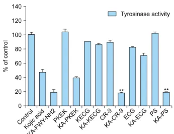
Fig. 3. Screening of newly synthesized Kojic acid peptides (KA-PKEK, KA-ECG, KA-CR9, KA-KECG, and KA-PS) through the invitro tyrosinase assay. Data are mean±standard deviation.
**p<0.001; t-test.
ΔAsample)/ΔAcontrol]×100). The IC50, defined as the concentration of KA-peptides required to inhibit tyrosinase activity by 50%, was determined for each sample. All ex- periments were performed at least three times, with sim- ilar consequences.
Cell viability assay
The viability of cells treated with respective KA-PS com- pounds was determined using an MTT assay. Briefly, 3×10⁴ B16F10 cells were seeded and adhered in 96-well plates. After 24 hours, DMEM was removed and 200 μl of various concentrations of fresh DMEM and different KA-PS solution were added, and the cells were incubated for 48 hours at 37oC in 5% CO2 incubator. After in- cubation, the medium was removed, and 100 μl of MTT in PBS solution (0.5 mg/ml) was added and incubated for 2 hours. Then blue crystalline precipitates were dissolved in 200 μl of 100% DMSO and viability was assessed by measuring absorbance at 540 nm using a molecular de- vice “Spectra Max 340 microplate reader”19. Untreated normal cells were used as a control group.
Measurement of melanin content
Highly pigmented B16F10 mouse melanoma cells were seeded into the 12-well plate, incubated for 24 hours, and then triplicate cultures were fed with fresh media and in- cubated for a further 48 hours. Then cells were detached by trypsin/EDTA solution and harvested. The harvested cells were washed twice with PBS for 5 minutes at 5,000 rpm, suspended in 100 μl of extraction buffer (1 N NaOH, 10% DMSO), heated at 80oC for 1 hour, and transferred to 96-well plate. Melanin content was de- termined by absorbance at 405 nm in ELISA microplate reader19,20. Normal untreated cells were used as a control.
Statistical analysis
All experiments were performed at least in triplicates and expressed as the means±standard deviation, and were an- alyzed using the unpaired t-test. A p-value of less than 0.05 was considered to be statistically significant com- pared with the control. SigmaPlot (Systat Software Inc., San Jose, CA, USA) was used to analyze the data.
RESULTS
Chemistry of KA-PS synthesis
By using the fomc chemistry and with the help of acti- vated KA, KA-peptides were synthesized and the yield and purity of KA-peptides were KA-peptide (95.2%, 27.5%), KA-ECG (98.5%, 36.1%), KA-KECG (99.3%, 42.0%), KA-PKEK (99.5%, 32.9%) and KA-CDPGYIGSR (KA-CR9) (98.0%, 22.4%) respectively shown in Table 1.
Determination of antityrosinase activity
Mushroom tyrosinase has been used for prescreening hy- popigmentation agents because it is commercially available.
The mushroom tyrosinase assay was performed in order to know the inhibitory activity of KA-peptides on tyrosinase and were observed that KA-CR-9 and KA-PS inhibit 81%
and 82% tyrosinase activity respectively at the concen- tration of 40 μM (Fig. 3). Fig. 4, 5 show the dose-depend- ent inhibition of mushroom tyrosinase activity at different concentration (5, 10, 20, 40, and 80 μM) of KA-CR-9 and KA-PS respectively.
Melanin content assay
Fig. 6A shows the inhibition of melanin by KA-PS in B16F10 mouse melanoma cell line and inhibition of mela- nin content was observed in α-melanocyte-stimulating hormone (αMSH) induced Melan-a mouse cell (Fig. 6B).
At the concentration of 5 mM, the melanin content was in- hibited by 42% in the B16F10 melanoma cell. Dose-de- pendent down-regulation was observed in Melan-a mouse cell.
Fig. 6. B16F10 melanoma cell and melan-a cells were cultured in the presence of Roswell Park Memorial Institute media supplemented with 10 fetal bovine serum. Arbutin and KA-PS acid inhibited melanin content of α-melanocyte-stimulating hormone (10 nM) induced B16F10 melanoma cells (A) and melan-a cells (B). Melanin content and cell viability (% of control) in both B16F10 cells (A) and melan-a melanocytes (B) are shown. Data are mean±standard deviation. *p<0.05, **p<0.001; t-test.
Fig. 4. Showing the dose dependent decrease in the mushroom tyrosinase activity by KA-CR-9. Data are mean±standard deviation.
*p<0.05, **p<0.001, ***p<0.0001; t-test.
Fig. 5. Showing the dose dependent inhibition of the mushroom tyrosinase activity by KA-PS. Data are mean±standard deviation.
*p<0.05, **p<0.001, ***p<0.0001; t-test.
Cell viability assay
The cytotoxicity of B16F10 and Melan-a cell was exam- ined using MTT assay and was observed that the KA-pep- tides were not toxic to the cell at the concentration of 5 mM (Fig. 6A) and at 3 mM in Melan-a cell (Fig. 6B).
DISCUSSION
KA was activated by CDI for coupling of KA to the N-ter-
minal of the peptides via urethane bond yielding KA-imi- dazole 3 (Fig. 1). The coupling, decoupling reaction was carried out at room temperature to restrict the decom- position of the desired product or from the oxidation of the product. The minimum quantity of DMF was added to the reaction as a cosolvent to improve the solubility of the KA in THF. With the subsequent reaction, the desired product precipitates and finally the yield was 77% (Fig. 1).
Using solid-phase Fmoc chemistry, the resin bound pep- tides were synthesized separately (Fig. 2). The reaction of coupling between activated KA and the resin was
time-consuming. Thus, to reduce the time and increase the purity of the final product the general catalyst HOBt was used (Fig. 2). HOBt has proven to be the most effec- tive catalyst for the coupling of the KA-imidazole to the resin-bound tripeptide2. HOBt donates the proton to the leaving group, imidazole and thus increasing the rate of reaction2.
Using solid phase parallel peptide synthesis method, we synthesized five compounds and all the compounds were purified by pre-cold ether precipitation under the high vacuum condition. The compounds synthesized were mentioned in Table 1. Purity and the correctness of the molecular weight value of peptide were analyzed by HPLC and MALDI-TOF-MS. Further, invitro tyrosinase as- say was performed to know the effect on tyrosinase. It was observed that the KA-PS and KA-CR9 show the maximum inhibitory effect of tyrosinase activity of 82%. 84%, respectively. The KA-PS, being the low molecular mole- cule, it was chosen for the further experiment. Therefore, with the help of HPLC crude KA-PS was purified and the purity was more than 95% (S1).
Subsequently, the KA-PS was subjected to the αMSH (10 nM) induced melanin assay and cell viability assay. The KA-PS inhibits the melanin content of B16F10 cells mel- an-a cell and no cytotoxicity was observed (Fig. 6).
Further, the stability of the KA-PS was analyzed by dissolv- ing KA-PS in DMSO at 37oC for 48 hours. Later on, mela- nin assay and cytotoxicity showed that KA-PS retains same activity (Data Not shown). Thus, this newly synthesized synthetic peptide (KA-PS) may be a promising ingredient in the cosmetic industry. It can be exploited as a new po- tent tyrosinase inhibitor, which reduces the melanin con- tent of B16F10 mouse melanoma cell (Fig. 6A). Dose-de- pendent down-regulation of melanin content was ob- served in Melan-a mouse cell (Fig. 6B). KA-PS can be used as a new depigmenting agent in the cosmetic industry.
However, before KA-PS is used in the cosmetic industry, it must progress through a series of in vivo trials to de- termine if it is safe and effective in intact skin system. It must be taken into consideration that sometimes the ab- sorbability of the compound differs between cell culture and natural skin21,22.
SUPPLEMENTARY MATERIALS
Supplementary data can be found via http://anndermatol.org/
src/sm/ad-28-555-s001.pdf.
ACKNOWLEDGMENT
This study was supported by a grant of the Korea Industrial Complex Corp. (KICOX, No. 1415136429).
REFERENCES
1. Jones G. Beauty imagined: a history of the global beauty industry. Oxford and New York: Oxford University Press, 2010.
2. Noh JM, Kwak SY, Kim DH, Lee YS. Kojic acid-tripeptide amide as a new tyrosinase inhibitor. Biopolymers 2007;
88:300-307.
3. Slominski A, Tobin DJ, Shibahara S, Wortsman J. Melanin pigmentation in mammalian skin and its hormonal regulation.
Physiol Rev 2004;84:1155-1228.
4. Breathnach AC, Nazzaro-Porro M, Passi S, Zina G. Azelaic acid therapy in disorders of pigmentation. Clin Dermatol 1989;7:106-119.
5. Verallo-Rowell VM, Verallo V, Graupe K, Lopez-Villafuerte L, Garcia-Lopez M. Double-blind comparison of azelaic acid and hydroquinone in the treatment of melasma. Acta Derm Venereol Suppl (Stockh) 1989;143:58-61.
6. Jimbow K. N-acetyl-4-S-cysteaminylphenol as a new type of depigmenting agent for the melanoderma of patients with melasma. Arch Dermatol 1991;127:1528-1534.
7. Neering H. Treatment of melasma (chloasma) by local application of a steroid cream. Dermatologica 1975;151:
349-353.
8. Griffiths CE, Finkel LJ, Ditre CM, Hamilton TA, Ellis CN, Voorhees JJ. Topical tretinoin (retinoic acid) improves melasma. A vehicle-controlled, clinical trial. Br J Dermatol 1993;129:415-421.
9. Kimbrough-Green CK, Griffiths CE, Finkel LJ, Hamilton TA, Bulengo-Ransby SM, Ellis CN, et al. Topical retinoic acid (tretinoin) for melasma in black patients. A vehicle-con- trolled clinical trial. Arch Dermatol 1994;130:727-733.
10. Lima LL, Lima RM, da Silva AF, do Carmo AM, da Silva AD, Raposo NR. Azastilbene analogs as tyrosinase inhibitors:
new molecules with depigmenting potential. Scientific- WorldJournal 2013;2013:274643.
11. Saghaie L, Pourfarzam M, Fassihi A, Sartippour B. Synthesis and tyrosinase inhibitory properties of some novel derivatives of kojic acid. Res Pharm Sci 2013;8:233-242.
12. Schurink M, van Berkel WJ, Wichers HJ, Boeriu CG. Novel peptides with tyrosinase inhibitory activity. Peptides 2007;
28:485-495.
13. Chen JS, Wei C, Marshall MR. Inhibition mechanism of kojic acid on polyphenol oxidase. J Agric Food Chem 1991;39:1897-1901.
14. Kobayashi Y, Kayahara H, Tadasa K, Nakamura T, Tanaka H. Synthesis of amino acid derivatives of kojic acid and their tyrosinase inhibitory activity. Biosci Biotech Biochem 1995;59:1745-1746.
15. Kadokawa J, Nishikura T, Muraoka R, Tagaya H, Fukuoka N. Synthesis of kojic acid derivatives containing phenolic
hydroxy groups. Commun 2003;33:1081-1086.
16. Kitano T, Tada H, Nishimura T, Teramukai S, Kanai M, Nishimura T, et al. Prevalence and incidence of anemia in Japanese cancer patients receiving outpatient chemotherapy.
Int J Hematol 2007;86:37-41.
17. Manga P, Orlow SJ. Inverse correlation between pink-eyed dilution protein expression and induction of melanogenesis by bafilomycin A1. Pigment Cell Res 2001;14:362-367.
18. Kubo I, Kinst-Hori I. Tyrosinase inhibitory activity of the olive oil flavor compounds. J Agric Food Chem 1999;47:
4574-4578.
19. Luo LH, Kim HJ, Nguyen DH, Lee HB, Lee NH, Kim EK.
Depigmentation of melanocytes by (2Z,8Z)-matricaria acid
methyl ester isolated from Erigeron breviscapus. Biol Pharm Bull 2009;32:1091-1094.
20. Park S, Morya VK, Nguyen DH, Singh BK, Lee HB, Kim EK.
Unrevealing the role of P-protein on melanosome biology and structure, using siRNA-mediated down regulation of OCA2. Mol Cell Biochem 2015;403:61-71.
21. Artursson P, Palm K, Luthman K. Caco-2 monolayers in experimental and theoretical predictions of drug transport.
Adv Drug Deliv Rev 2001;46:27-43.
22. Yoon M, Campbell JL, Andersen ME, Clewell HJ. Quantitative in vitro to in vivo extrapolation of cell-based toxicity assay results. Crit Rev Toxicol 2012;42:633-652.
Ann Dermatol Vol. 28, No. 5, 2016 http://dx.doi.org/10.5021/ad.2016.28.5.555
Supplementary Fig. 1. HPLC analysis chromatogram of crude (up) and purified (down) KA-PS peptide.
![Fig. 2. Synthesis of Kojic acid peptides (KA-peptides). Reagents and conditions; a) Treat with 2% 1,8-diazabicyclo[5.4.0]undec-7-ene in N,N’-dimethylformamide (DMF) for 10 min; b) Mixed Fomc-A.A2-OH (5.0 equivalent [eq.]), hydroxybenzotriazole (HOBt) (5.0](https://thumb-ap.123doks.com/thumbv2/123dokinfo/5168997.105601/3.892.165.722.929.1050/synthesis-peptides-reagents-conditions-diazabicyclo-dimethylformamide-equivalent-hydroxybenzotriazole.webp)