INTRODUCTION
The fetus differs from the adult in that it increases blood flow to different organs during growth by adjusting the dis- tribution of its cardiac output (1). During generalized fetal asphyxia, there is a significant redistribution of blood flow away from non-essential organs to maintain flow to the brain, the heart and the adrenals. This is thought to occur as a result of a altered response to sympathetic alpha receptors (2). Ani- mal experimental data have shown that in the presence of hypoxia, there is a reduction in blood flow to visceral organs, muscle and skin while preserving flow to the heart, the brain, and the adrenals and away from non-essential organs such as the carcass, the skin and limbs (2, 3). The exact mechanism is not fully understood. Blood flow could be redistributed as a result of a selectively increased sensitivity of different vascu- lar beds to vasoconstrictive agents, or a decrease response to vasodilatory agents. The aims of this study were: 1) to deter- mine if maternal oxygen modification using a maternal tra- cheal infusion of nitrogen could be carried out safely for a five day period, 2) to determine the effect of mild hypoxemia on fetal and maternal pH, blood gases, and hematocrit, as well
as fetal glucose, and lactate, 3) to determine if mild hypoxia alters the responsiveness to vasoconstrictive agents in the renal and the femoral arteries of the fetal sheep, and 4) to determine if this response is affected by the nitric oxide (NO) synthase inhibitor (N-nitro-L-arginine methyl ester, L-NAME).
MATERIALS AND METHODS Animal preparation
All the procedures for housing, handling, surgical implan- tation of catheters, and postoperative management were app- roved by Wake Forest University’s Institutional Care and Use Committee. Ten pregnant sheep (average weight 60 kg) were operated at 116 to 124 days’ gestation (term, 145 days) under 0.75% halothane general anesthesia. Polyvinyl catheters were placed in the maternal carotid artery, jugular vein, and trachea.
The tracheal catheter (3.5 mm outer diameter and 2.5 mm inner diameter) was introduced in the direction of the lungs to a distance of 15 cm into the trachea from a point approx- imately 2 to 3 cm below the cricoid (4). Polyvinyl catheters
Yoon Ha Kim, Jean-Claude Veille*, Moon Kyoung Cho, Myoung Seon Kang, Cheol Hong Kim, Tae-Bok Song, Jorge P. Figueroa�
Departments of Obstetrics and Gynecology, Chonnam National University Medical School, Gwangju, Korea; Albany Medical College*, Albany, New York 12208; Wake Forest University School of Medicine�, Winston-Salem, North Carolina 27157, U.S.A.
Address for correspondence Yoon Ha Kim, M.D.
Department of Obstetrics and Gynecology, Chonnam National University Medical School, 8 Hak-dong, Dong-gu, Gwangju 501-757, Korea Tel : +82.62-220-6375, Fax : +82.62-227-1637 E-mail : [email protected]
13
Chronic Hypoxia Alters Vasoconstrictive Responses of Femoral Artery in the Fetal Sheep
The purpose of this study was to determine if mild hypoxia alters the responsiveness to vasoactive agents in the renal and the femoral arteries in the fetal sheep. Ten preg- nant sheep were operated under halothane anesthesia at 116 to 124 days’ gesta- tion. A maternal tracheal catheter was placed for infusing compressed air (control group, n=5) or nitrogen (hypoxia group, n=5) starting on post operative day 6 and maintained for 5 days. Femoral and renal arteries were harvested from the fetus to study the constriction response to phenylephrine (PE 10-9to 10-5mol/L). To deter- mine the involvement of nitric oxide as a modulator of vessel constriction, N-nitro-L- arginine methyl ester (L-NAME) was used at a concentration of 10-4mol/L in parallel chambers. In the hypoxia group, maternal Pao2significantly decreased from a base- line of 110.4±1.4 to 80.5±1.6 (mmHg, p <0.01), fetal Pao2significantly decreased from a baseline of 20.9±0.3 to 15.5±0.1 (mmHg, p <0.01). Hypoxia was associ- ated with a significant increase in PE maximal response in the absence (184.5±6.6 vs. 146.2±4.3) and presence (166.9±6.3 vs. 145.0±4.5) of L-NAME, and a de- crease in EC50in the absence (6.0±1.1 vs. 27.0±4.1) of L-NAME of femoral arteries.
However, there were no significant differences in PE maximal response and EC50 in the absence and presence of L-NAME of renal arteries. We concluded that mild chronic hypoxia seems to increase the fetal femoral artery response to PE, but not in the fetal renal artery. This observation is consistent with a redistribution of cardiac output away from the carcass.
Key Words : Anoxia; Femoral artery; Blood Vessels; Phenylephrine
Received : 9 April 2004 Accepted : 13 September 2004
were introduced into the fetal carotid artery, jugular vein, and amniotic cavity as previously described (5). All catheters were exteriorized through the flank of the ewe. All the animals received ampicillin (1 g intravenously) and gentamicin (80 mg intravenously) once postoperatively and were allowed to recover for 5 days.
Experimental protocol
The experimental protocol began on the sixth day after surgery and continued for a period of 5 days. On the day before the experiment (day 0), no gas was infused into the maternal trachea. On the first experimental day (day 1), either com- pressed air (control, n=5) infused at a rate of 4.5 L/min or nitrogen (hypoxia, n=5) were infused through the tracheal catheter. Nitrogen flow was titrated (3-6 L/min) in order to achieve a 25% drop in the fetal Pao2without causing fetal aci- dosis. Maternal hypocarbia due to hyperventilation was avoid- ed by the simultaneous infusion of CO2at a rate of 0.25-0.5 L/min to the tracheal catheter. Blood samples were obtained at 30-min intervals for 3 hr and at 6 hr after initiation of nitro- gen gas infusion. Blood gases were measured in the morning and evening on each subsequent day. Throughout the exper- iment, fetal and maternal heart rate, fetal and maternal blood pressure, and amniotic cavity pressure were continuously moni- tored. Heparinized arterial blood samples (1 mL) were obtain- ed for measurement of pH, blood gases, and hematocrit. Addi- tional venous samples (1.5 mL) were obtained for measure- ment of plasma glucose and lactate. The blood gases were measured by means of a Radiometer (Copenhagen ABL5TM, Skanderborg, Denmark) maintained at a temperature of 39℃. Hematocrit was measured by an Adams Readacrit micro-he- matocrit centrifuge (Clay Adams, Inc., Chatsworth, Califor- nia, U.S.A.). Plasma glucose and lactate were measured by specific enzymatic spectrophotometric method using glucose and lactate reagent (Sigma Diagnostics, St. Louis, Missouri, U.S.A.).
Tissue collection
On experimental day 6, the fetuses were removed by cesare- an section under halothane anesthesia and the ewe euthanized.
The fetal sheep were sacrificed by injecting a lethal dose of pentobarbital sodium (50 mg) into the umbilical vein. Fetal renal and femoral arteries were collected, placed in ice-cold saline solution (4℃) and carefully dissected free of visible adventitia.
Arterial ring preparation
The femoral artery and the first branch of the renal artery proximal to the first bifurcation were cut into rings of 3 mm long and suspended by use of two stainless steel wires that were carefully inserted in the lumen of the ring. Each ring
was mounted in 20 mL organ chambers (Radnoti, Monrovia, CA, U.S.A.) filled with modified Krebs-Henseleit solution com- posed of (mM) 118 NaCl, 4.7 KCl, 1.2 MgSO4, 1.2 KH2PO4, 25.0 NaHCO3, 2.5 CaCl2, and 11.1 glucose, containing 10-5 mol/L indomethacin warmed at 37℃and gassed with 95%
oxygen and 5% carbon dioxide. In our experiments, we used indomethacin in the arterial ring to eliminate any contribu- tion of locally produced prostaglandins. Isometric tension was measured continuously with a force transducer and recorded on a Grass Instrument Co. (Quincy, Massachusetts, U.S.A.) No.7 polygraph. Before the start of the experiment each chamber was washed three times and the rings were allowed to equi- librate for at least 60 min. The rings were stretched to their optimum length-tension relationship by repeated stimulation with potassium chloride (40 mM/L) over a 1-hr period. To study the constriction response, the vessel was progressively constricted using phenylephrine (PE 10-9to 10-5mol/L). To determine the involvement of nitric oxide as a modulator of vessel constriction, L-NAME was used at a concentration of 10-4mol/L in parallel chambers. The femoral and renal arteries from the same animals were studied concurrently. Each ves- sels had a recorded response to KCl (KClmax) which was used to index the maximum response to PE. The percent maximal response to each dose of PE and the EC50(the PE dose yielding 50% of the maximum contraction) were calculated from com- puter generated log curves. EC50values were derived from nonlinear least square regression analysis. Vessels were ana- lyzed and grouped by normoxic or hypoxic conditions, and the presence or absence of L-NAME.
Statistical analysis
Data are presented as mean±SEM throughout the article.
Comparisons over the entire experimental time were performed by using repeated-measures analysis of variance and the Stu- dent-Newman-Keuls methods. The response to PE is express- ed as the percent of the KCl-induced maximum constriction.
EC50was derived from the ‘‘linear portion of the sigmoidal response curve’’ using Sigma Plot Scientific Graph System (Jandel Scientific, San Rafael, California, U.S.A.). Comparisons between hypoxia and control groups for all variables in vas- cular response were performed by using a paired t test. A p value of <.05 was considered statistically significant.
RESULTS Maternal blood gases, pH, and hematocrit
As shown in Table 1 there was no significant difference in maternal baseline Pao2between the ‘‘control’’ group and the
‘‘hypoxic’’ group (111.4±1.8 vs. 110±1.4 mmHg respec- tively). Maternal Pao2in the group receiving the continuous nitrogen infusion significantly decreased from a baseline value
of 110±1.4 to a value of 81.4±4.4 mmHg at day 3 (p<
0.01) and 76.0±3.9 mmHg at day 5 (p<0.01).
Maternal Paco2in the two groups was not different at base- line (36.5±0.8 vs. 35.7±0.6 mmHg) or during the study (Table 1). Similarly, maternal pH was not significantly dif- ferent at baseline between the two groups (7.47±0.01 vs.
7.47±0.01), at day 3 (7.46±0.02 vs. 7.47±0.02) or at day 5 (7.44±0.01 vs. 7.47±0.01) control versus hypoxia respectively.
No differences in maternal hematocrit between the two groups were observed at baseline (27.0±3.1 vs. 27.4±2.1%) or during the study period (Table 1).
Maternal plasma glucose and plasma lactate
Maternal plasma glucose levels were not different during the study period between groups at baseline (59.0±7.1 vs.
47.2±3.8 mg/dL). During the experimental period mater-
nal plasma glucose levels were not different at day 3 (59.8± 3.4 vs. 54.9±3.5 mg/dL) or at day 5 (60.7±4.6 vs. 56.4± 3.5 mg/dL). In either the controls or the hypoxic group mater- nal plasma glucose levels did not change significantly. Mater- nal plasma lactate levels were not different between the two groups at baseline (6.12±0.10 vs. 4.63±0.85 mg/dL) or during the study (Table 1).
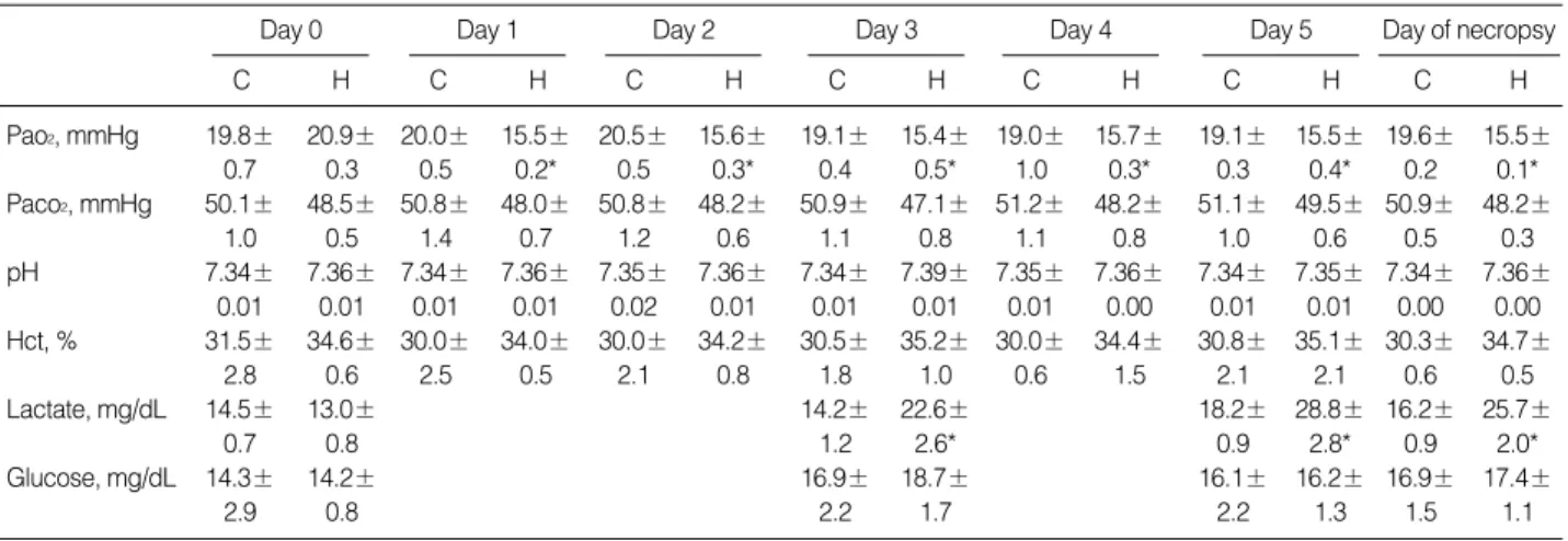
Fetal blood gases, pH and hematocrit
Fetal Pao2, Paco2, pH, and hematocrit were not significantly affected during the experimental period in control group. At baseline there was no difference in either the Pao2, Paco2, pH, or hematocrit between the controls and the hypoxic group. In the hypoxic group, values for fetal Pao2at day 3 (15.4±0.5 mmHg) and day 5 (15.5±0.4 mmHg) were significantly lower than fetal Pao2obtained at baseline for that group (20.9±0.3 mmHg; p<0.01). Fetal Paco2, pH, and hematocrit were not
Day 0
C H
Day 1
C H
Day 2
C H
Day 3
C H
Day 4
C H
Day 5
C H
Day of necropsy
C H
Pao2, mmHg 111.4± 110.4± 112.1± 80.1± 111.5± 83.4± 116.3± 81.4± 115.9± 83.0± 116.2± 76.0± 114.6± 80.5±
1.8 1.4 3.5 3.1* 3.2 3.3* 3.0 4.4* 3.8 3.7* 4.4 3.9* 1.5 1.6*
Paco2, mmHg 36.5± 35.7± 37.9± 35.7± 37.6± 35.5± 36.5± 35.6± 36.1± 34.8± 37.3± 35.2± 37.0± 35.4±
0.8 0.6 1.5 0.9 1.4 0.8 1.4 0.9 0.7 0.7 1.0 0.6 0.5 0.2
pH 7.47± 7.47± 7.46± 7.47± 7.46± 7.48± 7.46± 7.47± 7.45± 7.47± 7.44± 7.47± 7.45± 7.47±
0.01 0.01 0.02 0.01 0.02 0.01 0.02 0.02 0.01 0.02 0.01 0.01 0.01 0.01 Hct, % 27.0± 27.4± 27.0± 27.8± 26.5± 28.8± 26.3± 27.6± 25.7± 27.9± 25.5± 28.1± 26.3± 28.2±
3.1 2.1 2.5 2.0 2.6 1.8 2.7 1.8 3.2 2.2 3.0 1.9 1.0 0.8
Lactate, mg/dL 6.12± 4.63± 4.66± 6.85± 5.50± 5.75± 5.09± 6.30±
0.10 0.85 0.59 0.89 0.85 0.41 1.09 0.50
Glucose, mg/dL 59.0± 47.2± 59.8± 54.9± 60.7± 56.4± 60.3± 55.6±
7.1 3.8 3.4 3.5 4.6 3.5 2.8 2.4
Table 1.Maternal arterial blood gases, hematocrit, and metabolites in control and hypoxic fetal sheep
Values are mean±SE. C, Control; H, Hypoxic group; Hct, Hematocrit.
*p<0.05 repeated-measures analysis of variance and Student-Neuman-Keuls test.
Pao2, mmHg 19.8± 20.9± 20.0± 15.5± 20.5± 15.6± 19.1± 15.4± 19.0± 15.7± 19.1± 15.5± 19.6± 15.5±
0.7 0.3 0.5 0.2* 0.5 0.3* 0.4 0.5* 1.0 0.3* 0.3 0.4* 0.2 0.1*
Paco2, mmHg 50.1± 48.5± 50.8± 48.0± 50.8± 48.2± 50.9± 47.1± 51.2± 48.2± 51.1± 49.5± 50.9± 48.2±
1.0 0.5 1.4 0.7 1.2 0.6 1.1 0.8 1.1 0.8 1.0 0.6 0.5 0.3
pH 7.34± 7.36± 7.34± 7.36± 7.35± 7.36± 7.34± 7.39± 7.35± 7.36± 7.34± 7.35± 7.34± 7.36±
0.01 0.01 0.01 0.01 0.02 0.01 0.01 0.01 0.01 0.00 0.01 0.01 0.00 0.00 Hct, % 31.5± 34.6± 30.0± 34.0± 30.0± 34.2± 30.5± 35.2± 30.0± 34.4± 30.8± 35.1± 30.3± 34.7±
2.8 0.6 2.5 0.5 2.1 0.8 1.8 1.0 0.6 1.5 2.1 2.1 0.6 0.5
Lactate, mg/dL 14.5± 13.0± 14.2± 22.6± 18.2± 28.8± 16.2± 25.7±
0.7 0.8 1.2 2.6* 0.9 2.8* 0.9 2.0*
Glucose, mg/dL 14.3± 14.2± 16.9± 18.7± 16.1± 16.2± 16.9± 17.4±
2.9 0.8 2.2 1.7 2.2 1.3 1.5 1.1
Table 2.Fetal arterial blood gases, hematocrit, and metabolites in the control and hypoxic groups
Values are mean±SE. C, Control; H, Hypoxic group; Hct, Hematocrit.
*p<0.05 repeated-measures analysis of variance and Student-Neuman-Keuls test.
Day 0
C H
Day 1
C H
Day 2
C H
Day 3
C H
Day 4
C H
Day 5
C H
Day of necropsy
C H
significantly different at day 3 and at day 5 from baseline dur- ing maternal infusion of nitrogen (Table 2).
Fetal plasma glucose and plasma lactate
There were no significant differences in the fetal plasma
glucose and lactate levels between the control and the hypoxic groups at baseline (Day 0). In the control group, plasma glu- cose and lactate levels did not significantly change on day 3 or day 5. In the hypoxic group, fetal plasma lactate levels rose significantly from 13.0±0.7 at baseline to 22.6±2.6 (mg/
dL; p<0.01) on day 3 and continued to increase at day 5 (28.8
*p<0.01.
Artery L-NAME
Maximal response Control
(n=5)
Hypoxia (n=5)
EC50(×10-8M) Control
(n=5)
Hypoxia (n=5) Renal Without 110.9±2.5 119.7±2.5 29.1±3.1 35.5±3.0 Femoral Without 146.2±4.3 184.5±6.6* 29.8±3.9 34.4±4.7 Table 3.Responsiveness of isolated fetal sheep renal and femoral arteries to phenylephrine. Arteries were studied with and without L-NAME 10-4 M in the bath. Contractile response is expressed as percentage of KCl maximal response for each individual vessel
*p<0.05; �p<0.01.
Artery L-NAME
Maximal response Control
(n=5)
Hypoxia (n=5)
EC50(×10-8M) Control
(n=5)
Hypoxia (n=5) Renal With 134.5±3.7 133.4±7.3 14.9±3.3 7.5±1.8 Femoral With 145.0±4.5 166.9±6.3* 27.0±4.1 6.0±1.1� Table 4.esponsiveness of isolated fetal sheep renal and femoral arteries to phenylephrine. Arteries were studied with and without L-NAME 10-4 M in the bath. Contractile response is expressed as percentage of KCl maximal response for each individual vessel
Tension (%)
220 200 180 160 140 120 100 80 60 40 20 0
1e-9 1e-8 1e-7 1e-6 1e-5
Phenylephrine Dose (M) Renal
Fig. 1.Concentration-effect curves for phenylephrine (PE 10-9to 10-5mol/L) in isolated fetal sheep renal and femoral arteries obtained from normoxic (control group n=5) and hypoxic fetuses (hypoxia group n=5). Arteries were studied with and without L-NAME in the bath. Results are expressed as a percentage of the maximal response to KCl in each vessel and presented as mean±SEM.
Tension (%)
200 180 160 140 120 100 80 60 40 20 0
1e-9 1e-8 1e-7 1e-6 1e-5
Phenylephrine Dose (M)
Tension (%)
220 200 180 160 140 120 100 80 60 40 20 0
1e-9 1e-8 1e-7 1e-6 1e-5
Phenylephrine Dose (M) Femoral
Renal L-NAME
Tension (%)
200 180 160 140 120 100 80 60 40 20 0
1e-9 1e-8 1e-7 1e-6 1e-5
Phenylephrine Dose (M) Femoral L-NAME
A
B
C
D Control
Hypoxia
Control Hypoxia
Control Hypoxia
Control Hypoxia
±2.8 mg/dL; p<0.01). Fetal plasma glucose levels did not significantly change on day 3 or day 5 in the hypoxic group (Table 2).
Effects of hypoxia on fetal renal and femoral artery constric- tion by phenylephrine
There was a significant increase in the femoral artery max- imal response to PE in the absence (184.5±6.6 vs. 146.2± 4.3) and presence (166.9±6.3 vs. 145.0±4.5) of L-NAME in the hypoxic group (Table 3, 4). As shown in Fig. 1, this difference was evident whether or not the vessel had been pre- treated with the NOS inhibitor, L-NAME. However, there were no significant differences in PE maximal responses of renal arteries between the control and hypoxia groups in the presence or absence of L-NAME. A significant left-shift in the femoral artery dose response relationship to PE was also observed in the hypoxic group when pretreated with L-NAME in Fig. 1. As shown in Table 4, the PE EC50in the hypoxic group was significantly lower than that of the control group in the presence of L-NAME (6.0±1.1 vs. 27.0±4.1×10⁻⁸ M; p<0.01). There were no significant differences of PE EC50 in renal and femoral arteries in the absence of L-NAME bet- ween control and hypoxia group (Table 3).
DISCUSSION
We found that five days of mild fetal hypoxemia using maternal tracheal administration of nitrogen is well tolerat- ed by the mother and the fetus. At the onset of the nitrogen infusion, we observed a mild decrease in maternal Paco2as the mother hyperventilated in response to the fall in Pao2. This decrease was easily adjusted by adding CO2to the nitrogen infusion, and values were not different than those seen at base- line. In this protocol, we aimed to decrease fetal Pao2by 25%
which achieved and was well tolerated by the mother. Towell et al. (6) investigated the response to a decrease in fetal arte- rial Pao2of 5 mmHg or less sustained for a period of 24 hr in the chronically instrumented fetal lamb. They showed that the fetal lamb at 122 to 139 days’ gestation was highly sen- sitive to small decrements in Pao2of 5 mmHg or less. Fetal hypoxemia was not associated with any change in fetal pH.
During exposures to hypoxemia, fetal plasma lactate and cor- tisol were increased above preliminary values. The normal range of blood gases and acid-base balance in fetal sheep of 120-126 days’ gestation were Pao221.4±0.9 mmHg, Pco2 46.0±0.9 mmHg, pH 7.36±0.01 (7). We found that mild hypoxia did not significantly affect maternal and fetal pH, nor did it affect fetal Paco2. There was no significant change in fetal glucose plasma concentration during the entire exper- iment. Plasma lactate levels progressively increased in the hypoxic fetuses from baseline to a maximum on day 5. This finding is consistent with that of Towell et al. (6). Thus, despite
the absence of significant changes in fetal pH and Paco2, the fetus had a reduction in oxygen availability sufficient to cause anaerobic metabolism and lactate production. Maternal lac- tate plasma levels were not significantly increased in the con- trols or the hypoxic animals suggesting that the elevation in fetal plasma lactate levels was not due to transport of mater- nal lactate across the placenta. We cannot eliminate the pos- sibility that placental lactate production was increased and thus contributed to the observed increase in fetal lactate levels.
We found that this simple method has the major advantage of causing mild fetal hypoxia while the mother is eating and drinking freely without obvious discomfort during the entire course of nitrogen infusion. Selected specimens of maternal trachea were examined at the time of necropsy. There were only minimal areas of inflammation at the site of the catheter into the trachea and no signs of granulation tissue or obvious infection were noted. We consider that the tracheal nitrogen administration is a simple experimental tool to cause mild fetal hypoxia that can potentially be carried out for much longer study periods. We suggest that this model could be helpful to study the effect of long term mild hypoxemia on the cardiovascular system in studies related to fetal program- ming. Fetal programming is an area of great interest and this model of prolonged mild hypoxemia may well serve as a model to study the growth impaired fetus during infancy and dur- ing adult life.
Pregnancies that are complicated with chronic hypoxia are associated with several perinatal problems including fetal growth restriction and increased mortality and morbidity (8- 10). The growth restriction is asymmetric with brain and heart weight being reduced less than other organs. This asym- metry has been ascribed to redistribution of fetal cardiac out- put during hypoxia favoring the brain and heart. Redistribu- tion of blood flow may occur as a result of altered response to sympathetic alpha receptors (2). We found that mild hypoxia increased the sensitivity to PE in the femoral artery but not in the renal artery. This increase in contractile response seemed to be part due to alteration in NO production as the presence of the nitric oxide synthase inhibitor L-NAME did signifi- cantly affect the response to PE under basal and hypoxic con- dition. The contractile response to PE when L-NAME was added to the bath would suggest that NO production and or release may be involved in the fetal femoral arteries. The effect of hypoxia on the expression of the endothelial cell iso- form of NOS (eNOS) has been evaluated in cultured endothe- lial cells. The results of these studies are however conflictive.
In endothelial cells from human umbilical vein, hypoxic incu- bation inhibits eNOS expression (11) whereas it is associated with increase in both eNOS protein and mRNA (12) in endo- thelial cells from bovine aorta. Vascular reactivity to the sodi- um nitroprusside was increased by lowering Pao2in the tis- sue bath. Sensitivity also increased after endothelium denuda- tion and L-NAME treatment, suggesting that endothelium- derived nitric oxide inhibits nitrovasodilator relaxation in
isolated rat aortas (13). In fetal guinea pigs, acute hypoxia inhibited endothelium-dependent relaxation in response to acetylcholine and A23187, and increased sensitivity to sodi- um nitroprusside. Chronic hypoxia inhibited maximal relax- ation of arteries in response to acetylcholine but not relaxation of arteries in response to sodium nitroprusside with respect to relaxation seen in arteries from normoxic fetuses. They con- cluded that hypoxia may alter relaxation of fetal arteries by decreasing the availability of oxygen for nitric oxide produc- tion and causing vascular adaptations related to altered nitric oxide release (14).
The present results are consistent with the result found by Millard et al. (15) in the fetal lamb during acute hypoxia. In that experiment, acute fetal hypoxia was induced by admin- istering a mixture of 10% oxygen in nitrogen to the mother for 10 min. They showed that during hypoxia, there was an immediate vasoconstriction of the iliac vessels whereas the renal bed did not constrict initially, and in some experiments, the renal bed actually dilated in response to acute hypoxia.
After blockade of prostaglandin synthesis, the renal vascular bed behaved like the iliac bed, showing an immediate and marked vasoconstriction. Even under this protocol, 5 days of mild fetal hypoxia without hypocarbia and alkalosis, did not alter the responsiveness of these renal vessels to PE. It is possible that greater degree of hypoxia could change the res- ponsiveness of these vessels to vasoconstrictive agents such as PE. Other factors, such as endothelial-derived hyperpolariz- ing factors or endothelin-1 which we have not tested could potentially be involved and affect one particular vessel more than the others. Exposure to normobaric hypoxia has previ- ously been shown to increase levels of the endogenous vaso- constrictor endothelin in plasma and aorta of Sprague-Daw- ley rats (16).
In the present study, the increased contractile response to PE by the femoral artery in the hypoxic fetuses is different from what has been reported in nonpregnant rats exposed to chronic hypoxia (17). Studies of the effects of chronic hypoxia on in vivo systemic vasoreactivity have yielded conflicting data. Aoki and Robinson (18) found augmented pressor res- ponses to adrenoreceptor agonists in the hindlimb of chron- ically hypoxic rats. In contrast, Jin et al. (19) have demonstrat- ed impaired responses to vasopressin infusion in hypoxia-adapt- ed rats. Fukuda et al. (20) have reported that changes in sys- temic vascular resistance during infusion of phenylephrine and angiotensin II were not different in chronically hypoxic guinea pigs and normoxic controls. In these two studies chro- nic hypoxia decreased the contractile sensitivity to PE by do- wnregulating 1-adrenergic-receptors number and by alter- ing the excitation-contractile coupling in vascular smooth muscle (21). This difference may be due to the fact that these studies were done in adults and that these particular vascular beds were not studied.
In conclusions, five days of continuous maternal tracheal nitrogen infusion can induce fetal mild hypoxia without fetal
acidosis. Mild chronic hypoxia seems to increase vasoconstric- tive responses of femoral arteries in the fetal sheep. In addition, L-NAME pretreatment significantlly decreased EC50, along with the increased maximal contraction response. Therefore, the increase in contractile response may at least in part be due to alteration in NO production. Further studies are needed to confirm the effects of mild chronic hypoxia on renal and femoral vessel reactivity in the fetal sheep.
ACKNOWLEDGMENT
We are deeply grateful to all those who helped with this work, particularly Prof. Byun JS, Prof. E. Mueller-Heubach, Prof. Lee YI, Prof. Choi HS, Prof. Oh ST, Mr. H. Booze, and Mr. M. Dunlap.
REFERENCES
1. Rudolph AM, Heymann MA. Circulatory changes during growth in the fetal lamb. Circ Res 1970; 26: 289-99.
2. Campbell AG, Dawes GS, Fishman AP, Hyman AI. Regional redis- tribution of blood flow in the mature fetal lamb. Circ Res 1967; 21:
229-35.
3. Cohn HE, Sacks EJ, Heymann MA, Rudolph AM. Cardiovascular responses to hypoxemia and acidemia in fetal lambs. Am J Obstet Gynecol 1974; 120: 817-24.
4. Gleed RD, Poore ER, Figueroa JP, Nathanielsz PW. Modification of maternal and fetal oxygenation with the use of tracheal gas infusion.
Am J Obstet Gynecol 1986; 155: 429-35.
5. Nathanielsz PW, Abel MH, Bass FG, Krane EJ, Thomas AL, Liggins GC. Pituitary stalk-section and some of its effects on endocrine func- tion in the fetal lamb. Q J Exp Physiol 1978; 63: 211-9.
6. Towell ME, Figueroa J, Markowitz S, Elias B, Nathanielsz P. The effect of mild hypoxemia maintained for twenty-four hours on maternal and fetal glucose, lactate, cortisol, and arginine vasopressin in pregnant sheep at 122 to 139 days’ gestation. Am J Obstet Gynecol 1987; 157:
1550-7.
7. Jensen A, Berger R. Fetal circulatory responses to oxygen lack. J Dev Physiol 1991; 16: 181-207.
8. Kruger H, Arias-Stella J. The placenta and the newborn infant at high altitudes. Am J Obstet Gynecol 1970; 106: 586-91.
9. Mazess RB. Neonatal mortality and altitude in Peru. Am J Physiol Anthrop 1965; 23: 209-14.
10. McCullough RE, Reeves JT, Liljegren RL. Fetal growth retardation and increased infant mortality at high altitude. Obstet Gynecol Surg 1977; 32: 596-8.
11. McQuillan LP, Leung GK, Marsden PA, Kostyk SK, Kourembanas S. Hypoxia inhibits expression of eNOS via transcriptional and post- transcriptional mechanism. Am J Physiol 1994; 267: H1921-7.
12. Arnet UA, McMillan A, Dinerman JL, Ballermann B, Lowenstein CJ. Regulation of endothelial nitric-oxide synthase during hypoxia.
J Biol Chem 1996; 271: 15069-73.
13. Moncada S, Rees DD, Schulz R, Palmer RM. Development and mech- anism of specific supersensitivity to nitrovasodilators after inhibition of vascular nitric oxide synthesis in vivo. Proc Natl Acad Sci USA 1991; 88: 2166-70.
14. Thompson LP, Weiner CP. Effects of acute and chronic hypoxia on nitric oxide-mediated relaxation of fetal guinea pig arteries. Am J Obstet Gynecol 1999; 181: 105-11.
15. Millard RW, Baig H, Vatner SF. Prostaglandin control of the renal circulation in response to hypoxemia in the fetal lamb in utero. Circ Res 1979; 45: 172-9.
16. Elton TS, Oparil S, Taylor GR, Hicks PH, Yang RH, Jin H, Chen YF.
Normobaric hypoxia stimulates endothelin-1 gene expression in the rat. Am J Physiol 1992; 263: R1260-4.
17. Doyle MP, Walker BR. Attenuation of systemic vasoreactivity in chro- nically hypoxic rats. Am J Physiol 1991; 260: R1114-22.
18. Aoki VS, Robinson SM. Hindquarters vascular responses in chroni- cally hypoxic rats. Am J Physiol 1969; 217: 661-5.
19. Jin H, Yang RH, Chen YF, Thornton RM, Jackson RM, Oparil S. He- modynamic effects of arginine vasopressin in rats adapted to chronic hypoxia. J Appl Physiol 1989; 66: 151-60.
20. Fukuda S, Morioka M, Tanaka T, Taga K, Shimoji K. Endothelium dependence of effects of high PCO2on agonist-induced contractility of rat aorta. Am J Physiol 1993; 264: H512-9.
21. White MM, McCullough RE, Dyckes R, Robertson AD, Moore LG.
Effects of pregnancy and chronic hypoxia on contractile responsiveness to alpha 1 adrenergic stimulation. J Appl Physiol 1998; 85: 2322-9.