INTRODUCTION
The classification of Ehlers-Danlos syndrome (EDS) is based on the extent of clinical and pathological features. Vascular EDS (type IV EDS, MIM #130050) is the most malignant form and owes its bad reputation to a susceptibility to sud- den death from spontaneous catastrophic bleeding or the rupture of blood vessels or hollow organs (1). Type IV EDS is characterized by its clinical manifestations: easy bruising, thin skin with visible veins, acrogeria, and the rupture of arteries, uterus, or intestines (2). Vasculopathy involves the rupture, dissection, or aneurysms of the aorta or large arter- ies, carotid-cavernous fistula (CCF), and intracranial aneu- rysms. Arterial complications are the leading cause of death in vascular EDS because they are unpredictable and surgical repair is difficult because of tissue fragility (3). In a recent review of 419 patients, 70% of deaths were the result of arte- rial rupture, mostly of thoracic and abdominal vessels, but also of cerebral vessels (1). However, bruisability is very strik- ing, and ‘ecchymotic-type EDS’ is a synonym for type IV EDS. Type IV EDS is inherited in an autosomal dominant fashion but approximately 50% of patients represent new mutations. Most patients have a defect in either the synthe- sis or structure of type III procollagen, with a genetic muta-
tion in COL3A1. However, it has been suggested that EDS type IV is a genetically heterogeneous disease. Pepin et al.
have reported that only 61% of patients with abnormal type III procollagen molecules have mutations in the COL3A1 gene, whereas the others have no mutation in COL3A1 (1).
In a recent study, mutations in the genes encoding transform- ing growth factor receptors 1 and 2 (TGFBR1 and TGF- BR2, respectively) have been identified in patients with a provisional diagnosis of EDS type IV without the character- istic type III collagen abnormalities. This new syndrome was designated Loeys-Dietz syndrome, type 2 (4, 5).
Here, we report our experience of three Korean patients, in whom a clinical diagnosis of EDS type IV was made based on clinical findings. However, a genetic study identified CO- L3A1 mutations in two of these three patients. In the other one patient, no mutations were observed in COL3A1, TG- FBR1, or TGFBR2.
MATERIALS AND METHODS
The diagnoses of these patients were made based on clini- cal presentation, physical examinations, and radiological find- ings. Information about deceased family members and other
Jeong Hoon Yang, Seung-Tae Lee*, Jee-Ah Kim�, Sung Hae Kim�, Shin-Yi Jang, Chang-Seok Ki*, Duk-Kyung Kim
Departments of Internal Medicine, Laboratory Medicine*, Samsung Medical Center, Sungkyunkwan University School of Medicine, Seoul; Garak High School�, Seoul; Department of Cardiology�, Konkuk University, College of Medicine, Seoul, Korea
Address for correspondence Duk-Kyung Kim, M.D.
Department of Internal Medicine, Samsung Medical Center, Sungkyunkwan University School of Medicine, 50 Irwon-dong, Gangnam-gu, Seoul 135-710, Korea Tel : +82.2-3410-3419, Fax : +82.2-3410-3849 E-mail : [email protected]
698
Genetic Analysis of Three Korean Patients with Clinical Features of Ehlers-Danlos Syndrome Type IV
Ehlers-Danlos syndrome (EDS) is a hereditary disorder of the connective tissue.
EDS type IV (EDS IV), the vascular type of the disease, is characterized by easy bruising, thin skin with visible veins, and spontaneous rupture of the large arteries, uterus, or bowel. EDS IV is caused by mutations in the gene for type III procollagen (COL3A1). However, recent studies suggest that the causative mutation of EDS IV is not homogeneous. We report our experience with three patients presenting with clinical features of type IV EDS. A 48-yr-old woman presented with acute aor- tic dissection (patient 1) and 36-yr-old and 21-yr-old women presented with carotid- cavernous fistula (patients 2 and 3, respectively). All three patients bruised easily.
Two patients (patients 1 and 3) had thin transparent skin with visible veins. Genetic analysis of COL3A1 revealed a Gly732Val (c.2195G>T) mutation in patient 1 and a duplication of 15 base pairs (c.3221_3235dup) which resulted in an interposition of five amino acids (p.Gly1074_Pro1078dup) in patient 2. However, no mutations were observed in COL3A1 or transforming growth factor receptors 1 and 2 in patients 3, which might be either due to a deletion of single or multiple exons in the COL3A1 gene or due to a genetic heterogeneity. This is the first report of geneti- cally confirmed cases of EDS IV in Korea.
Key Words : Ehlers-Danlos Syndrome Type IV; COL3A1; Mutation; Dissection; Carotid-cavernous Sinus Fistula
Received : 8 November 2006 Accepted : 15 January 2007
Genetic Analysis of Ehlers-Danlos Syndrome Type IV 699
family members who were suspected of being affected was obtained from the probands. Informed consent was obtained.
Genomic DNA was extracted from peripheral blood leuko- cytes with the Wizard Genomic DNA Purification Kit (Pro- mega, Madison, WI, U.S.A.), following the manufacturer’s instructions. The coding exons and their flanking introns of the COL3A1 (total 51 exons) on chromosome 2q31, and TGFBR1 and TGFBR2 on chromosome 9q33-q34 and
3p22, respectively, were amplified using primers designed by the authors (available upon request). Direct sequencing was performed with the BigDye Terminator Cycle Sequenc- ing Ready Reaction kit (Applied Biosystems, Foster City, CA, U.S.A.) on the ABI 3100 Genetic Analyzer (Applied Biosystems).
RESULTS
The clinical and pathological findings for the three patients are shown in Table 1.
Patient 1
A 48-yr-old woman was admitted with sudden severe epi- gastric pain radiating to the back one day before admission.
Her medical history revealed two uncomplicated cesarean sections. A spontaneous retroperitoneal hematoma had been treated with percutaneous catheter drainage three years pre- viously. On first presentation, her blood pressure was 121/78 mmHg, her pulse rate was 80 beats per minute, and her
Fig. 1. (A) The patient’s skin was very thin with visible vessels over the upper thorax. A minor trauma caused by the detachment of the elec- trocardiographic electrode elicited excessive bruising. (B, C). The patient’s skin over the hands and feet was thin and finely wrinkled (acroge- ria). Equinus deformity was noted in both feet. (D) An radiography of the hands showed osteoporosis and absorption of the midshafts of the finger bones (acroosteolysis).
A B
C D
Variables Patient 1 Patient 2 Patient 3
Sex Female Female Female
Age at examination (yr) 48 36 21
Mutation of COL3A1 Yes Yes No
Family history of sudden death Yes No No
Vascular involvement Yes Yes Yes
Bowel involvement No No No
Easy bruising Yes Yes Yes
Thin skin with visible veins Yes No Yes
Acrogeria Yes Suspicious Suspicious
Hypermobility of small joints No Yes No
Joint dislocations No Yes No
Table 1.Clinical and pathological findings
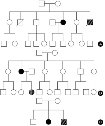
heartbeat was regular. A clinical examination showed ten- derness in the left lower abdominal quadrant, very thin skin with visible vessels on the thorax, which bruised easily with minimal trauma (Fig. 1A), and acrogeria (Fig. 1B, C). Equi- nus deformity was noted in both feet (Fig. 1C). Acroosteol- ysis was present (Fig. 1D). The results of a neurological exam- ination were normal. Computed tomography (CT) demon- strated an abdominal aortic dissection with extension from the proximal superior mesenteric artery to both common iliac arteries (Fig. 2A, B). A provisional diagnosis of EDS IV was made. Because of the high surgical mortality rate in patients with EDS type VI, conservative medical treatment was given. Three days later, she suddenly developed back pain with shock. Follow-up CT angiography revealed a pro- gression of the aortic dissection with hemothorax, hemoperi- toneum, and retroperitoneal hemorrhage (Fig. 2C, D). The patient died from uncontrolled bleeding before surgical inter- vention. A family history revealed that her younger brother had died suddenly from cerebral hemorrhage (Fig. 3A).
Genetic analysis revealed a G to T transition (c.2195G>T) leading to the substitution of the glycine at position 732
(p.Gly732Val) of the triple helix of type III protocollagen with valine (Fig. 4).
Patient 2
A 36-yr-old woman presented with left ocular pain, head- ache, diplopia, and tinnitis, which had started one month before presentation. On physical examination, her skin showed a bruise at a trauma site (Fig. 5A). The patient’s foot showed chronic dislocation of the first metatarsal joint (Fig. 5B). Hy- permobility of the distal interphalangeal joints of the hands was observed (Fig. 5C). Kyphosis of the lumbar and thoracic vertebrae was noted. A family history revealed that her daugh- ter had a chronic subluxation of the proximal interphalangial joint of the left hand (Fig. 3B). Magnetic resonance imaging revealed a large CCF, and the diagnosis was confirmed by angiography (Fig. 5D). The CCF was successfully treated with detachable balloons using a femoral arterial approach.
During the procedure, a pseudoaneurysm of the left proximal internal carotid artery occurred, and a stent was inserted. A large subcutaneous hematoma occurred at the puncture site
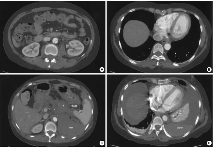
Fig. 2. Computed tomography (CT) of the thoracoabdominal aorta. (A, B) Initial CT demonstrated aortic dissection, which involved the ab- dominal aorta and spared the thoracic aorta. (Cand D) Seven days later, the dissection showed retrograde progression and rupture, which produced hemoperitoneum (*), retroperitoneal hemorrhage (**), and hemothorax (***). Intramural hematoma was noted in the descend- ing thoracic aorta (D).
A B
C D
*
** ***
Genetic Analysis of Ehlers-Danlos Syndrome Type IV 701
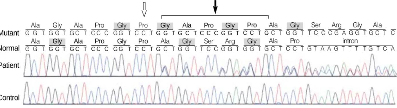
after angiography. Then, surgical evacuation of a massive hematoma was performed, followed by the insertion of a stent graft into the punctured femoral artery, with arteriotomy to control bleeding. The postoperative course was uneventful, except for ophthalmoplegia and the loss of visual acuity due to a neurotropic corneal ulcer. The patient is alive and doing well at the seven-year follow up. A mutation analysis by DNA sequencing of the COL3A1 gene revealed a duplication of 15 base pairs (c.3221_3235dup), which resulted in an inter- position of five amino acids (p.Gly1074_Pro1078dup) and a subsequent formation of a repeat unit composed of only two amino acids (Gly-X) (Fig. 6).
Patient 3
Some of the clinical findings of this patient have been report- ed previously (6). A 21-yr-old woman presented with left ocular pain, severe pulsatile headache, and vomiting after her left eye was hit with a champagne cork. She had bruised easily since she was young. On physical examination, her skin was so thin that the subcutaneous blood vessels were visible (Fig. 7A, B). Echymoses and bruises were noted at trauma sites (Fig. 7B). There was no significant hyperelasticity of the skin or hypermobility of the joints of the hand. Her fam-
ily history revealed that her younger brother had bruised eas- ily (Fig. 3C). Carotid angiography showed a large CCF and an aneurysm in the cervical portion of the left internal carotid artery (Fig. 7C). Computed tomography of the thoracoab- dominal aorta demonstrated a large ovoid aneurysm of the left renal artery and a medium-sized aneurysm of the splenic artery (Fig. 7D). The CCF was successfully occluded with detachable balloons using a femoral arterial approach. Huge groin hematomas and echymoses occurred at the puncture sites after angiography.
Seven years later, she developed a spontaneous hematoma of the left calf, which was relieved by conservative manage- ment. The patient is alive and doing well at the eight-year follow up. Genetic analysis revealed no mutation of either COL3A1, TGFBR1, or TGFBR2.
DISCUSSION
Here, we report three patients with a provisional diagno- sis of EDS type IV. The first and second patients had muta- tions of the gene for type III procollagen (COL3A1). Altho- ugh the other patient showed clinical features compatible with EDS type IV, genetic analysis of COL3A1 did not reveal a mutation. Also, additional genetic analysis of TGFBR1 and TGFBR2 did not show a mutation. Absence of mutations
A
B
C
Fig. 3.Pedigrees of the families. Squares indicate male family mem- bers, circles indicate female family members, and symbols with slashes indicate deceased family members. Open symbols indi- cate unaffected family members, filled symbols indicate probands, and shaded symbols indicate family members who are or were probably affected. (A) patient 1, (B) patient 2, (C) patient 3.
Fig. 4.Direct sequencing of COL3A1 gene revealed a G-to-T tran- sition at cDNA position 2195 (arrow; c.2195G>T) resulting in a Gly- 732Val missense mutation. Wild type alleles in her siblings sug- gested a de novo mutation.
Ref cDNA seq Ref amino acid seq
Elder brother
Elder sister
Patient
Younger sister
in patients 3 might be either due to a deletion of single or multiple exons in the COL3A1 gene or due to a genetic het- erogeneity. Description of patients with EDS type IV-like features in Korea is quite rare (7). To the best of our knowl- edge, patient 1 and 2 are the first Korean patients with a molecular diagnosis of type IV EDS.
The mutation found in patient 1 is a novel mutation rather than a genetic variation because most of known missense mutations of COL3A1 involves glycine residue and the same variation of the patient was not found in either other family members with normal phenotype or fifty control subjects.
Also, any genetic alteration at the duplicated location of pa-
Fig. 5. (A) The skin showed a bruise at the trauma site. (B) The patient’s foot showed chronic dislocation of the first metatarsal joint. (C) The patient had mild hyperextensibility of the fingers. (D) Left internal carotid angiogram, lateral view, showed a high-flow, direct carotid- cavernous fistula in the early arterial phase (arrow).
Fig. 6. Direct sequencing of the COL3A1 gene revealed a duplication of 15 base pairs (c.3221_3235dup), which results in an interposi- tion of five amino acids (p.Gly1074_Pro1078dup). The duplicated segment of nucleotides and amino acids in mutant allele and its origi- nal position in normal allele were marked in bold characters. The critical glycine residues were marked in gray square boxes. The solid arrow points to the additional interposed amino acids, and the open arrow indicates a subsequently formed repeat unit composed of only two amino acids (Gly-X).
A B
C D
Mutant Ala
Normal
G G T
Patient
Control
Gly G G T
Ala G C T
Pro C C C
Gly G G T
Pro C C T
Gly G G T
Ala G C T
Pro C C C
Gly G G T
Pro C C T
Ala GC T
Gly G G T
Ser T C C
Arg C G A
Gly G G T
Ala G C T C Pro
C C T
intron
G T A A G T T T T G T C A Ala
G C T Gly G G T Arg G G T Ser T C C Gly G G T Ala GC T Pro C C T Gly G G T Pro C C C Ala G C T Gly G G T Ala G G T
Genetic Analysis of Ehlers-Danlos Syndrome Type IV 703
tient 2 had not been previously reported in EDS type IV. The nature of the mutation found in patient 2 was quite different from the vast majorities of other previously reported muta- tions on the COL3A1 gene, which usually were point muta- tions affecting the glycine residue of the normal Gly-X-Y repeats and caused classical EDS type IV (1). As a result of duplication of 15 base pairs, an interposition of five amino acids among the triple repeats has occurred, and a subsequent formation of a repeat unit consisting of two amino acids (Gly- X) just before the duplicated segment was observed. These atypical properties of the mutation in this patient might have had an effect on her phenotypic features that lacked
one of the cardinal manifestations of classical EDS type IV, i.e. thin transparent skin. We previously reported patient 3 as having EDS type IV (6), demonstrating that a genetic diagnosis is mandatory for a final diagnosis in patients with the clinical features of EDS type IV.
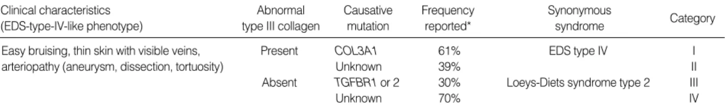
Recent developments in genetic diagnosis have broadened the clinical spectrum of type IV EDS. In the largest series of patients with EDS IV, Pepin et al. analyzed the COL3A1 gene in 220 index patients who had biochemical defects in type III collagen (1). They identified causative mutations in the COL3A1 gene in only 135 index cases (61%). In other words, 39% of patients with EDS IV, with abnormal type III colla-
Fig. 7. (A) The patient’s skin over the upper thorax was very thin and translucent with visible vessels. (B) The patient’s skin showed a bruise and visible veins in the leg. (C) Left internal carotid angiogram, lateral view, showed a high-flow, direct carotid-cavernous fistula in the early arterial phase (arrow) and an aneurysm in the cervical portion of the left internal carotid artery (arrowhead). (D) A computed tomography scan demonstrated a large ovoid aneurysm of the left renal artery (arrow) and a medium-sized aneurysm of the splenic artery (arrowhead).
A B
C D
Clinical characteristics (EDS-type-IV-like phenotype)
Abnormal type III collagen
Causative mutation
Frequency reported*
Synonymous
syndrome Category
Easy bruising, thin skin with visible veins, Present COL3A1 61% EDS type IV I
arteriopathy (aneurysm, dissection, tortuosity) Unknown 39% II
Absent TGFBR1 or 2 30% Loeys-Diets syndrome type 2 III
Unknown 70% IV
Table 2.Newly proposed categories of patients with EDS-type-IV-like phenotypes
COL3A1, gene for type III procollagen; TGFBR1 and 2, genes for transforming growth factor receptor 1 and 2, respectively. *References, 1, 4.
gen, had a mutation in a gene other than COL3A1, which indicates that even EDS IV with a proven defect in type III collagen is a genetically heterogeneous disease. Furthermore, in a recent study, mutations in the TGFBR1 and TGFBR2 genes have been reported in Loeys-Dietz syndrome type 1, which is characterized by the triad of arterial tortuosity and aneurysms, hypertelorism, and bifid uvula or cleft palate (5, 6, 8). Mutations in the gene encoding TGFBR1 or TGFBR2 were also found in 12 (30%) of 40 patients with EDS type IV without the characteristic type III collagen abnormalities.
All 12 patients had at least two of the findings associated with EDS IV. They had previously received a provisional diagno- sis of EDS IV by a medical geneticist. This new disease was designated Loeys-Diets syndrome type 2, distinguishing it from type 1, which has additional craniofacial abnormalities.
Based on these findings, we propose new categories of patients who have EDS-type-IV-like phenotypes (Table 2). Patient 1 and 2 belongs to category I. Patients 3 could be classified into category II or IV. However, there is a possibility that a causative genetic abnormality in patients 3 could be a dele- tion of single or multiple exons in the COL3A1, TGFBR1 or TGFBR2 gene.
The first patient had the most severe phenotype of the three patients and died of rapidly progressive aortic dissection with rupture. Some authors have suggested that the nature of a mutation relates to the patient’s phenotype (9). Mutations in the middle or at the 5′end of a gene produce more vari- able phenotypes and milder variants compared with those caused by mutations around the 3′end, which have severe manifestations. However, Pepin et al. (1) demonstrated no apparent relationship between the type of complication and the locations of mutations. Even though patient 1 had a sub- stitution in the middle part of the COL3A1 gene product (732nd codon of 1,466 amino acids) and patient 2 had a duplication in the 3’ part of the COL3A1 gene (1,074th to 1,078th residues of 1,466 amino acids), patient 1 had rela- tively severe and full-blown clinical manifestations compar- ed to those of patient 2. The patient 1 survived two cesarean sections. Pepin et al. reported that, among 183 pregnancies with 167 deliveries of live-born infants at term, 12 women died (1). The overall risk of death during delivery was 7%.
However, it is not known whether the use of elective cesare- an section reduces mortality. The age at death of our patient was 48, which is consistent with a previous report in which the calculated median survival of the entire cohort of 220 index patients was 48 yr (1).
Patients 2 and 3 presented with CCF. CCF is exceedingly rare in the general population, among whom the vast major- ity is posttraumatic. Spontaneous CCF is one of the most common central nervous system vascular complications of EDS IV. North et al. reviewed these cerebral vascular com- plications in 202 individuals with biochemically confirmed type IV EDS (10). Of these, six individuals (3%) experienced spontaneous CCF.
One of the striking features of patients 2 and 3 was their vascular complications after invasive vascular procedures.
The arteries of both patients were so fragile that they were easily dissected during catheter and guidewire interventions or the ballooning of arteries. They also showed severe com- plications at transfemoral puncture sites, including giant groin hematoma or severe ecchymosis, which required sur- gical evacuation of the hematoma and insertion of a stent graft into the injured artery. Therefore, as far as possible, con- ventional arteriography should be avoided because of the high risk of massive hematoma and/or arterial dissection, as noted in our patients. In an attempt to avoid arterial dissec- tion or rupture, CCF could be closed via a venous rather than the more standard arterial route (11).
In conclusion, we identified three patients with EDS-IV- like phenotypes. Two patients were shown to have mutations in COL3A1. Based on our experience, we emphasize that patients with a presentation suggestive of EDS IV should undergo biochemical analysis of their type III collagen and DNA analysis of the COL3A1, TGFBR1, and TGFBR2 genes. However, these laboratory tests cannot identify the genes causing the category II or IV disease, which await fur- ther progress and understanding of the molecular pathogen- esis of patients with EDS-IV-like phenotypes.
REFERENCES
1. Pepin M, Schwarze U, Superti-Furga A, Byers PH. Clinical and genet- ic features of Ehlers-Danlos syndrome type IV, the vascular type. N Engl J Med 2000; 342: 673-80.
2. Beighton P, De Paepe A, Steinmann B, Tsipouras P, Wenstrup RJ.
Ehlers-Danlos syndromes: revised nosology, Villefranche, 1997.
Ehlers-Danlos National Foundation (USA) and Ehlers-Danlos Sup- port Group (UK). Am J Med Genet 1998; 77: 31-7.
3. Germain DP, Herrera-Guzman Y. Vascular Ehlers-Danlos syndrome.
Ann Genet 2004; 47: 1-9.
4. Loeys BL, Schwarze U, Holm T, Callewaert BL, Thomas GH, Pannu H, De Backer JF, Oswald GL, Symoens S, Manouvrier S, Roberts AE, Faravelli F, Greco MA, Pyeritz RE, Milewicz DM, Coucke PJ, Cameron DE, Braverman AC, Byers PH, De Paepe AM, Dietz HC.
Aneurysm syndromes caused by mutations in the TGF-beta receptor.
N Engl J Med 2006; 355: 788-98.
5. Loeys BL, Chen J, Neptune ER, Judge DP, Podowski M, Holm T, Meyers J, Leitch CC, Katsanis N, Sharifi N, Xu FL, Myers LA, Spe- vak PJ, Cameron DE, De Backer J, Hellemans J, Chen Y, Davis EC, Webb CL, Kress W, Coucke P, Rifkin DB, De Paepe AM, Dietz HC.
A syndrome of altered cardiovascular, craniofacial, neurocognitive and skeletal development caused by mutations in TGFBR1 or TGF- BR2. Nat Genet 2005; 37: 275-81.
6. Koh JH, Kim JS, Hong SC, Choe YH, Do YS, Byun HS, Lee WR, Kim DK. Skin manifestations, multiple aneurysms, and carotid-cav- ernous fistula in Ehlers-Danlos syndrome type IV. Circulation 1999;
100: 57-8.
Genetic Analysis of Ehlers-Danlos Syndrome Type IV 705
7. Jeong DS, Kim KH, Ahn H. Surgical treatment of acute type A aor- tic dissection in Ehlers-Danlos syndorme. Korean J Thorac Cardio- vasc Surg 2000; 33: 910-4.
8. Ki CS, Jin DK, Chang SH, Kim JE, Kim JW, Park BK, Choi JH, Park IS, Yoo HW. Identification of a novel TGFBR2 gene mutation in a Korean patient with Loeys-Dietz aortic aneurysm syndrome; no muta- tion in TGFBR2 gene in 30 patients with classic Marfan’s syndrome.
Clin Genet 2005; 68: 561-3.
9. de Paepe A. The Ehlers-Danlos syndrome: a heritable collagen dis-
order as cause of bleeding. Thromb Haemost 1996; 75: 379-86.
10. North KN, Whiteman DA, Pepin MG, Byers PH. Cerebrovascular complications in Ehlers-Danlos syndrome type IV. Ann Neurol 1995;
38: 960-4.
11. Chuman H, Trobe JD, Petty EM, Schwarze U, Pepin M, Byers PH, Deveikis JP. Spontaneous direct carotid-cavernous fistula in Ehlers- Danlos syndrome type IV: two case reports and a review of the litera- ture. J Neuroophthalmol 2002; 22: 75-81.