INTRODUCTION
The evaluation of acute chest pain is a challenging task for physicians who work in emergency departments (EDs). Al- though complex diagnostic algorithms that combine multi- ple diagnostic modalities were developed to minimize error (1- 3), a high portion (2-6%) of acute coronary syndrome (ACS) patients are being mistakenly discharged from EDs (4-6). This is related to adverse clinical outcomes (4) and responsible for the largest portion of malpractice suits (7).
The recent development of computed tomography coronary angiography (CTCA) provides a high spatial and temporal resolution images non-invasively (8). It provides comprehen- sive information about the patency of coronary artery, the integri- ty of great vessels and the lungs (triple rule out) (9, 10). In many comparative studies, targeting a non-ED based popu- lation, CTCA already showed its high diagnostic accuracy and high negative predictive value (8).
The application to the patients with an acute chest pain in
ED environment is also being actively evaluated. Some recent studies had shown that CTCA is reliable for ruling out an ACS in low risk group. However, previous studies are mostly small observational studies and/or have a narrow spectrum of tar- get population (11-17).
We tried to evaluate the safety and efficacy of the CTCA- based approach for the patients with acute chest pain in real world ED situation.
MATERIALS AND METHODS Study population
This study was performed at the urban teaching hospital with more than 44 thousand visits per year (2007) in ED. A total of 1,125 adult patients who visited the ED with acute chest pain was screened from November 2005 to February 2007. Initial clinical evaluation included history taking, phys-
466
Joonghee Kim1, Hwijae Lee1, Sungwook Song1, Jinsik Park2, Hwanjun Jae3, Whal Lee3, Sangdo Shin1, Sungkoo Jung1, Youngho Kwak1, Giljoon Suh1, and Jaehyung Park3
Departments of Emergency Medicine1and Internal Medicine, Divisions of Cardiology2and Radiology3, Seoul National University Hospital, Seoul, Korea
Address for Correspondence Jinsik Park, M.D.
Current address: Department of Cardiology, Sejong General Hospital, 91-121 Sosabon 2-dong, Sosa-gu, Bucheon 422-711, Korea
Tel : +82.32-1599-6677, Fax : +82.32-349-3005 E-mail : [email protected]
Efficacy and Safety of the Computed Tomography Coronary
Angiography Based Approach for Patients with Acute Chest Pain at an Emergency Department: One Month Clinical Follow-up Study
To evaluate the safety and efficacy of the computed tomography coronary angiog- raphy (CTCA) for evaluation of acute chest pain in real world population, we prospec- tively enrolled 296 patients with acute chest pain at emergency department (ED) from November 2005 to February 2007. The patients were grouped based on the clinical information and CTCA result. The patients with a low risk profile and no sig- nificant coronary stenosis (>50%) in CTCA were discharged immediately (Group 1, n=103). On the other hand, the patients with an intermediate risk profile without sig- nificant stenosis were observed in ED for 24 hr (Group 2, n=104). The patients with significant stenosis underwent further coronary evaluation and management accord- ingly (Group 3, n=89). While no false negative case was found in Group 1, seven cases (6.73%) were found in Group 2, mostly during the observation period. In Group 3, there were 54 (60.67%) cases of acute coronary syndrome including 10 myocar- dial infarctions. The overall accuracy of CTCA for acute coronary syndrome was 88.5% (sensitivity), 85.1% (specificity), 60.7% (positive predictive value) and 96.6%
(negative predictive value). In conclusion, clinical decision based on CTCA is safe and effective for low risk patients. Further validation is needed in patients with inter- mediate risk profile.
Key Words : Angina; Diagnosis; Imaging; Angiography; Coronary Disease; Tomography
Received : 10 July 2008 Accepted : 20 April 2009
ⓒ 2010 The Korean Academy of Medical Sciences.
This is an Open Access article distributed under the terms of the Creative Commons Attribution Non-Commercial License (http://creativecommons.org/licenses/by-nc/3.0) which permits unrestricted non-commercial use, distribution, and reproduction in any medium, provided the original work is properly cited.
ical examination, chest radiography, electrocardiogram (ECG), cardiac biomarker and creatinine level. The patients with high risk clinical profile were excluded after initial evaluation. High risk clinical profile was defined to the following criteria: 1) ST- segment elevation or new onset LBBB, 2) Increased level of serum troponin or CK-MB, 3) Typical chest pain with ische- mic ST-T change (ST segment depression >1 mm or T-wave inversion >3 mm), 4) Overt congestive heart failure, recur- rent ventricular arrhythmia, 5) Ongoing or recurrent angina at rest or minimal effort, and 6) Proven significant coronary artery disease.
After exclusion of these high risk patients, we determined whether each patient has intermediate or low risk profile. Inter- mediate risk profile was defined as having any of the follow- ing characteristics: 1) Atypical chest pain with any possible ischemic ECG change (mainly ST-segment depression or T- wave inversion), 2) Typical chest pain with normal ECG. And all patients who do not have possible ischemic ECG change nor typical chest pain were considered to have low risk pro- file.
The cases with definite evidence of non-coronary etiology of chest pain (e.g., newly diagnosed pneumothorax, overt her- pes zoster or evident history of acute chest trauma) or which are not eligible for the test (significant arrhythmia, uncontrol- lable heart rate, contraindication to beta-blocker or hypersen- sitivity to radiocontrast agent) were also excluded.
Initial evaluation, enrollment and data collection were per- formed prospectively by trained emergency physician. The data also were reviewed by a cardiologist every week.
CTCA scanning and analysis
The patients with low to intermediate clinical profile and without other exclusion criteria underwent CTCA evaluation with 16 or 64-channel multi-detector computed tomogra- phy (MDCT) with ECG gating capability. Heart rate was controlled under 65 bpm with metoprolol. Scan results were analyzed by a radiologist specialized in cardiovascular imag- ing. CTCA criterion of significant stenosis was defined as more than 50% diameter stenosis of any major coronary arteries.
Management protocol at ED for the patients with acute chest pain
The patients were classified into three groups according to their clinical characteristics and CTCA reading (Fig. 1): 1) Group I (Immediate discharge group)-No significant coro- nary stenosis in the CTCA with low risk profile, 2) Group II (ED observation group)-No significant coronary stenosis in the CTCA with intermediate risk profile, 3) Group III (Fur- ther work up group)-Any CTCA evidence of significant coro- nary stenosis with low to intermediate risk profile.
The patients in Group I were immediately discharged from ED with a short-term out-patient department (OPD) follow-
up arrangement. The patients in Group II were observed in ED chest pain unit with serial ECG and cardiac biomarker assay. During the observation period, if a patient had recur- rent chest pain or dynamic ST-T change or rising cardiac bio- marker level, he was admitted and had coronary angiography.
When a patient showed no above-mentioned sign and ACS still could not be ruled out, patients underwent further diag- nostic test such as treadmill test (TMT) or myocardial single photon emission computed tomography (SPECT). The pati- ents in Group III underwent the coronary angiography in most cases. However, in the cases with intermediate coronary lesions in CTCA, the myocardial SPECT or TMT was also considered.
Clinical follow-up
The follow-up evaluation was done by a cardiologist at out- patient department within one week for all patients discharg- ed from ED. On the follow-up visit, the cardiologist review- ed every detail about ED evaluation and clinical courses after the discharge. If there was any doubt about patient’s diag- nosis, additional diagnostic modalities were followed accord- ingly. Any occurrence of major adverse cardiovascular events (MACE), which is defined as death, myocardial infarction, unplanned coronary revascularization, and repeat ED visits or hospitalization for unstable angina, were also monitored during one month of clinical follow-up. After this period, the cardiologist determined whether the patient had ACS or not.
Statistical analysis
The statistical analysis was performed with the use of STA- TA 10 software package (StataCorp LP, College Station, TX, USA). We calculated sensitivity, specificity, and positive and negative predictive values of MDCT findings for diagnosis of ACS in whole study group and each clinical risk group.
Categorical variables were analyzed with chi-square test. Con- tinuous variables were analyzed with ANOVA. A P value
<0.05 was considered significant.
RESULTS
From November 2005 to February 2007, a total of 1,125 patients visited our ED with chief complaint of acute chest pain. High risk patients (n=271), evident non-cardiac etiolo- gy of chest pain (n=358) and patients having contraindication for CTCA (n=195) were excluded. As a result, 301 patients were initially enrolled in the registry and underwent CTCA scan. Five (1.7%) cases were excluded from the final analysis according to the poor image quality of CTCA (Fig. 1).
Demographics
Of the 296 enrolled patients, 103 (34.8%) were classified
as Group I, 104 (35.1%) classified as Group II, and 89 (30.0
%), as Group III. Table 1 shows the demographic informa- tion of enrolled patients. There was little difference in the baseline characteristics between the three groups except mean
age which was significantly higher in Group III.
Diagnostic accuracy of CTCA
The prevalence of ACS was 20.6%. Diagnostic accuracy of CTCA for ACS was 88.5% (sensitivity), 85.1% (specifici-
ED acute chest pain (n=1,125)
Initial clinical assessment 1. History, Physical exam 2. Electrocardiogram 3. Cardiac biomarkers 4. Chest radiography
Excluded patients, n=824 1) High risk profile, n=271 2) Evident non-coronary origin,
n=358
3) Not eligible for CTCA, n=195 Exclusion criteria?
CT quality assessment
Image analysis (Study population,
n=296)
Clinical risk profile
Poor image quality (n=5)
Group I (Immediately discharged, n=103)
Group II (Observation for 24 hr,
n=104)
Significant lesion
Intermediate risk profile Low risk profile
No significant lesion No
Yes
Uninterpretable
Group III (Further work-up,
n=89) CTCA scanning
(n=301)
Fig. 1. Study protocol of CTCA based approach for acute chest pain pati- ents. This flowchart shows CTCA bas- ed approach for patients with acute chest pain in our ED. Every patient without contraindication who visited our ED had CTCA and managed ac- cording to clinical risk profile and result of CTCA analysis.
CTCA, computed tomography coro- nary angiography; ED, emergency department.
*Age showed statistically significant difference between three groups (P<0.001).
DM, diabetes mellitus; Previous CAD, previous coronary revascularization, myocardial infarction; Previous CVA, previous cerebrovascular accident.
Group I (n=103)
Group II (n=104)
Group III (n=89) Age* (yr) 53.8±12.8 56.9±10.6 64.5±11.1 Male (%) 50 (49.0%) 46 (44.2%) 50 (56.2%)
DM (%) 7 (6.8%) 13 (12.5%) 13 (14.6%)
Hypertension (%) 42 (40.8%) 47 (45.2%) 45 (50.6%) Dysplipidemia (%) 15 (14.6%) 12 (11.5%) 13 (13.5%) Previous CAD 5 (4.9%) 12 (11.5%) 13 (14.6%)
Previous CVA 5 (4.9%) 3 (2.9%) 7 (7.9%)
Smoker, current 15 (14.6%) 12 (11.5%) 14 (15.7%) Table 1. Baseline characteristics according to the discharge group
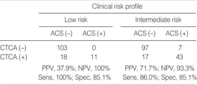
ACS, final diagnosis of acute coronary syndrome; PPV, positive predic- tive value; NPV, negative predictive value; Sens, Sensitivity; Spec, Speci- ficity.
Clinical risk profile Low risk
ACS (-) ACS (+)
Intermediate risk ACS (-) ACS (+)
CTCA (-) 103 0 97 7
CTCA (+) 18 11 17 43
PPV, 37.9%; NPV, 100% PPV, 71.7%; NPV, 93.3%
Sens, 100%; Spec, 85.1% Sens, 86.0%; Spec, 85.1%
Table 2. Diagnostic accuracy of CTCA according to clinical risk profile
ty), 60.7% (positive predictive value) and 96.6% (negative predictive value). However, according to the risk profile, the positive and negative predictive values were 37.9% and 100%
in the low risk profile group, while 71.7% and 93.3% in inter- mediate risk profile group (Table 2).
Safety of CTCA based approach to the patients with acute chest pain
Table 3 shows the result of clinical follow-up for each group.
None of the Group I patients had MACE during follow-up period. In some cases additional tests were performed during follow-up period, but no ACS case was found in this group.
Among Group II patients, seven (6.73%) were finally cate- gorized as ACS, although there was no case of myocardial infarction or mortality (Table 4). Six of the seven were iden- tified during the ED observation period and were admitted directly from ED. Among the six was a case of 65 yr old male who was diagnosed with microvascular angina. He had repro- ducible ischemic symptom and ECG change during a tread- mill test which was performed during his ED observation period. He was admitted and his invasive coronary angiog- raphy showed no significant stenosis. Although he had no definite lesion found, he was considered as one of the seven
CAG, Coronary angiography; PCI, Percutaneous coronary intervention;
CABG, Coronary artery bypass graft; ACS, Acute coronary syndrome.
Group I (n=103)
Group II (n=104)
Group III (n=89)
CAG (Positive/All tested) 0/13 5/21 43/56
Revascularization (PCI/CABG) 0 5 33
Myocardial infarction 0 0 10
Death 0 0 0
ACS as final diagnosis 0 7 54
Table 3. Result of clinical follow-up
*He had reproducible exertion-related dynamic ST-T change with chest pain without coronary lesion; �The Lesion was located at the distal part of the coronary artery.
LAD, Left anterior descending coronary artery; LCx, Left circumflex coronary artery; RCA, Right coronary artery; PCI, Percutaneous coronary intervention.
Reason for invasive angiography MDCT finding
Age (yr)
Sex Angiography
finding Treatment Final diagnosis
F 59 Motion artifact, Poor image quality, 3 VD STENT Unstable angina
almost non-calcified lesion Positive SPECT
F 61 Small calcified plaque, mid LAD, Dynamic ST-T change 1 VD No PCI Stable angina
Diagonal branch, RCA (LAD 50%)
F 52 Mid-LAD calcification Dynamic ST-T change 1 VD STENT Unstable angina
(LAD 90%)
M 65 No significant stenosis, Dynamic ST-T change, Normal* No PCI Microvascular angina
calcified plaque at proximal LAD Positive treadmill test
F 78 Calcified plaque Blooming effect, 1VD STENT Unstable angina
at proximal and mid LAD Positive Myocardial SPECT (LAD 80%)�
M 42 Multiple calcified stenosis Blooming effect, Unknown 1 VD STENT Unstable angina
significance of LCx. Lesion (LCx 75%)
F 73 Normal, multiple tiny calcified Recurrent symptom, 1 VD STENT Unstable angina
plaque at LAD Positive Myocardial SPECT (LCx 95%)� Table 4. Seven false negative cases of Group II
MDCT, Multi-detector computed tomography; Sens, Sensitivity; Spec, Specificity; PPV, Positive predictive value; NPV, Negative predictive value; ACS, Acute coronary syndrome.
Modality Sens Spec PPV NPV
No. of cases Reported
year Authors
(Ref. No.)
ACS prevalence
Inadequate images excluded
STENT evaluation
White et al. (11) 2005 69 16-MDCT 83% 96% 83% 96% 18.84% No No
Ghersin et al. (12) 2006 59 16-MDCT 80% 89% 52% 97% n/a Yes Yes
Hoffmann et al. (13) 2006 40 16/64-MDCT 100% 74% 38% 100% 12.50% No Yes
Hoffmann et al. (14) 2006 103 64-MDCT 100% 82% 47% 100% 14% No Yes
Olivetti et al. (15) 2006 31 MSCT 71.4% 99.6% 93.7% 97.7% 58.06% No No
Gallagher et al. (16) 2007 85 64-MDCT 86% 92% 50% 99% 8% Yes No
Rubinstein et al. (17) 2007 58 64-MDCT 92% 76% 52% 97% 34.48% No Yes
Table 5. Diagnostic accuracies of CTCA for patients with acute chest pain syndromes
false negative cases. The one missed ACS case was about a 73 yr old female who was discharged from ED after 24 hr observation period and had recurrent exertional chest pain at home. She was admitted and had coronary angiography (CAG) which showed 75% stenosis at the left circumflex artery. However there was no evidence that she suffered myo- cardial infarction. Neither myocardial infarction nor mortal- ity case was missed in Group II.
In Group III (n=89), 53 patients (59.6%) had coronary angiography of which 43 patients (48.3%) showed signifi- cant stenosis and 33 (37.1%) were treated with interventions including PCI or CABG. Of the 53 patients examined by coronary angiography, 10 (18.9%) patients were diagnosed as myocardial infarction. There was no mortality.
DISCUSSION
Previous non ED-based studies of 64-channel MDCT re- ported very high diagnostic accuracy of CTCA (8). Both sen- sitivity and specificity were about 95% and negative predic- tive value was about 99% (8). In two meta-analyses, 64-chan- nel MDCT showed sensitivity and specificity of 92% and 95%
respectively for segment analysis. For patient analysis, both sensitivity and specificity reached 100% (18, 19). These excel- lent results of CTCA diagnosis, however, may not be appli- cable in patients with acute chest pain in ED. In recent small studies, 64 channel MDCT showed rather low discrimina- tory power compared to the previous non-ED based studies (Table 5) (13, 14, 16, 17). And the American College of Car- diology Foundation guideline 2006 mentioned that the clini- cal application of CTCA for acute chest pain can only be con- sidered ‘‘appropriate’’ when its application is limited to pati- ents with intermediate pre-test probability without ECG and serial biomarker change (20).
Our intention in this study was testing the clinical appli- cability of CTCA in the real world ED situation. We had to exclude high risk patients from the study population because application of CTCA to diagnose or exclude an ACS for the population is considered to be neither safe nor efficient. And we also excluded patients whose diagnoses were overtly evi- dent at the beginning of clinical evaluation.
The remaining ‘low to intermediate risk’ patients group underwent rapid diagnostic evaluation by CTCA. Conven- tional stress tests require 4 to 8 hr of pre-test observation peri- od and impose cardiovascular loading to patients which can be potential danger to them occasionally. In contrast, we could obtain CTCA image right after the brief initial clinical eval- uation without causing the potentially undesirable cardio- vascular loading. And immediate discharge from ED was safe in low risk profile group which was also reported to be safe in one previous study (21). In terms of time and effort to eval- uate the patients with acute chest pain at ED, the CTCA- based algorithm we applied in this study can be considered
more efficient than conventional approaches based on the stress tests. And it was also safe at least for low risk patients. How- ever, there was seven false negative cases found in the inter- mediate risk group even though six out of the seven were suc- cessfully identified during observation period. It signifies the importance of clinical risk profiling. Until further technical advancement is to be made, judicious selection of patients and proper period of observation will remain important for most clinicians.
In terms of ‘cost-effectiveness’ there is possibility that CTCA may lead additional healthcare cost when used as a primary evaluation tool for acute chest pain. Otero and Rybicki eval- uated cost-effectiveness of CTCA, stress echocardiography and myocardial SPECT for 10,000 simulated patients using reported imaging test characteristics, prevalence and risk of coronary heart disease, and medicare reimbursement sched- ules (22). They reported that the clinical application of CTCA may significantly reduced the overall observation period, neg- ative CAG rate and total healthcare cost (22). However, inju- dicious use of CTCA can result in increased healthcare cost and develop undesirable health effect, such as radiation expo- sure, anaphylaxis and nephropathy that can also increase over- all cost.
This study has some limitations. This study was conduct- ed in a single, tertiary referral hospital. And more than 70%
of initially screened patients were excluded. The results of this study could be applicable only for the ‘‘CTCA eligible’’
patients with ‘‘low to intermediate clinical risk profile’’. It is impossible to compare the result of this study with that of previous studies involving other popular diagnostic meth- ods (e.g., myocardial SPECT, TMT), as this study is a single arm observational study. And the aspect of cost-effectiveness was not assessed in this study.
REFERENCES
1. Lau J, Ioannidis JP, Balk EM, Milch C, Terrin N, Chew PW, Salem D. Diagnosing acute cardiac ischemia in the emergency department:
a systematic review of the accuracy and clinical effect of current tech- nologies. Ann Emerg Med 2001; 37: 453-60.
2. Selker HP, Zalenski RJ, Antman EM, Aufderheide TP, Bernard SA, Bonow RO, Gibler WB, Hagen MD, Johnson P, Lau J, McNutt RA, Ornato J, Schwartz JS, Scott JD, Tunick PA, Weaver WD. An eval- uation of technologies for identifying acute cardiac ischemia in the emergency department: a report from a National Heart Attack Alert Program Working Group. Ann Emerg Med 1997; 29: 13-87.
3. Zalenski RJ, Selker HP, Cannon CP, Farin HM, Gibler WB, Gold- berg RJ, Lambrew CT, Ornato JP, Rydman RJ, Steele P. National Heart Attack Alert Program position paper: chest pain centers and programs for the evaluation of acute cardiac ischemia. Ann Emerg Med 2000; 35: 462-71.
4. Pope JH, Aufderheide TP, Ruthazer R, Woolard RH, Feldman JA, Beshansky JR, Griffith JL, Selker HP. Missed diagnoses of acute car-
diac ischemia in the emergency department. N Engl J Med 2000;
342: 1163-70.
5. Christenson J, Innes G, McKnight D, Boychuk B, Grafstein E, Thomp- son CR, Rosenberg F, Anis AH, Gin K, Tilley J, Wong H, Singer J.
Safety and efficiency of emergency department assessment of chest discomfort. Canadian Med Ass J 2004; 170: 1803-7.
6. Collinson PO, Premachandram S, Hashemi K. Prospective audit of incidence of prognostically important myocardial damage in patients discharged from emergency department. BMJ 2000; 320: 1702-5.
7. Vukmir RB. Medical malpractice: managing the risk. Med Law 2004;
23: 495-513.
8. Budoff MJ, Achenbach S, Blumenthal RS, Carr JJ, Goldin JG, Green- land P, Guerci AD, Lima JA, Rader DJ, Rubin GD, Shaw LJ, Wiegers SE; Intervention AHACoCIa, Intervention AHACoCRa, American Heart Association Committee on Cardiac Imaging CoCC. Assessment of coronary artery disease by cardiac computed tomography: a sci- entific statement from the American Heart Association Committee on Cardiovascular Imaging and Intervention, Council on Cardiovascu- lar Radiology and Intervention, and Committee on Cardiac Imaging, Council on Clinical Cardiology. Circulation 2006; 114: 1761-91.
9. Schussler JM, Smith ER. Sixty-four-slice computed tomographic coro- nary angiography: will the ‘‘triple rule out’’ change chest pain eval- uation in the ED? Am J Emerg Med 2007; 25: 367-75.
10. Gaspar T, Halon DA, Lewis BS, Adawi S, Schliamser JE, Rubinshtein R, Flugelman MY, Peled N. Diagnosis of coronary in-stent resteno- sis with multidetector row spiral computed tomography. J Am Coll Cardiol 2005; 46: 1573-9.
11. White CS, Kuo D, Kelemen M, Jain V, Musk A, Zaidi E, Read K, Sliker C, Prasad R. Chest pain evaluation in the emergency depart- ment: can MDCT provide a comprehensive evaluation? AJR Am J Roentgenol 2005; 185: 533-40.
12. Ghersin E, Litmanovich D, Dragu R, Rispler S, Lessick J, Ofer A, Brook OR, Gruberg L, Beyar R, Engel A. 16-MDCT coronary angiog- raphy versus invasive coronary angiography in acute chest pain syn- drome: a blinded prospective study. AJR Am J Roentgenol 2006; 186:
177-84.
13. Hoffmann U, Pena AJ, Moselewski F, Ferencik M, Abbara S, Cury RC, Chae CU, Nagurney JT. MDCT in early triage of patients with acute chest pain. AJR Am J Roentgenol 2006; 187: 1240-7.
14. Hoffmann U, Nagurney JT, Moselewski F, Pena A, Ferencik M, Chae CU, Cury RC, Butler J, Abbara S, Brown DF, Manini A, Nichols JH, Achenbach S, Brady TJ. Coronary multidetector computed tomog- raphy in the assessment of patients with acute chest pain. Circula- tion 2006; 114: 2251-60.
15. Olivetti L, Mazza G, Volpi D, Costa F, Ferrari O, Pirelli S. Multislice
CT in emergency room management of patients with chest pain and medium-low probability of acute coronary syndrome. Radiol Med 2006; 111: 1054-63.
16. Gallagher MJ, Ross MA, Raff GL, Goldstein JA, O’Neill WW, O’Neil B. The diagnostic accuracy of 64-slice computed tomography coro- nary angiography compared with stress nuclear imaging in emergen- cy department low-risk chest pain patients. Ann Emerg Med 2007;
49: 125-36.
17. Rubinshtein R, Halon DA, Gaspar T, Jaffe R, Karkabi B, Flugelman MY, Kogan A, Shapira R, Peled N, Lewis BS. Usefulness of 64-slice cardiac computed tomographic angiography for diagnosing acute coronary syndromes and predicting clinical outcome in emergency department patients with chest pain of uncertain origin. Circulation 2007; 115: 1762-8.
18. Schuijf JD, Bax JJ, Shaw LJ, de Roos A, Lamb HJ, van der Wall EE, Wijns W. Meta-analysis of comparative diagnostic performance of magnetic resonance imaging and multislice computed tomography for noninvasive coronary angiography. Am Heart J 2006; 151: 404-11.
19. Stein PD, Beemath A, Kayali F, Skaf E, Sanchez J, Olson RE. Mul- tidetector computed tomography for the diagnosis of coronary artery disease: a systematic review. Am J Med 2006; 119: 203-16.
20. Hendel RC, Patel MR, Kramer CM, Poon M, Hendel RC, Carr JC, Gerstad NA, Gillam LD, Hodgson JM, Kim RJ, Kramer CM, Lesser JR, Martin ET, Messer JV, Redberg RF, Rubin GD, Rumsfeld JS, Taylor AJ, Weigold WG, Woodard PK, Brindis RG, Hendel RC, Douglas PS, Peterson ED, Wolk MJ, Allen JM, Patel MR; Group ACoCFQSDCACW, Radiology ACo, Tomography SoCC, Reso- nance SfCM, Cardiology ASoN, Imaging NASfC, Interventions SfCAa, Radiology SoI. ACCF/ACR/SCCT/SCMR/ASNC/NASCI/
SCAI/SIR 2006 appropriateness criteria for cardiac computed tomog- raphy and cardiac magnetic resonance imaging: a report of the Ameri- can College of Cardiology Foundation Quality Strategic Directions Committee Appropriateness Criteria Working Group, American Col- lege of Radiology, Society of Cardiovascular Computed Tomogra- phy, Society for Cardiovascular Magnetic Resonance, American Soci- ety of Nuclear Cardiology, North American Society for Cardiac Imag- ing, Society for Cardiovascular Angiography and Interventions, and Society of Interventional Radiology. J Am Coll Cardiol 2006; 48:
1475-97.
21. Hollander JE, Litt HI, Chase M, Brown AM, Kim W, Baxt WG. Com- puted tomography coronary angiography for rapid disposition of low-risk emergency department patients with chest pain syndromes.
Acad Emerg Med 2007; 14: 112-6.
22. Otero HJ, Rybicki FJ. Reimbursement for chest-pain CT: estimates based on current imaging strategies. Emerg Radiol 2007; 13: 237-42.