A Prediction Rule to Identify Severe Cases among Adult Patients Hospitalized with Pandemic Influenza A (H1N1) 2009
The purpose of this study was to establish a prediction rule for severe illness in adult patients hospitalized with pandemic influenza A (H1N1) 2009. At the time of initial presentation, the baseline characteristics of those with severe illness (i.e., admission to intensive care unit, mechanical ventilation, or death) were compared to those of patients with non-severe illnesses. A total of 709 adults hospitalized with pandemic influenza A (H1N1) 2009 were included: 75 severe and 634 non-severe cases. The multivariate analysis demonstrated that altered mental status, hypoxia (PaO2/FiO2
≤ 250), bilateral lung infiltration, and old age (≥ 65 yr) were independent risk factors for severe cases (all P < 0.001). The area under the ROC curve (0.834 [95% CI, 0.778–
0.890]) of the number of risk factors were not significantly different with that of APACHE II score (0.840 [95% CI, 0.790-0.891]) (P = 0.496). The presence of ≥ 2 risk factors had a higher sensitivity, specificity, positive predictive value and negative predictive value than an APACHE II score of ≥ 13. As a prediction rule, the presence of ≥ 2 these risk factors is a powerful and easy-to-use predictor of the severity in adult patients hospitalized with pandemic influenza A (H1N1) 2009.
Key Words: pandemic influenza A (H1N1) 2009; Severity; Risk Factors; Prediction Rule
Won Sup Oh1,*, Seung-Joon Lee1,*,
Chang-Seop Lee2, Ji-An Hur3, Ae-Chung Hur4, Yoon Seon Park4, Sang-Taek Heo5, In-Gyu Bae5, Sang Won Park6, Eu Suk Kim7, Hong Bin Kim8, Kyoung-Ho Song8, Kkot Sil Lee9, Sang-Rok Lee10, Joon Sup Yeom11, Su Jin Lee12, Baek-Nam Kim13, Yee Gyung Kwak14, Jae Hoon Lee15,
Yong Keun Kim16, Hyo Youl Kim16, Nam Joong Kim17, and Myoung-Don Oh17
1Department of Internal Medicine, Kangwon National University School of Medicine, Chucheon; 2Department of Internal Medicine, Chonbuk National University Medical School, Jeonju; 3Department of Internal Medicine, Yeungnam University College of Medicine, Daegu; 4Department of Internal Medicine, National Health Insurance Corporation Ilsan Hospital, Goyang; 5Department of Internal Medicine, Gyeongsang National University School of Medicine, Jinju;
6Department of Internal Medicine, Boramae Medical Center, Seoul National University College of Medicine, Seoul;
7Department of Internal Medicine, Dongguk University Ilsan Hospital, Dongguk University College of Medicine, Goyang;
8Department of Internal Medicine, Seoul National University Bundang Hospital, Seoul National University College of Medicine, Seongnam; 9Department of Internal Medicine, Kwandong University Myongji Hospital, Kwandong University College of Medicine, Goyang; 10Department of Internal Medicine, Cheongju St. Mary’s Hospital, Cheongju;
11Department of Internal Medicine, Kangbuk Samsung Hospital, Sungkyunkwan University School of Medicine, Seoul; 12Department of Internal Medicine, Pusan National University Yangsan Hospital, Pusan National University College of Medicine, Yangsan; 13Department of Internal Medicine, Inje University Sanggye-Paik Hospital, Inje University College of Medicine, Seoul; 14Department of Internal Medicine, Inje University Ilsan-Paik Hospital, Inje University College of Medicine, Goyang; 15Department of Internal Medicine, Wonkwang University College of Medicine, Iksan; 16Department of Internal Medicine, Wonju Christian Hospital, Yonsei University Wonju College of Medicine, Wonju; 17Department of Internal Medicine, Seoul National University Hospital, Seoul National University College of Medicine, Seoul, Korea
*Won Sup Oh and Seung-Joon Lee contributed equally to this work.
Received: 3 December 2010 Accepted: 17 February 2011 Address for Correspondence:
Myoung-Don Oh, MD
Department of Internal Medicine, Seoul National University College of Medicine, 101 Daehak-ro, Jongno-gu, Seoul 110-744, Korea Tel: +82.2-2072-2945, Fax: +82.2-765-6354
E-mail: [email protected]
This study was supported by a 2010 Research Grant from Kangwon National University (Grant number: C1007258-01-01 (120100335)).
DOI: 10.3346/jkms.2011.26.4.499 • J Korean Med Sci 2011; 26: 499-506
INTRODUCTION
While most pandemic influenza A (H1N1) 2009 infections were mild or subclinical, early reports suggested that clinical courses of pandemic influenza A (H1N1) 2009 were somewhat different from that of seasonal influenza (1, 2). Although individuals with comorbid conditions were at high risk, small subsets of previ- ously healthy people developed rapidly progressive disease. In severe cases, patients generally began to deteriorate 3-5 days after the onset of symptom, with rapid progression to respirato- ry failure within 24 hr. Most of these required immediate life sup- port with mechanical ventilation, renal replacement therapy, or extracorporeal membrane oxygenation.
Although most studies have evaluated risk factors for mortal- ity (3-5), a few have focused on those associated with severe pan- demic influenza A (H1N1) 2009 (6, 7). From the viewpoint of clinical practice, knowledge on risk factors for the severity in- cluding the mortality and the prediction of severe cases is more crucial for decisions regarding hospitalization, treatment, or in- tensive care of these patients. Therefore, we evaluated the base- line characteristics of adult patients hospitalized with pandem- ic influenza A (H1N1) 2009 to identify risk factors associated with severity. Eventually, a rule comprising these risk factors was es- tablished to predict the severity of illness in adult patients hos- pitalized with pandemic influenza A (H1N1) 2009.
MATERIALS AND METHODS Patients
This study was conducted at 17 teaching hospitals in Korea:
Kangwon National University Hospital, Chuncheon; Gyeong- sang National University Hospital, Jinju; Kwandong University Myongji Hospital, Goyang; Dongguk University Ilsan Hospital, Goyang; Boramae Medical Center, Seoul; Seoul National Uni- versity Bundang Hospital, Seongnam; Seoul National Universi- ty Hospital, Seoul; Pusan National University Yangsan Hospital, Yangsan; Wonju Christian Hospital, Wonju; Yeungnam Univer- sity Medical Center, Daegu; Wonkwang University Hospital, Ik- san; Inje University Sanggye-Paik Hospital, Seoul; Inje Univer- sity Ilsan-Paik Hospital, Goyang; National Health Insurance Cor- poration Ilsan Hospital, Goyang; Chunbuk National University Hospital, Jeonju; Cheongju St. Mary’s Hospital, Cheongju; and Kangbuk Samsung Hospital, Seoul.
All adult patients hospitalized with the laboratory-confirmed, pandemic influenza A (H1N1) 2009 from September 1, 2009 to February 28, 2010 were included in the study. Laboratory-con- firmed cases were defined as the presence of influenza-like ill- ness with pandemic influenza A (H1N1) virus infection con- firmed by real-time or multiplex reverse-transcriptase polymerase chain reaction assays. Patients younger than 18 yr were exclud- ed.
Data collection and definitions
Demographic, clinical, laboratory and radiographic data were collected from all patients. Obesity was defined as a body mass index (BMI) of more than 25 kg/m2. Comorbid conditions in- cluded chronic lung diseases (chronic obstructive pulmonary disease, asthma, pneumoconiosis, or bronchopulmonary dys- plasia), cardiovascular diseases (congestive heart failure, isch- emic heart disease, or cyanotic congenital heart disease), cere- brovascular diseases (stroke or cerebral hemorrhage), malig- nancy, immunosuppression (HIV infection, asplenia or hy- posplenia, transplantation, anticancer chemotherapy, cortico- steroid, or other immunosuppressants), diabetes mellitus, chron- ic renal diseases (nephrotic syndrome or chronic renal failure), chronic liver diseases (liver cirrhosis or chronic active hepati- tis), and neurocognitive diseases (mental retardation, demen- tia, or seizure).
To identify risk factor(s) associated with severe pandemic in- fluenza A (H1N1) 2009 at the time of initial presentation, we compared the baseline characteristics of patients with severe illness to those of patients with non-severe illness. Severe cases were defined as those who had been admitted to intensive care unit (ICU), mechanically ventilated, or died of influenza itself or related complications; other cases were considered to have a non-severe illness. Clinical or laboratory parameters derived from Pneumonia Severity Index (PSI) for community-acquired pneumonia or the diagnostic criteria for sepsis or severe sepsis were used in part to evaluate the baseline characteristics of pa- tients (8, 9). The presence of the following complications due to infection was also determined: pneumonia (defined as new or progressive infiltrate(s) on chest radiography), acute respiratory distress syndrome (PaO2/FiO2 ≤ 200 in the presence of bilateral alveolar infiltrates in chest radiography), septic shock (systolic blood pressure < 90 mm Hg despite adequate fluid resuscita- tion), acute renal failure (serum creatinine ≥ 2.0 mg/dL without previous renal disease), rhabdomyolysis (profound muscle weak- ness and tenderness, brown-colored urine, and CPK ≥ 5 times the upper normal limit), and exacerbation of underlying dis- eases (worsening of the patient’s condition such that additional treatment is required).
Statistical analysis
A Pearson’s chi-square or Fisher’s exact test was used for cate- gorical variables, and the Student’s t-test was used for continu- ous variables. A multivariate logistic regression analysis using baseline characteristics seen only at the time of initial presenta- tion was performed to identify risk factors associated with se- vere pandemic influenza A (H1N1). A level of significance of less than 0.10 was required for inclusion and greater than 0.05 meant exclusion. The goodness-of-fit for this regression model was verified by the Hosmer-Lemeshow test (10).
As a prediction rule for severe pandemic influenza A (H1N1),
both the number of risk factors, derived from the logistic regres- sion model, and the Acute Physiology and Chronic Health Eval- uation (APACHE) II score were calculated for each patient. To validate their discriminatory power, the sensitivity and specific- ity of the risk factors were compared to those of the APACHE II score by means of the receiver operating characteristic (ROC) curves (11). We also determined their cut-points defining severe pandemic influenza A (H1N1) to maximize the sum of sensitiv- ity and specificity.
A two-tailed P < 0.05 was considered to be statistically signifi- cant for all analyses. IBM SPSS Statistics 18 (SPSS Inc., Chicago, IL, USA) and dBSTAT for Windows version 5.0 (dBSTAT, Seoul, Korea) were used.
Ethics statement
This study protocol was approved by the institutional review board (IRB) of Kangwon National University Hospital (IRB ap- proval number: 09-22). Informed consent was waived in this study by the board.
RESULTS
A total of 709 adult patients hospitalized with the laboratory- confirmed, pandemic influenza A (H1N1) 2009 were included in the study. There were 280 (39.5%) males and 429 (60.5%) fe- males, and the median age was 50 yr (interquartile range [IQR], 31-66 yr). A total of 75 (10.6%) patients had severe illness: 23 were admitted to ICU but not ventilated, 35 were mechanically ventilated in the ICU, 5 were mechanically ventilated in emer- gency departments or general wards, and 12 died without ei- ther admission to ICU or mechanical ventilation.
Baseline characteristics of severe pandemic influenza A (H1N1)
The demographic, clinical, laboratory and radiographic findings of patients at the time of initial presentation are summarized in Table 1. The median age of severe cases was higher than that of non-severe cases (65 yr [IQR, 50-75 yr] vs 49 yr [IQR, 30-63 yr];
P < 0.001). The proportions of males and nursing home residents were also higher in severe cases (P = 0.01 and 0.002, respective- ly). Of the total, 367 (52.1%) had one or more comorbidities: 62 (82.7%) severe cases vs 305 (48.1%) non-severe cases (P < 0.001).
Chronic lung disease (21.7%) was the most common comorbid- ity, followed by diabetes mellitus (13.6%), cardiovascular dis- ease (9.4%), malignancy (9.4%), immunosuppression (8.4%), chronic liver disease (5.5%), and chronic renal diseases (5.0%).
Of these, chronic lung disease, cardiovascular disease, and im- munosuppression were more frequently observed in severe cases than in non-severe cases (P < 0.001, < 0.001, and 0.011, respectively). However, there were no significant differences in the proportions of pregnancy and obesity between severe and
non-severe cases (P = 0.11 and 0.499, respectively).
While fever was the most common symptom (78.3%) at ini- tial presentation, it was less frequent in severe than in non-se- vere cases (65.3% vs 79.8%; P = 0.004). On the contrary, dyspnea, purulent sputum, altered mental status, and cyanosis were more frequent in severe than in non-severe cases (P < 0.001, 0.018,
< 0.001, and < 0.001, respectively). However, the median interval from symptom onset to initial presentation in severe cases was not significantly different than that in non-severe cases (2 days [IQR, 1-4 days] vs 2 days [IQR, 1-4 days]; P = 0.619). With respect to initial laboratory findings, anemia (hematocrit < 30%), azote- mia (BUN ≥ 30 mg/dL), hyperglycemia (serum glucose ≥ 250 mg/dL), hyponatremia (serum sodium < 130 mEq/L), acidosis
Table 1. Baseline characteristics of adults hospitalized with pandemic influenza A (H1N1) 2009 (N = 709)
Baseline characteristics
No. (%) of pandemic influenza A (H1N1)
P value Severe
cases (n = 75)
Non-severe cases (n = 634) Demography
Age ≥ 65 yr Male
Nursing home resident Pregnancy
Obesity (body mass index ≥ 25 kg/m2)
38 (50.7) 40 (53.3) 7 (9.3)
0 15 (20.0)
150 (23.7) 240 (37.9) 16 (2.5) 21 (3.3) 36 (5.8)
< 0.001 0.010 0.002 0.110 0.499 Comorbid conditions
Chronic lung disease Cardiovascular disease Malignancy Immunosuppression Diabetes mellitus Chronic liver disease Chronic renal disease
31 (41.3) 17 (22.7) 10 (13.3) 12 (16.0) 10 (13.3) 3 (4.0) 4 (5.3)
122 (19.2) 49 (7.7) 56 (8.8) 47 (7.4) 86 (13.6) 36 (5.7) 31 (4.9)
< 0.001
< 0.001 0.205 0.011 0.956 0.547 0.867 Symptoms
Fever (oral temperature ≥ 37.8°C) Duration of fever > 48 hr Cough
Purulent sputum Dyspnea Sore throat Rhinorrhea
49 (65.3) 19 (25.3) 49 (66.2) 42 (56.0) 51 (68.0) 11 (14.9) 14 (18.9)
506 (79.8) 198 (19.0) 473 (74.6) 264 (41.6) 178 (28.1) 216 (34.1) 175 (27.6)
0.004 0.295 0.085 0.018
< 0.001 0.001 0.098 Signs
Systolic blood pressure < 90 mmHg Heart rate ≥ 125 beats/min Respiratory rate ≥ 30 breaths/min Temperature < 35°C or ≥ 40°C Altered mental status Cyanosis
11 (14.7) 11 (14.7) 7 (9.3) 2 (2.7) 23 (30.7)
9 (12.0)
8 (1.3) 25 (3.9) 15 (2.4) 3 (0.5) 17 (2.7) 5 (0.8)
< 0.001
< 0.001 0.001 0.032
< 0.001
< 0.001 Laboratory findings
WBC < 4,000 cell/µL Hematocrit < 30%
BUN ≥ 30 mg/dL Serum glucose ≥ 250 mg/dL Serum sodium < 130 mEq/L Arterial pH < 7.35 PaO2/FiO2 ≤ 250
11 (14.7) 20 (26.7) 20 (26.7) 9 (12.0) 13 (17.3) 22 (29.3) 34 (45.3)
91 (14.4) 72 (11.4) 48 (7.6) 22 (3.5) 30 (4.7) 18 (2.8) 45 (7.1)
0.942
< 0.001
< 0.001 0.001
< 0.001
< 0.001
< 0.001 Radiographic findings
Presence of pneumonic infiltration Bilateral lung infiltration
53 (70.7) 46 (61.3)
209 (33.0) 135 (21.3)
< 0.001
< 0.001
(arterial pH < 7.35), and hypoxia (PaO2/FiO2 ≤ 250) were more frequent in severe than in non-severe cases (P < 0.001, < 0.001, 0.001, < 0.001, and < 0.001, respectively). Of all patients, 262 (37.0%) had pneumonia at the time of initial presentation: 53 (70.7%) severe vs 209 (33.0%) non-severe cases (P < 0.001). Both lungs were more frequently involved in severe cases than in non- severe cases (61.3% vs 21.3%; P < 0.001).
Clinical course of severe pandemic influenza A (H1N1) The clinical courses of patients with pandemic influenza A (H1N1) are outlined in Table 2. Of the total, 692 (97.6%) patients were treated with one or more antiviral agents: 670 (94.5%) with oseltamivir, 19 (2.7%) with zanamivir, 1 (0.1%) with peramivir, and 2 (0.3%) with a combination of oseltamivir, amantadine, and ribavirin. There was no significant difference in antiviral regimens between severe and non-severe cases (P = 0.362). The median interval from symptom onset to initiation of antiviral therapy in the 692 treated cases was 2 days (IQR, 1-3 days); there was no significant difference between severe and non-severe
cases (1 day [IQR, 1-3 days] vs 2 days [IQR, 1-3 days]; P = 0.734).
However, the median duration of fever after initiation of antivi- ral therapy was longer in severe than in non-severe cases (2 days [IQR, 1-5 days] vs 1 day [IQR, 1-2 days]; P = 0.001). Antibacterial agent(s), vasopressor(s), corticosteroid(s), and supplementary oxygen therapy were more frequently given to severe than non- severe cases (all P < 0.001). The median length of hospital stay in all patients was 5 days (IQR, 3-9 days): 11 days (IQR, 7-25 days) in severe vs 5 days (IQR, 3-8 days) in non-severe cases (P < 0.001).
Influenza-related complications were observed in 110 (15.5%) patients: 54 (72.0%) severe vs 56 (8.8%) non-severe cases (P <
0.001). Pneumonia (37.0%) was the most common complica- tion, followed by exacerbation of underlying lung disease (8.3%), acute respiratory distress syndrome (3.9%), acute renal failure (2.4%), septic shock (1.8%), and exacerbation of underlying heart disease (1.8%).
A total of 37 severe cases died during hospitalization (in-hos- pital case-fatality rate, 5.2%), and nine (24.3%) of these died with- in 3 days of initial presentation. The median age of fatal cases was 68 yr (IQR 59-79 yr), which was higher than that of non-fa- tal cases (49 yr, [IQR 30-64 yr]; P < 0.001). Of the 37 fatalities, only three (8.1%) were under 65 yr of age and none had underlying comorbidities. There was no significant difference in the medi- an interval from symptom onset to initiation of antiviral agent(s) between fatal and non-fatal cases (1 day, IQR 0-4 days) vs 2 days (IQR 1-3 days; P = 0.918). Either influenza itself or pneumonia (40.5%) was the most common cause of in-hospital death, fol- lowed by exacerbation of underlying lung disease (13.5%), ex- acerbation of underlying heart disease (10.8%), exacerbation of other underlying disease (18.9%), nosocomial infection (5.4%), and unknown causes (10.8%).
Table 2. Clinical course of adults hospitalized with pandemic influenza A (H1N1) 2009 (N = 709)
Clinical course
No. (%) of pandemic influenza A (H1N1)
P value Severe
cases (n = 75)
Non-severe cases (n = 634)
Use of antiviral(s) 71 (94.7) 621 (97.9) 0.079
Interval from symptom onset to antiviral(s) > 2 days
15 (20.0) 165 (26.0) 0.257 Duration of fever after antiviral(s)
> 3 days
26 (34.7) 75 (11.8) < 0.001 Use of antibacterial(s) 69 (92.0) 397 (62.6) < 0.001 Use of vasopressor(s) 38 (50.7) 4 (0.6) < 0.001 Use of corticosteroid(s) 26 (34.7) 45 (7.1) < 0.001 Use of supplemental oxygen 35 (46.7) 64 (10.1) < 0.001 Length of hospital stay > 14 days 27 (36.0) 79 (12.5) < 0.001 Complications
Septic shock 13 (17.3) 0 < 0.001
Acute renal failure 12 (16.0) 5 (0.8) < 0.001
Rhabdomyolysis 3 (4.0) 1 (0.2) < 0.001
Exacerbation of underlying lung disease 23 (30.7) 36 (5.7) < 0.001 Exacerbation of underlying heart disease 4 (5.3) 9 (1.4) 0.017
Table 3. Multivariate analysis of risk factors associated with severity at initial pre- sentation (N = 709)
Variables Adjusted OR (95% CI) P value
Altered mental status 14.918 (6.716-33.139) < 0.001 Hypoxia (PaO2/FiO2 ≤ 250) 7.094 (3.750-13.420) < 0.001 Bilateral lung infitration 3.459 (1.909-6.268) < 0.001
Old age (≥ 65 yr) 3.082 (1.718-5.528) < 0.001
OR, odds ratio; CI, confidence interval.
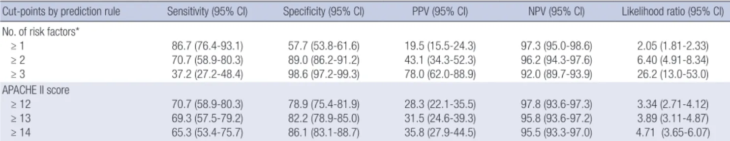
Table 4. Performance of number of risk factors and APACHE II score in predicting severe pandemic influenza A (H1N1) 2009
Cut-points by prediction rule Sensitivity (95% CI) Specificity (95% CI) PPV (95% CI) NPV (95% CI) Likelihood ratio (95% CI) No. of risk factors*
≥ 1 ≥ 2 ≥ 3
86.7 (76.4-93.1) 70.7 (58.9-80.3) 37.2 (27.2-48.4)
57.7 (53.8-61.6) 89.0 (86.2-91.2) 98.6 (97.2-99.3)
19.5 (15.5-24.3) 43.1 (34.3-52.3) 78.0 (62.0-88.9)
97.3 (95.0-98.6) 96.2 (94.3-97.6) 92.0 (89.7-93.9)
2.05 (1.81-2.33) 6.40 (4.91-8.34) 26.2 (13.0-53.0) APACHE II score
≥ 12 ≥ 13 ≥ 14
70.7 (58.9-80.3) 69.3 (57.5-79.2) 65.3 (53.4-75.7)
78.9 (75.4-81.9) 82.2 (78.9-85.0) 86.1 (83.1-88.7)
28.3 (22.1-35.5) 31.5 (24.6-39.3) 35.8 (27.9-44.5)
97.8 (93.6-97.3) 95.8 (93.6-97.2) 95.5 (93.3-97.0)
3.34 (2.71-4.12) 3.89 (3.11-4.87) 4.71 (3.65-6.07)
*Risk factors included altered mental status, hypoxia (PaO2/FiO2 ≤ 250), bilateral lung infiltration, and old age (≥ 65 yr). PPV, positive predictive value; NPV, negative predictive value; CI, confidence interval.
Risk factors and the prediction of severe pandemic influenza A (H1N1) 2009
In the multivariate logistic regression model using baseline char- acteristics seen only at the time of initial presentation, altered mental status, hypoxia (PaO2/FiO2 ≤ 250), bilateral lung infiltra- tion, and old age (≥ 65 yr) were independent risk factors associ- ated with severe pandemic influenza A (H1N1) (all P < 0.001;
Table 3). The Hosmer-Lemeshow test did not show statistical significance (P = 0.88), indicating the goodness of fit of this lo- gistic regression model.
The median number of risk factors in severe cases was higher than that in non-severe cases (2, IQR 1-3, vs 0, IQR 0-1) (P < 0.001).
The median APACHE II score was also higher in severe cases than in non-severe cases (17, IQR 10-22, vs 6, 3-11) (P < 0.001).
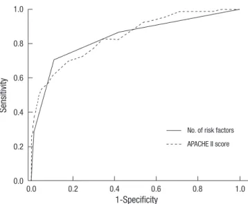
Both the number of risk factors and APACHE II scores were sig- nificantly correlated with patient age (Pearson’s correlation co- efficients > 0.5; all P < 0.001). The ROC curve for the number of risk factors, which were derived from the logistic regression mod- el, was compared to that for the APACHE II scores (Fig. 1). The areas under the ROC curves were 0.834 (95% confidence inter- val [CI], 0.778-0.890) for the number of risk factors and 0.840 (95% CI, 0.790-0.891) for APACHE II score, but there was no sig- nificant difference between both areas under the ROC curves (P
= 0.496). When the cut-points were determined to maximize the sum of the sensitivity and specificity by means of the ROC curves, they were 2 for the number of risk factors and 13 for the APACHE II score (Table 4). As a prediction rule for severe pan- demic influenza A (H1N1) 2009, the sensitivity, specificity, pos- itive predictive value (PPV), and negative predictive value (NPV) of the number of risk factors ≥ 2 were all higher than those of the APACHE II score ≥ 13.
DISCUSSION
During an influenza A (H1N1) 2009 pandemic, the determina- tion of the severity of illness played an important role in early detection and proper management of severe cases and, eventu- ally, improvement of clinical outcome. Although many studies about risk factors for death in patients with pandemic influenza A (H1N1) 2009 has been published, no prediction rule for the severe case has yet been established. In this study, the number of risk factors (i.e., altered mental status, hypoxia, bilateral lung infiltration, and old age) as a prediction rule seems to be ade- quate for the detection of severe cases among adult patients hospitalized with pandemic influenza A (H1N1) 2009. While APACHE II has been widely used to measure the severity of ill- ness in critically ill patients, its application to all patients with pandemic influenza A (H1N1) can be limited in clinical prac- tice because of the complexity of its parameters. When the cut- points were determined to maximize the sum of the sensitivity and specificity, the area under the ROC curve of the number of risk factors was comparable with that of the APACHE II score.
More precisely, the presence of ≥ 2 risk factors had a higher sen- sitivity, specificity, PPV, and NPV than an APACHE II score ≥ 13.
Therefore, we think that the presence of ≥ 2 risk factors is a more useful predictor for severe pandemic influenza A (H1N1) 2009, especially during a pandemic when the burden of care for the patients is too heavy for the physicians.
This study also highlights several clinical features observed in adult patients hospitalized with severe pandemic influenza A (H1N1) 2009 infection. Early case reports indicated that the in- fection in previously healthy young individuals might often be associated with serious complications or death (4, 12). Most of the serious illnesses occurred among children and young adults, and approximately 90% of deaths were observed in those < 65 yr of age. However, the overall case fatality rate among hospital- ized patients appeared to be highest among those ≥ 50 yr of age.
In this study, which included only adult patients hospitalized with pandemic influenza A (H1N1) 2009, there was a tendency for the severity of illness to increase with patient age. Most deaths occurred in patients who were ≥ 65 yr of age or had one or more comorbidities, but the only death attributed directly to influen- za occurred in a previously healthy young individual. Although determination of the true case-fatality rate of pandemic influ- enza A (H1N1) 2009 is particularly challenging, it is thought to be less than 0.5%, with a broad range of estimates (13, 14). In a surveillance data (15), a total of 740,835 patients were reported to be infected with pandemic influenza A (H1N1) 2009 and 225 of them were reported to have died during 2009-2010 influenza season. The incidence was calculated as 1,493 per 100,000 pop- ulation and the case fatality rate was 30 per 100,000 cases. In an early report (16) that included 272 patients hospitalized with pandemic influenza A (H1N1) 2009, the in-hospital case-fatali- Fig. 1. Receiver operating characteristic (ROC) curves for the number of risk factors
and APACHE II score. The areas under the ROC curves are 0.834 (95% confidence interval [CI], 0.778-0.890) for the number of risk factors and 0.840 (95% CI, 0.790- 0.891) for the APACHE II score (P = 0.496 for each pairwise comparison).
Sensitivity
1-Specificity
0.0 0.2 0.4 0.6 0.8 1.0
1.0
0.8
0.6
0.4
0.2
0.0
No. of risk factors APACHE II score
ty rate was -7%; it was -5% in the present study.
Previous studies have suggested that pregnant women are at increased risk for complication and death from pandemic in- fluenza A (H1N1) infection, and that this risk is highest in the third trimester (17-19). In a cohort study (20), however, there was no death among 211 pregnant women, suggesting that the prevention of disease progression with early treatment might account for the cohort of mild cases. In the present study, preg- nancy itself was not significantly associated with complications (spontaneous abortion, preterm labor, or fetal distress), the se- verity of illness, or death. This may be attributable to the small number of pregnant women as well as the early treatment with antiviral agent(s) in this study.
Several studies have suggested that fever is one of the most common symptoms (> 80%) at initial presentation (12, 21). In this study, approximately 80% of all hospitalized patients pre- sented initially with fever; however, more than one-third of pa- tients with severe illness had no fever at initial presentation, re- gardless of use of antipyretic agent(s). This finding suggests that pandemic influenza A (H1N1) 2009 should not be excluded in patients with severe influenza-like illness, even if they have nei- ther fever nor a history of fever. Furthermore, a confirmatory test for pandemic influenza A (H1N1) 2009 may be necessary in such patients.
In recent studies (6, 7, 21), the presence of one or more co- morbidities was found to be associated with both admission to ICU and death in patients with pandemic influenza A (H1N1) 2009. Comorbidities associated with complications of seasonal influenza are also risk factors for complications related to this virus. Although chronic lung disease, cardiovascular disease, and immunosuppression were more frequently detected in se- vere than in non-severe cases in the bivariate analysis of this study, they were not found to be independent risk factors for severe illness in the multivariate logistic regression analysis. In- stead, the multivariate logistic regression analysis demonstrat- ed that altered mental status, hypoxia, bilateral lung infiltration, and old age were independent risk factors for severe illness in adult patients hospitalized with pandemic influenza A (H1N1) 2009.
The use of neuraminidase inhibitor within 48 hr of symptom onset may reduce the risk of progression to severe illness or death (16, 21, 22). In an early report (23), the use of neuraminidase in- hibitor even 48 hr after symptom onset was associated with re- duced rates of death. In this study, however, more than 70% of infected patients received neuraminidase inhibitor within 48 hr after symptom onset, so that their clinical outcome was not re- lated with the timing of antiviral administration.
The strengths of this study include it being a nationwide mul- ticenter study with a large number of laboratory-confirmed cas- es, relatively little missing data, careful control of confounding factors in the analyses, and the establishment of a prediction
rule for severe pandemic influenza A (H1N1) 2009 with large sample size (e.g., the number of events per each variable ≥ 40).
This study has several limitations. Because only adult patients hospitalized with pandemic influenza A (H1N1) 2009 were in- cluded in this study, the derived prediction rule cannot be ap- plied to estimate the severity of illness in children or adult out- patients. In addition, final outcome was measured as in-hospi- tal mortality because many of the patients were lost to follow-up.
Finally, a prediction rule derived from this study need be exter- nally validated in other larger-scaled studies for testing accura- cy and generalizability and studying the clinical impact of a rule on physician’s behavior and patient’s outcome (24).
In summary, although clinical features of adult patients hos- pitalized with pandemic influenza A (H1N1) 2009 can be diverse, altered mental status, hypoxia, bilateral lung infiltration, and old age are independent risk factors for severe illness. As a pre- diction rule, furthermore, the presence of ≥ 2 of these risk fac- tors can be used to determine the likely severity of illness.
REFERENCES
1. Novel Swine-Origin Influenza A (H1N1) Virus Investigation Team, Da- wood FS, Jain S, Finelli L, Shaw MW, Lindstrom S, Garten RJ, Gubareva LV, Xu X, Bridges CB, Uyeki TM. Emergence of a novel swine-origin in- fluenza A (H1N1) virus in humans. N Engl J Med 2009; 360: 2605-15.
2. World Health Organization. Clinical features of severe cases of pandemic influenza. Available at http://www.who.int/csr/disease/swineflu/notes/
h1n1_clinical_features_20091016/en/print.html. [accessed on 10 Mar 2010].
3. ANZIC Influenza Investigators, Webb SA, Pettilä V, Seppelt I, Bellomo R, Bailey M, Cooper DJ, Cretikos M, Davies AR, Finfer S, Harrigan PW, Hart GK, Howe B, Iredell JR, McArthur C, Mitchell I, Morrison S, Nichol AD, Paterson DL, Peake S, Richards B, Stephens D, Turner A, Yung M. Criti- cal care services and 2009 H1N1 influenza in Australia and New Zealand.
N Engl J Med 2009; 361: 1925-34.
4. Kumar A, Zarychanski R, Pinto R, Cook DJ, Marshall J, Lacroix J, Stelfox T, Bagshaw S, Choong K, Lamontagne F, Turgeon AF, Lapinsky S, Ahern SP, Smith O, Siddiqui F, Jouvet P, Khwaja K, McIntyre L, Menon K, Hutchi- son J, Hornstein D, Joffe A, Lauzier F, Singh J, Karachi T, Wiebe K, Olaf- son K, Ramsey C, Sharma S, Dodek P, Meade M, Hall R, Fowler RA; Ca- nadian Critical Care Trials Group H1N1 Collaborative. Critically ill pa- tients with 2009 influenza A (H1N1) infection in Canada. JAMA 2009;
302: 1872-9.
5. Echevarría-Zuno S, Mejía-Aranguré JM, Mar-Obeso AJ, Grajales-Muñiz C, Robles-Pérez E, González-León M, Ortega-Alvarez MC, Gonzalez- Bonilla C, Rascón-Pacheco RA, Borja-Aburto VH. Infection and death from influenza A H1N1 virus in Mexico: a retrospective analysis. Lancet 2009; 374: 2072-9.
6. Campbell A, Rodin R, Kropp R, Mao Y, Hong Z, Vachon J, Spika J, Pelle- tier L. Risk of severe outcomes among patients admitted to hospital with pandemic (H1N1) influenza. CMAJ 2010; 182: 349-55.
7. Subramony H, Lai FY, Ang LW, Cutter JL, Lim PL, James L. An epidemi- ological study of 1348 cases of pandemic H1N1 influenza admitted to Sin- gapore Hospitals from July to September 2009. Ann Acad Med Singapore
2010; 39: 283-8.
8. Fine MJ, Auble TE, Yealy DM, Hanusa BH, Weissfeld LA, Singer DE, Coley CM, Marrie TJ, Kapoor WN. A prediction rule to identify low-risk patients with community-acquired pneumonia. N Engl J Med 1997; 336:
243-50.
9. Dellinger RP, Levy MM, Carlet JM, Bion J, Parker MM, Jaeschke R, Rein- hart K, Angus DC, Brun-Buisson C, Beale R, Calandra T, Dhainaut JF, Gerlach H, Harvey M, Marini JJ, Marshall J, Ranieri M, Ramsay G, Sevran- sky J, Thompson BT, Townsend S, Vender JS, Zimmerman JL, Vincent JL; International Surviving Sepsis Campaign Guidelines Committee;
American Association of Critical-Care Nurses; American College of Chest Physicians; American College of Emergency Physicians; Canadi- an Critical Care Society; European Society of Clinical Microbiology and Infectious Diseases; European Society of Intensive Care Medicine; Eu- ropean Respiratory Society; International Sepsis Forum; Japanese As- sociation for Acute Medicine; Japanese Society of Intensive Care Medi- cine; Society of Critical Care Medicine; Society of Hospital Medicine;
Surgical Infection Society; World Federation of Societies of Intensive and Critical Care Medicine. Surviving Sepsis Campaign: international guidelines for management of severe sepsis and septic shock: 2008. Crit Care Med 2008; 36: 296-327.
10. Hosmer DW, Hosmer T, Le Cessie S, Lemeshow S. A comparison of good- ness-of-fit tests for the logistic regression model. Stat Med 1997; 16: 965-80.
11. Hanley JA, McNeil BJ. A method of comparing the areas under receiver operating characteristic curves derived from the same cases. Radiology 1983; 148: 839-43.
12. Perez-Padilla R, de la Rosa-Zamboni D, Ponce de Leon S, Hernandez M, Quiñones-Falconi F, Bautista E, Ramirez-Venegas A, Rojas-Serrano J, Ormsby CE, Corrales A, Higuera A, Mondragon E, Cordova-Villalobos JA; INER Working Group on Influenza. Pneumonia and respiratory fail- ure from swine-origin influenza A (H1N1) in Mexico. N Engl J Med 2009;
361: 680-9.
13. Nguyen-Van-Tam JS, Openshaw PJ, Hashim A, Gadd EM, Lim WS, Sem- ple MG, Read RC, Taylor BL, Brett SJ, McMenamin J, Enstone JE, Arm- strong C, Nicholson KG; Influenza Clinical Information Network (FLU- CIN). Risk factors for hospitalization and poor outcome with pandemic A/H1N1 influenza: United Kingdom first wave (May-September 2009).
Thorax 2010; 65: 645-51.
14. Garske T, Legrand J, Donnelly CA, Ward H, Cauchemez S, Fraser C, Fer- guson NM, Ghani AC. Assessing the severity of the novel influenza A/
H1N1 pandemic. BMJ 2009; 339: b2840.
15. Kim JH, Yoo HS, Lee JS, Lee EG, Park HK, Sung YH, Kim S, Kim HS, Shin SY, Lee JK. The spread of pandemic H1N1 2009 by age and region and the comparison among monitoring tools. J Korean Med Sci 2010; 25: 1109-12.
16. Jain S, Kamimoto L, Bramley AM, Schmitz AM, Benoit SR, Louie J, Sug-
erman DE, Druckenmiller JK, Ritger KA, Chugh R, Jasuja S, Deutscher M, Chen S, Walker JD, Duchin JS, Lett S, Soliva S, Wells EV, Swerdlow D, Uyeki TM, Fiore AE, Olsen SJ, Fry AM, Bridges CB, Finelli L; 2009 Pan- demic Influenza A (H1N1) Virus Hospitalizations Investigation Team.
Hospitalized patients with 2009 H1N1 influenza in the United States, April-June 2009. N Engl J Med 2009; 361: 1935-44.
17. Louie JK, Acosta M, Jamieson DJ, Honein MA; California Pandemic (H1N1) Working Group. Severe 2009 H1N1 influenza in pregnant and postpartum women in California. N Engl J Med 2010; 362: 27-35.
18. Jamieson DJ, Honein MA, Rasmussen SA, Williams JL, Swerdlow DL, Biggerstaff MS, Lindstrom S, Louie JK, Christ CM, Bohm SR, Fonseca VP, Ritger KA, Kuhles DJ, Eggers P, Bruce H, Davidson HA, Lutterloh E, Harris ML, Burke C, Cocoros N, Finelli L, MacFarlane KF, Shu B, Olsen SJ; Novel Influenza A (H1N1) Pregnancy Working Group. H1N1 2009 in- fluenza virus infection during pregnancy in the USA. Lancet 2009; 374:
451-8.
19. World Health Organization. Transmission dynamics and impact of pan- demic influenza A (H1N1) 2009 virus. Wkly Epidemiol Rec 2009; 84: 481-4.
20. Lim ML, Chong CY, Tee WS, Lim WY, Chee JJ. Influenza A/H1N1 (2009) infection in pregnancy-an Asian perspective. BJOG 2010; 117: 551-6.
21. Cao B, Li XW, Mao Y, Wang J, Lu HZ, Chen YS, Liang ZA, Liang L, Zhang SJ, Zhang B, Gu L, Lu LH, Wang DY, Wang C; National Influenza A Pan- demic (H1N1) 2009 Clinical Investigation Group of China. Clinical fea- tures of the initial cases of 2009 pandemic influenza A (H1N1) virus in- fection in China. N Engl J Med 2009; 361: 2507-17.
22. Libster R, Bugna J, Coviello S, Hijano DR, Dunaiewsky M, Reynoso N, Cavalieri ML, Guglielmo MC, Areso MS, Gilligan T, Santucho F, Cabral G, Gregorio GL, Moreno R, Lutz MI, Panigasi AL, Saligari L, Caballero MT, Egües Almeida RM, Gutierrez Meyer ME, Neder MD, Davenport MC, Del Valle MP, Santidrian VS, Mosca G, Garcia Domínguez M, Alva- rez L, Landa P, Pota A, Boloñati N, Dalamon R, Sanchez Mercol VI, Espi- noza M, Peuchot JC, Karolinski A, Bruno M, Borsa A, Ferrero F, Bonina A, Ramonet M, Albano LC, Luedicke N, Alterman E, Savy V, Baumeister E, Chappell JD, Edwards KM, Melendi GA, Polack FP. Pediatric hospital- izations associated with 2009 pandemic influenza A (H1N1) in Argenti- na. N Engl J Med 2010; 362: 45-55.
23. Domínguez-Cherit G, Lapinsky SE, Macias AE, Pinto R, Espinosa-Perez L, de la Torre A, Poblano-Morales M, Baltazar-Torres JA, Bautista E, Mar- tinez A, Martinez MA, Rivero E, Valdez R, Ruiz-Palacios G, Hernández M, Stewart TE, Fowler RA. Critically Ill patients with 2009 influenza A (H1N1) in Mexico. JAMA 2009; 302: 1880-7.
24. Toll DB, Janssen KJ, Vergouwe Y, Moons KG. Validation, updating and impact of clinical prediction rules: a review. J Clin Epidemiol 2008; 61:
1085-94.
AUTHOR SUMMARY
A Prediction Rule to Identify Severe Cases among Adult Patients Hospitalized with Pandemic Influenza A (H1N1) 2009
Won Sup Oh, Seung-Joon Lee, Chang-Seop Lee, Ji-An Hur, Ae-Chung Hur, Yoon Seon Park, Sang-Taek Heo, In-Gyu Pai,
Sang Won Park, Eu Suk Kim, Hong Bin Kim, Kyoung-Ho Song, Kkot Sil Lee, Sang-Rok Lee, Joon Sup Yeom, Su Jin Lee, Baek-Nam Kim, Yee Gyun g Kwak, Jae Hoon Lee, Yong Keun Kim, Hyo Youl Kim, Nam Joong Kim, and Myoung-don Oh
During a pandemic, it is important to establish a prediction rule for detecting severe cases among patients with pandemic influenza A (H1N1) 2009. Data from this study showed that altered mental status, hypoxia (PaO2/FiO2 ≤ 250), bilateral lung infiltration, and old age (≥ 65 yr) were independent risk factors for severe cases among patients with pandemic influenza A (H1N1) 2009. For detecting severe cases, the presence of ≥ 2 risk factors had a higher sensitivity, specificity, positive predictive value and negative predictive value than an APACHE II score of ≥ 13. As a prediction rule, the presence of ≥ 2 these risk factors can be easily used to determine the severity in adult patients hospitalized with pandemic influenza A (H1N1) 2009.