Journal of Bacteriology and Virology 2014. Vol. 44, No. 1 p.84 – 94 http://dx.doi.org/10.4167/jbv.2014.44.1.84
Regulation of microRNA-7-5p and LRP6 by Epstein-Barr Virus-Encoded RNAs in Burkitt's Lymphoma Cell Line Akata
Ji Won Son, Ho Yun Choi, Han Na Lee, Min Koo Seo and Suk Kyeong Lee
* Department of Medical Lifescience, College of Medicine, The Catholic University Seoul, KoreaEpstein-Barr virus (EBV)-encoded small non-coding RNAs (EBERs) are abundantly expressed in various EBV- associated malignancies, and play critical roles in cell proliferation, tumorigenesis, and apoptosis resistance. However, the mechanism how EBERs regulate cell function awaits further clarification. In this study, we investigated the effect of EBERs on the expression of cellular microRNA (miRNA) and mRNA expression. To test the effect of EBERs while unaffected by other EBV genes, we used EBERs-deleted recombinant EBV infected Burkitt's lymphoma cell line (Akata(+)EBERs(-)) as well as EBV-infected (Akata(+)) and EBV uninfected (Akata(-)) cell lines. They all have the same genetic backgrounds. First, 15 different cellular miRNAs which have reverse complementary sequences to EBERs and have reported targets were selected by bioinformatics analysis. When RT-PCR was carried out for the 16 miRNAs using RNAs from Akata(+), Akata(-), and Akata(+)EBERs(-) cells, hsa-miR-7-5p was the only one showing down- regulated expression in Akata(+) than in Akata(-) and Akata(+)EBERs(-) cells. Bioinformatics and mRNA microarray analyses for Akata(+), Akata(-), and Akata(+)EBERs(-) cell lines were then carried out to predict putative targets of hsa-miR-7-5p. Among the 6 predicted targets of hsa-miR-7-5p, only low density lipoprotein receptor-related protein 6 (LRP6) was up-regulated in EBERs-expressing cells when tested by RT-PCR and Western blot. However, luciferase reporter assay showed that the 3'-UTR of LRP6 was not directly targeted by hsa-miR-7-5p. Our data suggest that both hsa-miR-7-5p and LRP6 are regulated by EBERs in Akata cells, and these genes may partly mediate the tumorigenic function of EBERs in Burkitt's lymphoma.
Key Words: Epstein-Barr virus, EBERs, microRNA, Burkitt's lymphoma cell, LRP6, hsa-miR-7-5p
INTRODUCTION
Epstein-Barr virus (EBV)는 172 kbp의 이중 나선 DNA 를 게놈으로 가지고 있는 감마 허피스바이러스로 전세계 90% 이상의 인구에 감염되어 있다. EBV는 대부분의 사 람들에게 무증상의 잠복감염을 일으키나 버키트 림프종,
호즈킨 림프종, 비인강암, 위암과 같은 여러 악성 종양 발생과 관련이 있다고 알려져 있다 (1, 2). EBV는 잠복감 염 시 6종의 EBV nuclear antigen (EBNA-1, -2, -3A, -3B, -3C, -LP)과 3종의 latent membrane protein (LMP1, 2A, 2B), 2종의 EBV-encoded small non-coding RNA (EBER1, EBER2) 및 BamHI A rightward transcripts (BARTs) 등이 발 현될 수 있으며, EBV 유전자의 발현 양상에 따라 3가지
84
Received: January 23, 2014/ Revised: February 6, 2014/ Accepted: February 14, 2014
*Corresponding author: Suk Kyeong Lee. Department of Medical Lifescience, College of Medicine, The Catholic University, 222 Banpo-daero, Seocho-gu, Seoul, 137-701, Korea.
Phone: +82-2-2258-7480, Fax: +82-2-535-7481, e-mail: [email protected]
**This work was supported by the National Research Foundation of Korea (NRF) grant funded by the Korea government (MEST) (No. 5-2011- A0154-00072) and by grants from the Gyeonggi Regional Research Center (GRRC) of the Catholic University of Korea [(GRRC Catholic 2013-B06), RNA-Based Development of Biopharmaceutical Lead Molecules].
○CCThis is an Open Access article distributed under the terms of the Creative Commons Attribution Non-Commercial License (http://creativecommons.org/license/by-nc/3.0/).
Original Article
의 잠복감염 상태(latency I, II, III)로 나뉘어진다.
EBER1 (167 nt)과 EBER2 (172 nt)는 RNA polymerase III에 의해 만들어지는 헤어핀 구조의 non-translated RNA 로서 (3), EBV에 잠복감염된 모든 세포들에서 매우 높게 발현된다(106 copies/cell) (4). EBER1과 EBER2는 서열상으 로는 유사성이 높지 않으나 구조적으로는 매우 높은 유 사성을 보인다 (3). 여러 다른 EBV strain에서 EBER1과 EBER2 서열이 잘 보존되어 있어서 (5), 이들이 EBV 생 활사에 중요한 역할을 할 가능성을 보여준다.
MicroRNA (miRNA)는 세포 내에서 만들어지는 19~25 nt 길이의 작은 단일 가닥 RNA이다 (6). RNA polymerase II에 의해 전사된 primary microRNA (pri-miRNA)는 Drosha와 Dicer 효소에 의해 잘려 약 21 nt 길이의 성 숙한 miRNA로 만들어지게 된다 (7). 성숙한 miRNA는 RNA-induced silencing complex (RISC)와 결합한 후 (8), 타 겟 유전자 mRNA의 3'-untranslated region (3'-UTR)에 상보 적으로 결합하여 번역과정을 억제하거나 타겟 RNA의 분해를 유도한다 (9). miRNA는 성장, 분화 (10), 증식, 사 멸 (11) 같은 다양한 세포 기능을 조절하며, 일부 miRNA 는 종양 유전자 (12, 13) 또는 종양억제자 (14, 15)로 기능 하기도 한다.
최근 miRNA와 결합할 수 있는 상보적인 서열을 가지 는 non-coding RNA가 miRNA의 타겟 mRNA와 경쟁하여 sponge와 같이 작용함으로써, miRNA를 모두 격리시키고 miRNA의 기능을 저해할 수 있다는 사실이 보고되었다 (16). 예를 들어, EBV와 같은 감마 허피스바이러스에 속하는 Herpesvirus saimiri (HSV)에서 발현되는 7종의 Herpesvirus saimiri U-rich RNA (HSUR) 중 하나인 HSUR1 은 miR-27과 서열 상보적으로 결합하여 분해시킴으로써, miR-27의 타겟 유전자인 Forkhead box O 1 (FOXO1) 단백 질의 발현을 증가시키는 miRNA sponge 기능을 한다 (17).
HSUR들은 small non-coding RNA로 헤어핀 형태의 RNA 로 만들어지며 (18), in vitro에서 바이러스의 복제와 세포 의 형질전환에는 필수적이지 않은 것으로 보고되었다 또한, 잠복감염 상태의 세포에서 다량 발현되고 서열이 strain 간에 매우 잘 보존되어 있다는 특징을 가지고 있다 (18, 19). 이러한 HSUR의 특징은 EBER의 특징과 매우 유사한 것으로 확인된다 (3~5).
본 연구에서는 EBV 연관 종양 세포에서 항상 다량으 로 발현되는 EBER의 발현 여부에 따른 세포 miRNA 발현 변화를 조사하여, EBER에 의해 발현이 저해되는
miRNA와 그 예상 타겟 유전자를 찾고, EBER 발현이 세 포의 생장 및 세포 사멸을 변화시켜 종양 발생을 촉진하 는 기전을 이해하고자 하였다.
MATERIALS AND METHODS
세포 배양
EBV 음성 버키트 림프종 세포인 Akata(-)와 EBV 양성 버키트 림프종 세포인 Akata(+) (20)는 10% 우태아 혈청 (Fetal Bovine Serum, Hyclon, Logan, USA), 항생제(100 unit /ml penicillin 및 100 μg/ml streptomycin, Gibco BRL, Grand Island, NY, USA), fungizone (250 μg/ml, Gibco BRL)이 포 함된 RPMI 1640 배지(Gibco BRL)로, 37℃, 5% CO2가 유 지되는 세포배양기에서 배양하였다. 자궁경부암 세포인 HeLa (ATCC CCL-2)는 DMEM 배지(Gibco BRL)로 배양 하였다. 본 연구에서는 EBV가 발현하는 다른 단백질이나 RNA의 영향 없이 EBER에 의한 miRNA 조절 가능성을 조사하기 위해, EBER 서열만 결손되어 있는 재조합 EBV 를 Akata(-) 세포에 감염시켜 생성된 Akata(+)EBERs(-)도 함께 이용하였다 (21). Akata(+)EBERs(-)는 Akata(+) 세 포와 같은 조건의 RPMI 1640 배지에 G418 (900 μg/ml, Gibco BRL)을 첨가하여 배양하였다.
Quantitative RT-PCR
Akata(-)와 Akata(+), Akata(+)EBERs(-) 세포를 수확한 후 RNAzolTM B reagent (Tel-Test, Friendswood, TX, USA)를 사용하여 RNA를 추출하였다. miRNA 확인에 필요한 cDNA는 Mir-X miRNA First Strand Synthesis Kit (Takara, Tokyo, Japan)를 사용하여 합성하였고, mRNA 확인에 필 요한 cDNA는 oligo (dT) (Ahram Biosystems, Seoul, Korea) 와 M-MLV reverse transcriptase (Invitrogen, Carlsbad, CA, USA)를 사용하여 합성하였다. Real-time PCR은 SYBR green Premix Ex TaqTM II (Takara)와 Mx3000PTM Real-Time PCR System (Stratagene, La Jolla, CA, USA)을 이용해서 확 인하였다. miRNA 발현 확인을 위한 PCR 과정은 95℃에 서 10초 반응 후 95℃에서 5초, 60℃에서 20초를 35회 반복하고, 해리곡선(dissociation curve) 분석을 위해 95℃
에서 1분, 55℃에서 30초, 95℃에서 30초로 반응을 진행 하였다. EBV DNA의 존재나 세포 mRNA 발현 확인을 위한 PCR 과정은 95℃에서 5분 반응 후 95℃에서 30초, 60℃에서 30초, 72℃에서 30초를 35회 반복하고, 해리곡
선 분석을 위해 95℃에서 1분, 55℃에서 30초, 95℃에서 30초로 반응을 진행하였다. PCR 반응에 사용한 primer 서열들은 Table 1에 정리하였으며, 상대적인 miRNA와 mRNA 발현량은 각각 U6 (miRNA)와 GAPDH (mRNA)의 Ct 값과 비교 정량하여 계산하였다.
Northern blotting
Akata(+)EBERs(-)에서 EBER의 결손을 확인하기 위하 여, Akata(-), Akata(+), Akata(+)EBERs(-)에서 RNA를 추출 한 후에 1 μg RNA를 15% polyacrylamide-urea 겔에 전기 영동하고 Zeta-Probe blotting membrane (Bio-Rad Laboratories, Hercules, CA, USA)으로 transfer하였다. 확인하고자 하는 probe 5' 말단에 [γ-32P] ATP로 표식하였으며, ExpressHyb hybridization solution (Clontech, Palo Alto, CA, USA)을 사 용하여 hybridization을 수행하고 X-ray 필름에 감광시켜 현상하였다. 실험에 사용한 probe 서열은 EBER1: 5'- CCAGCTGGTACTTGACCCAAGAC-3', EBER2: 5'-ATTA- GAGAATCCTGACTTGCAAATGCTCT-3', U6: 5'-GCAGG- GGCCATGCTAATCTTCTCTGTATCG-3'이며, 내부 대조군 으로 U6 snRNA를 사용하였다.
Western blotting
Low density lipoprotein receptor-related protein 6 (LRP6) 단백질을 검출하기 위해, Akata(-)와 Akata(+), Akata(+)- EBERs(-) 세포를 수확한 후 세포 용해액(20 mM Tris [pH 8.0], 137 mM NaCl, 10% glycerol, 2 mM EDTA, 1% Triton X-100)과 protease inhibitor를 사용하여 단백질을 추출하 였다. 단백질을 50 μg씩 8% polyacrylamide 겔에 전기영
Table 1. Continued
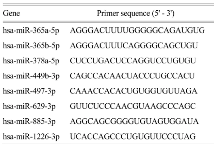
Gene Primer sequence (5' - 3')
hsa-miR-365a-5p AGGGACUUUUGGGGGCAGAUGUG hsa-miR-365b-5p AGGGACUUUCAGGGGCAGCUGU hsa-miR-378a-5p CUCCUGACUCCAGGUCCUGUGU hsa-miR-449b-3p CAGCCACAACUACCCUGCCACU hsa-miR-497-3p CAAACCACACUGUGGUGUUAGA hsa-miR-629-3p GUUCUCCCAACGUAAGCCCAGC hsa-miR-885-3p AGGCAGCGGGGUGUAGUGGAUA hsa-miR-1226-3p UCACCAGCCCUGUGUUCCCUAG Table 1. Primer sequences.
Gene Primer sequence (5' - 3') EBER1 TTTGCTAGGGAGGAGACGTGTGT
(F)
AAGCAGAGTCTGGGAAGACAACCA (R)
EBER2 AGGACAGCCGTTGCCCTAGTGGTT TCGGA (F)
AAAACAGCGGACAAGCCGAATACC (R)
EBNA2 AAAACAGCGGACAAGCCGAATACC (F)
GCCACCTGGCAGCCCTAAAG (R)
EBNA3B CCCTTGCGGATGCAGCCAAT (F)
GGCTGATATGGAATGTGCCC (R)
EBNA3C AGAAGGGGAGCGTGTGT (F)
CGGCTCGTTTTTGACGTC (R)
ARL15 TTTGCTAGGGAGGAGACGTGTGT (F)
AAGCAGAGTCTGGGAAGACAACCA (R)
EXOSC2 TTGCACACGAGGAGCCTGAAA (F)
GAGGCACCACATGGCAAATCA (R)
LRP6 GCTTGGGCTCAACCGTGAAGT (F)
TGAATGATGGTGCGGTTT (R)
TAPBP GCTGCAACTCCTGGGCTGAAT (F)
AGAAGGTCCCATTTCCGGTCC (R)
TMEM48 AATGGGAATGGTGATGGCCTG (F)
AATGGGAATGGTGATGGCCTG (R)
VPS13D ATATGGAAATGTCGGGGGCCT (F)
CCGATTGTCCATCGTCTTCCC (R)
GAPDH ATGCCATCACTGCCACCCAG (F)
GTCGTTGAGGGCAATGCCAG (R)
miRNA Primer sequence (5' - 3')
hsa-miR-7-5p UGGAAGACUAGUGAUUUUGUUGU hsa-miR-92b-5p AGGGACGGGACGCGGUGCAGUG hsa-miR-106b-3p CCGCACUGUGGGUACUUGCUGC hsa-miR-140-3p UACCACAGGGUAGAACCACGG hsa-miR-197-3p UUCACCACCUUCUCCACCCAGC hsa-miR-204-3p GCUGGGAAGGCAAAGGGACGU hsa-miR-337-5p GAACGGCUUCAUACAGGAGUU
동한 후, nitrocellulose membrane (Invitrogen)으로 transfer하 고 5% skim milk로 blocking 하였다. 항체 처리는 rabbit anti-LRP6 (1:1,000, Cell Signaling Technologies, Danvers, MA, USA)을 4℃에서 24시간 반응시켰으며, 2차 항체로 horseradish peroxidase (HRP)가 결합된 goat anti-rabbit (1:
3,000, Santa Cruz Biotechnology)을 사용하여 상온에서 2시 간 반응시켰다. 단백질 확인을 위해 ECL Western blotting analysis system 용액(Amersham Life Sciences, Bucking- hamshire, UK)을 처리하여 X-ray 필름에 감광시키고 현 상하였다. 동량의 단백질이 겔에 loading 되었는지 확인 하기 위해 동일 membrane을 anti-β-actin 항체(1:2,000, Cell Signaling Technology, Danvers, MA, USA)와도 반응시켜 정량하였으며, 감광하여 얻은 결과는 FUJIFILM Multi Gauge 프로그램을 이용하여 정량 분석하였다 (22).
Plasmid construction
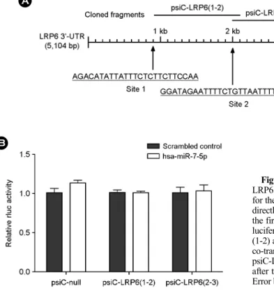
Akata(+) 세포에서 DNA를 추출한 후 hsa-miR-7-5p seed 서열과 상보적인 LRP6 3'-UTR (5,104 base pairs)의 세 부 위 중, 첫 번째와 두 번째 부위 혹은 두 번째와 세 번째
부위가 포함되도록 각각 증폭하였다. 그 다음 이들을 psiCHECK-2 reporter 벡터(Promega, Madison, WI)에 있는 Renilla luciferase 코딩 서열과 poly(A) 사이의 XhoI/NotI 위 치에 클로닝하여 psiC-LRP6(1-2)과 psiC-LRP6(2-3)를 제작 하였다(Fig. 4A). 증폭에 사용한 primer 서열은 LRP6(1-2):
5'-TCGACTCGAGAGCTCCATTCCCCAGTAGGCTT-3'; 5'- GGCCGCGGCCGCCCATTTTGTCTGACACTGTGGGC-3', LRP6(2-3): 5'-TCGACTCGAGTGTTGGAGACTGTAAGA- ATCAGGTGC-3'; 5'-GGCCGCGGCCGCTGGCAGAAAGT- GCCAAAGGC-3'이다.
Luciferase reporter assay
HEK293T 세포를 96-well plate에 5 × 10³ cells/well로 24시간 배양 후 hsa-miR-7-5p mimic과 함께 psiC-LRP6 (1-2)나 psiC-LRP6(2-3)를 co-transtection 하였다. Renilla luciferase 활성은 48시간 후에 Dual-Glo luciferase reporter assay system (Promega)을 이용하여 측정하였으며, 내부 대조군으로 psiC-LRP6(1-2)나 psic-LRP6(2-3)에 내재된 firefly luciferase 활성을 측정하여 사용하였다.
RESULTS
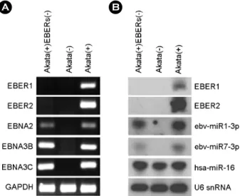
Akata(+)EBERs(-) 세포에서 EBER 발현 확인 실험에 사용한 Akata(+)EBERs(-) 세포에 EBER만 결손 되어 있는지 확인하기 위해, EBER1, EBER2 및 EBV 잠 복 유전자(EBNA2, EBNA3B, EBNA3C)의 존재 여부를 PCR로 조사하였다. 실험 결과 Akata(+)EBERs(-)에서는 Akata(-)와 동일하게 EBER1과 EBER2 유전자가 모두 존재하지 않았으나, EBV 잠복유전자와 GAPDH 유전자 의 존재는 명확히 확인되었다(Fig. 1A). 또한, Akata(+)- EBERs(-)에서 EBER1, EBER2 및 몇몇 EBV miRNA (ebv- miR-BART1-3p, ebv-miR-BART7-3p)의 발현도 Northern blot으로 조사하였다(Fig. 1B). 실험 결과, Akata(+)EBERs(-) 에서 예상대로 EBER1과 EBER2는 발현되지 않았으며, EBV miRNA들은 Akata(+) 세포에서와 유사한 수준으로 발현되었다. Akata(-)와 Akata(+), Akata(+)EBERs(-)에서 cellular miRNA인 hsa-miR-16도 정도에 큰 차이 없이 잘 발현되었다. 이로써 Akata(+)EBERs(-) 세포는 EBER1과 EBER2의 결손이 있는 것을 제외하고 Akata(+)와 유사한 특징을 나타냄을 확인할 수 있었다(Fig. 1B).
A B
Figure 1. Verification of EBER deletion in Akata(+)EBERs(-) cell line. (A) PCR analysis was carried out to examine the absence of EBER (EBER1, 2) sequences in the EBV genome of Akata(+)- EBERs(-). For comparison, Akata(-) and Akata(+) cells were used.
The presence of EBV latent genes (EBNA2, 3B, and 3C) was also examined. GAPDH was used as an internal control. (B) Expression of EBERs and two EBV BART miRNAs in Akata(-), Akata(+), and Akata(+)EBERs(-) cells were analyzed by Northern blot analysis. The expression of human miR-16 (hsa-miR-16) was also assessed as a reference. The quality and quantity of the loaded RNA were examined by reprobing the blot for U6 snRNA.
EBER와 결합하여 발현이 조절될 것으로 예상되는 human cellular miRNA 선정
EBER가 cellular miRNA에 sponge로 작용하여 발현을 억제시킬 가능성이 있는지 PITA 예측 프로그램(http://
bibiserv.techfak.uni-bielefeld.de/rnahybrid/)을 사용하여 조 사하였다. 본 연구에서는 EBER가 cellular miRNA에 결 합함으로써 miRNA의 기능을 억제할 것으로 가정하고, EBER1이나 EBER2와 결합 가능성이 높은 cellular miRNA (Version 11.0)를 각각 25개씩 찾은 후 문헌조사를 통해 이들 중 타겟이 보고된 바 있는 15종의 miRNA를 선별 하였다(Table 2).
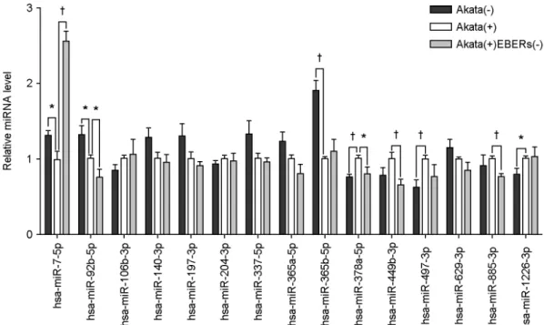
EBER와 결합 가능성이 있는 cellular miRNA의 발현 분석
선별된 15종의 human cellular miRNA (Table 2)들이 EBER에 의해 발현이 감소될 가능성을 확인하기 위해, Akata(-)와 Akata(+), Akata(+)EBERs(-) 세포에서 정제한
RNA를 사용하여 quantitative real-time PCR (qRT-PCR)을 실시하였다. 내부 대조군으로 U6 snRNA를 사용하였고, 독립적인 3번의 실험을 수행하였다. 실험 결과, 15종의 miRNA 중 3종의 miRNA (hsa-miR-7-5p, hsa-miR-92b-5p, and hsa-miR-365b-5p)가 Akata(+)에서 Akata(-)에 비해 발 현이 감소하는 것으로 나타났으나(p < 0.05), 이 중 hsa- miR-7-5p만이 Akata(+)EBERs(-)에서 Akata(+)에 비해 유 의적으로 발현이 증가된 양상을 보여주었다(p < 0.01).
즉, EBER와 서열 상보성이 있는 15종의 miRNA 중 hsa- miR-7-5p가 EBER에 의한 sponge 작용을 받을 가능성이 있다고 생각되었다(Fig. 2).
Hsa-miR-7-5p에 의해 조절될 것으로 예상되는 타겟 mRNA 후보군 선정
EBER 발현과 반비례하여 발현이 변화되었던 hsa- miR-7-5p가 타겟할 가능성이 있는 유전자들을 TarBase program (Version 6.0)을 이용하여 검색하였다. 검색된 415개의 유전자들 중 mRNA 마이크로어레이 실험 결과, Table 2. Cellular miRNAs which are expected to be targeted by EBERs.
EBERs microRNA Positiona Seedb ΔdGduplexc
EBER 1 hsa-miR-7-5p 91 7:0:0 -15.40
hsa-miR-92b-5p 47 7:0:0 -32.90
hsa-miR-140-3p 83 7:0:0 -18.90
hsa-miR-197-3p 67 8:0:0 -22.00
hsa-miR-204-3p 93 8:0:0 -31.00
hsa-miR-365a-5p 38 7:0:0 -28.30
hsa-miR-365b-5p 46 7:0:0 -29.30
hsa-miR-449b-3p 20 7:0:0 -27.60
hsa-miR-885-3p 1 7:0:0 -24.40
hsa-miR-1226-3p 131 7:0:0 -14.20
EBER 2 hsa-miR-105-5p 75 8:0:0 -25.20
hsa-miR-106b-3p 4 8:0:0 -24.90
hsa-miR-337-5p 2 7:0:0 -16.60
hsa-miR-378a-5p 104 8:0:0 -22.00
hsa-miR-497-3p 2 7:0:0 -18.50
hsa-miR-629-3p 139 7:0:0 -18.70
aDistance in base pairs from the beginning of EBER1 or EBER2.
bPITA's "X:Y:Z" notation for describing the seed represents the size of the seed (X), the number of mismatches (Y) and the number of G:U wobble pairs (Z).
cThe binding energy of the microRNA-EBER duplex.
Akata(+) 세포에서 Akata(-)나 Akata(+)EBERs(-) 세포에서 발현이 증가된 유전자를 조사하여, 총 6종 LRP6, (ADP- ribosylation factor-like 15 (ARL15), exosome component 2 (EXOSC2), TAP binding protein (TAPBP), transmembrane protein 48 (TMEM48), vacuolar protein sorting 13 homolog D (VPS13D))의 hsa-miR-7-5p 후보 유전자를 최종 선별하였 다(Table 3).
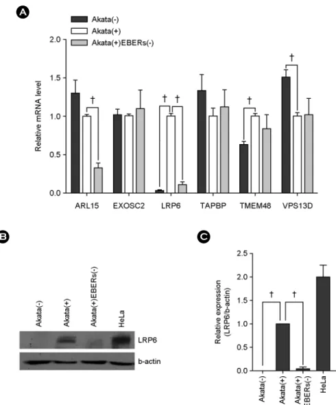
EBER의 발현이 hsa-miR-7-5p의 타겟 후보유전 자 발현에 미치는 영향
EBER가 발현될 때 선별된 6종의 hsa-miR-7-5p 잠재 타 겟 mRNA의 발현이 증가할 가능성을 Akata(-)와 Akata(+), Akata(+)EBERs(-)의 RNA로 qRT-PCR을 실시하여 조사하 였다. 내부 대조군으로 GAPDH를 사용하였으며 독립적 인 3번의 실험을 수행한 결과, 6종의 유전자 중 LRP6와 TMEM48 mRNA가 Akata(+)에서 Akata(-)에 비해 유의적 으로 증가하였으나(p < 0.01), LRP6 mRNA만이 Akata(+)에 서 Akata(+)EBERs(-)에 비해 유의적으로 증가되는 양상 을 보였다(p < 0.01) (Fig. 3A). LRP6가 단백질 수준에서도
EBER 발현 시 증가하는 양상을 보이는지 알아보기 위 해 Western blot을 수행하였다. LRP6 항체에 대한 양성 대 조군으로 자궁경부암 세포인 HeLa를 사용하였고 loading control로 β-actin을 사용하였다. 실험 결과, mRNA의 결 과와 마찬가지로, Akata(-)나 Akata(+)EBERs(-)에 비해
Table 3. Putative target genes of hsa-miR-7-5p miRNA microarray
Akata(+)/Akata(-) Akata(+)/Akata(+)EBERs(-) Gene
Fold
change P-value Fold
change P-value ARL15 1.842 0.013 1.838 0.029 EXOSC2 1.522 0.017 1.168 0.062
LRP6 3.745 0.025 3.649 0.032
TAPBP 1.339 0.068 1.376 0.055 TMEM48 3.236 0.001 1.589 0.036 VPS13D 1.264 0.026 1.193 0.063 Figure 2. Down-regulated hsa-miR-7-5p expression by EBERs in Akata cells. Real-time RT-PCR was performed for 15 different cellular miRNAs to test whether EBERs can affect the expression of them. Total RNA was extracted from Akata(-), Akata(+), and Akata(+)EBERs(-) cells, and miRNA qRT-PCR was conducted using Mir-X miRNA First Strand Synthesis and SYBR qRT-PCR Kit.
Relative gene expression was calculated according to the comparative CT method using U6 snRNA as a loading control. The ratio of each miRNA to U6 in Akata(+) cells was arbitrarily set as 1. (n=3, †; p < 0.01, *; p < 0.05)
Akata(+) 세포에서 LRP6 단백질의 발현이 증가하는 것으 로 확인되었다(Fig. 3B). 3번의 독립적인 실험 결과에 대 해 Multi Gauge 프로그램을 이용하여 정량 분석을 실시한 결과 Akata(+)에서 Akata(-)에 비해 약 99% (p < 0.01)의 증 가를 보였으며, Akata(+)에서는 Akata(+)EBERs(-)에서보다 약 96%(p < 0.01) 증가하였다(Fig. 3C). 결과적으로 hsa- miR-7-5p의 타겟 mRNA 후보군 6종 중 LRP6가 EBER가
발현할 때 증가되는 발현 양상을 보임으로써, EBER 발현 시 감소되는 hsa-miR-7-5p에 의해 영향을 받을 가능성이 있는 타겟 후보로 최종 선별되었다.
Hsa-miR-7-5p가 LRP6의 3'-UTR과 직접 결합하 여 발현을 조절하는지 확인
EBER 발현 시 증가되는 양상을 보인 LRP6가 hsa-
β-actin
Relative expression (LRP6/β-actin)
A
B C
Figure 3. Up-regulated LRP6 expression by EBERs in Akata cells. (A) Real-tizme RT-PCR was performed for six different genes to test whether EBERs can affect the expression of them. Total RNA was extracted from Akata(-), Akata(+), and Akata(+)EBERs(-) cells and quantitative real-time RT-PCR were carried out using SYBR Premix Ex Taq. Relative gene expression was calculated according to the comparative CT method using GAPDH as a loading control. The ratio of each mRNA to GAPDH in Akata(+) cells was arbitrarily set as 1.
(B) The effect of EBERs on the expression of LRP6 at protein level was analyzed. Western blot analysis was carried out using cell lysate from in Akata(-), Akata(+), and Akata(+)EBERs(-) cells. β-actin was used as a loading control and HeLa cell line was used as a positive control for LRP6 expression. (C) Results similar to those in panel B were obtained in two more independent experiments, and the means and standard deviation (SD) from all three independent experiments are plotted. (n=3, †; p < 0.01)
miR-7-5p의 직접적인 타겟인지 확인하기 위해, 우선 PITA 예측 프로그램을 사용하여 hsa-miR-7-5p seed 부분과 LRP6 3'-UTR의 상보적 결합 예상 부위를 조사하였으며, 그 결과 LRP6 3'-UTR에서 3 개(site1, site2, site3)의 예상 부위가 존재함을 확인하였다(Fig. 4). 확인된 3개의 결합 부위 중 각각 두 곳씩이 포함하도록 psiCHECK-2 reporter 벡터에 클로닝한 psiC-LRP6(1-2)과 psiC-LRP6(2-3) 각각 을 HEK293T 세포에 hsa-miR-7-5p mimic과 co-transfection 시켜 luciferase activity를 측정하였다. Luciferase reporter assay 결과에 대한 내부 대조군으로 firefly luciferase 활성 을 사용하였으며, 독립적인 3번의 실험을 수행하였다. 실 험 결과, psiC-LRP6(1-2)와 psiC-LRP6(2-3) 모두 hsa-miR- 7-5p mimic에 의해 luciferase 활성에 변화를 보이지 않는 것으로 확인되었다(p < 0.05).
EBER의 발현이 LRP6 발현을 증가시키는 유전자 발 현에 어떠한 영향을 주는지 확인
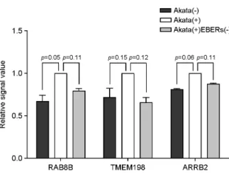
EBER 발현 시 증가되는 양상을 보인 LRP6가 miRNA 가 아닌 다른 요인에 의해 조절될 수 있는 가능성을 확인하기 위하여, LRP6 발현을 증가시킴으로써 Wnt/
β-catenin 신호전달을 활성화시키는 것으로 보고된 유전자 들을 조사하였다. 그 결과 3종의 유전자(Transmembrane 198 (TMEM198), β-arrestin 2 (ARRB2), APC membrane recruitment protein 1 (AMER1))가 LRP6의 발현을 증가시 키는 것으로 보고되었다 (23~25). 보고된 3종의 유전자가 Akata 세포에서 EBER 발현 시 LRP6 발현 조절자로 기 능을 할 가능성을 확인하기 위해, Akata mRNA 마이크로 어레이 결과를 분석하니 이들 3종의 유전자 모두 EBER 가 있을 때 발현이 증가하는 경향을 보였으나 통계적 유 의성은 보이지 않았다(Fig. 5).
DISCUSSION
본 논문에서 사용한 Akata 세포는 latency I을 보이는 버킷트 림프종 세포이다 (26). 버킷트 림프종 세포에서 EBER의 발현은 interferon regulatory factor 3 (IRF-3) 신호 전달을 조절하는 RIG-I을 활성화시켜 interferon (27)과 IL-10 (28)의 발현을 유도하고, PKR의 인산화를 억제하여 세포 사멸 감소와 (29, 30) 세포성장, 세포 골격 재구성에 관여하는 유전자들을 조절하는 것으로 알려져 있다 (31, A
B
Figure 4. No direct targeting of has-miR-7 for the 3'-UTR of LRP6. (A) Three seed-matched regions in the 3'-UTR of LRP6 for the has-miR-7 are shown. To test whether LRP6 expression is directly regulated by has-miR-7, the 3'-UTR fragments containing the first two or last two seed matched regions were cloned into a luciferase reporter (psiCHECK-2) vector to product psiC-LRP6 (1-2) and psiC-LRP6(2-3), respectively. (B) HEK293T cells were co-transfected with has-miR-7-5p mimic, and psiC-LRP6(1-2) or psiC-LRP6(2-3). Renillar luciferase activity was analyzed 48 h after transfection and normalized using firefly luciferase activity.
Error bars indicate SD. (n=3, †; p < 0.01, *; p < 0.05).
32). 또한 EBV에 감염되지 않은 버킷트 림프종 세포에 EBER를 발현시킬 경우 세포 증식이 증가되며 종양 형성, 세포 사멸 내성을 증가시키는 것으로 보고되었다 (29). 하 지만 이러한 EBER와 관련된 세포 기능 조절의 상세한 기전에 대해서는 아직 명확하게 밝혀지지 않았다.
본 논문에서는 EBER1과 EBER2만 제거된 recombinant EBV에 감염된 Akata(+)EBERs(-)를 Akata(-)와 Akata(+) 세포와 함께 사용하여 다른 EBV 유전자의 존재나 발현 여부와 상관없이 EBER 발현에 따른 세포 miRNA와 유 전자 발현의 변화를 조사하였다. 여러 암세포에서 다양한 종양 형성유전자를 억제하는 것으로 알려져 있는 hsa- miR-7-5p (33~36)의 발현이 EBER 발현에 의해 크게 감 소한 우리의 결과는(Fig. 2), EBV 연관 버킷트 림프종 세 포에서 EBER 발현에 의해 hsa-miR-7-5p 발현이 감소되 고, 이에 의해 종양 형성 유도와 증식 촉진이 일어날 가 능성을 제시한다.
LRP6는 Wnt/β-catenin 신호전달에서 Wnt와 결합하는 frizzled (37)와 같은 공수용체로써, LRP6를 과발현시킬 경우 Wnt/β-catenin 신호전달이 활성화되고 세포 증식과 종양 형성이 일어난다 (38, 39). 따라서 EBER에 의한 LRP6의 발현 증가(Fig. 3)는 β-catenin의 분해를 막고 Wnt/
β-catenin 신호전달을 활성화시켜, 결과적으로 EBER에 의해 세포 증식이 증가되는 기전에서 중요한 역할을 할 가능성을 보여준다.
본 연구에서 우리는 B 세포주인 Akata 세포에서 EBER
가 발현될 경우 hsa-miR-7-5p의 발현이 감소되는 것을 확 인하였다. Hsa-miR-7-5p는 여러 암세포에서 다양한 종양 형성 유전자를 억제하는 것으로 알려져 있다 (33~36). 역 으로 LRP6는 EBER를 발현하는 Akata(+) 세포에서 그렇 지 않은 Akata(-) 및 Akata(+)EBERs(-) 세포에 비해 발 현이 크게 증가하는 것을 확인하였다. LRP6의 3'-UTR 에 hsa-miR-7-5p와 seed match 가능한 서열이 있으나 luciferase assay 결과 예상했던 것과는 달리 LRP6의 발현 이 hsa-miR-7-5p에 의해서 직접적으로 조절되지는 않았 다. 따라서 LRP6와 hsa-miR-7-5p는 EBER에 의해 각각 독립적인 기전에 의해 조절되는 것으로 생각된다.
최근, 여러 논문에서 다양한 유전자들이 LRP6의 발현 및 인산화를 촉진시킴으로써 Wnt/β-catenin 신호전달을 활성화시키는 것으로 보고되었다 (23~25). 예를 들어, Ras GTPase family인 RAB8B는 LRP6와 결합하여 RAB8B가 감소할 경우 인산화된 LRP6와 세포질 내 β-catenin 축 적을 감소시키며, 과발현할 경우 LRP6 발현과 inter- nalization을 촉진시켜 Wnt/β-catenin 신호전달의 활성화를 증가시킨다. Transmembrane 198 (TMEM198)은 LRP6와 특이적으로 결합하여 인산화와 발현을 촉진시키며 (25), β-arrestin 2 (ARRB2)는 APC membrane recruitment protein 1 (AMER1)과의 상호작용을 통해 LRP6의 인산화를 촉진시 킨다 (23). 따라서 Akata 세포에서 EBER가 발현될 경우 RAB8B, TMEM198, ARRB2의 발현 증가(Fig. 5)는 LRP6 의 발현과 인산화를 촉진시킴으로써, β-catenin의 인산화 억제와 안정화를 유도하고 Wnt/β-catenin 신호전달의 활 성화를 돕는 것으로 보여진다. EBER가 어떤 기전에 의 해 hsa-miR-7-5p의 발현을 감소시키는지와 EBER가 hsa- miR-7-5p에 대해 sponge로 작용하는지에 관해서 보다 상 세한 연구가 필요할 것이다.
본 연구의 결과 EBER에 의한 hsa-miR-7-5p와 LRP6의 조절을 확인하였으며, 향후 그에 따른 세포 내 여러 생리 학적 조절 기전에 대한 추가적인 연구가 필요하다. EBER 와 관련된 EBV 감염 세포의 성장 기전을 밝힌다면 EBV 양성 관련 질병들에 대한 치료에 도움을 줄, 효과적인 대 안책을 제시할 수 있게 될 것으로 기대된다.
REFERENCES
1) Thompson MP, Kurzrock R. Epstein-Barr virus and cancer.
Clin Cancer Res 2004;10:803-21.
Figure 5. Validation of selected mRNA from microarray data.
Microarray analysis was performed for Akata(-), Akata(+), and Akata(+)EBERs(-) cells. Relative mRNA intensity was normalized by the average intensity of Akata(+) cell.
2)Kieff E, Rickinson. A. Epstein-Barr virus. In: Knipe D, Howley P, editors. Fields virology. Philadelphia: Raven Press;
2007. p. 2603-54.
3)Rosa MD, Gottlieb E, Lerner MR, Steitz JA. Striking similarities are exhibited by two small Epstein-Barr virus- encoded ribonucleic acids and the adenovirus-associated ribonucleic acids VAI and VAII. Mol Cell Biol 1981;1:785 -96.
4) Rymo L. Identification of transcribed regions of Epstein-Barr virus DNA in Burkitt lymphoma-derived cells. J Virol 1979;
32:8-18.
5) Arrand JR, Young LS, Tugwood JD. Two families of sequences in the small RNA-encoding region of Epstein-Barr virus (EBV) correlate with EBV types A and B. J Virol 1989;63:
983-6.
6) Ambros V. MicroRNA pathways in flies and worms: growth, death, fat, stress, and timing. Cell 2003;113:673-6.
7)Bartel DP. MicroRNAs: genomics, biogenesis, mechanism, and function. Cell 2004;116:281-97.
8) Meister G, Tuschl T. Mechanisms of gene silencing by double- stranded RNA. Nature 2004;431:343-9.
9) Mack GS. MicroRNA gets down to business. Nat Biotechnol 2007;25:631-8.
10)Chen JF, Mandel EM, Thomson JM, Wu Q, Callis TE, Hammond SM, et al. The role of microRNA-1 and microRNA- 133 in skeletal muscle proliferation and differentiation. Nat Genet 2006;38:228-33.
11)Cheng AM, Byrom MW, Shelton J, Ford LP. Antisense inhibition of human miRNAs and indications for an involvement of miRNA in cell growth and apoptosis. Nucleic Acids Res 2005;33:1290-7.
12) Takamizawa J, Konishi H, Yanagisawa K, Tomida S, Osada H, Endoh H, et al. Reduced expression of the let-7 microRNAs in human lung cancers in association with shortened postoperative survival. Cancer Res 2004;64:3753-6.
13) Eis PS, Tam W, Sun L, Chadburn A, Li Z, Gomez MF, et al.
Accumulation of miR-155 and BIC RNA in human B cell lymphomas. Proc Natl Acad Sci U S A 2005;102:3627-32.
14) He L, Thomson JM, Hemann MT, Hernando Monge E, Mu D, Goodson S, et al. A microRNA polycistron as a potential human oncogene. Nature 2005;435:828-33.
15) Donnell CA, Pollock WJ, Sybers WA. Thyroid carcinosarcoma.
Arch Pathol Lab Med 1987;111:1169-72.
16)Ebert MS, Sharp PA. Emerging roles for natural microRNA
sponges. Curr Biol 2010;20:R858-61.
17)Cazalla D, Yario T, Steitz JA. Down-regulation of a host microRNA by a Herpesvirus saimiri noncoding RNA. Science 2010;328:1563-6.
18) Cook HL, Lytle JR, Mischo HE, Li MJ, Rossi JJ, Silva DP, et al. Small nuclear RNAs encoded by Herpesvirus saimiri upregulate the expression of genes linked to T cell activation in virally transformed T cells. Curr Biol 2005;15:974-9.
19)Murthy S, Kamine J, Desrosiers RC. Viral-encoded small RNAs in herpes virus saimiri induced tumors. EMBO J 1986;
5:1625-32.
20)Takada K. Cross-linking of cell surface immunoglobulins induces Epstein-Barr virus in Burkitt lymphoma lines. Int J Cancer 1984;33:27-32.
21)Yajima M, Kanda T, Takada K. Critical role of Epstein-Barr Virus (EBV)-encoded RNA in efficient EBV-induced B- lymphocyte growth transformation. J Virol 2005;79:4298-307.
22)Kim SY, Lee SK. Inhibition of transfer infection of Epstein- Barr virus to epithelial cells by integrin beta6 siRNA. J Bacteriol Virol 2012;42:346-52.
23)Kriz V, Pospíchalová V, Masek J, Kilander MB, Slavík J, Tanneberger K, et al. beta-arrestin promotes Wnt-induced Low Density Lipoprotein Receptor-related Protein 6 (Lrp6) phosphorylation via increased membrane recruitment of Amer1 Protein. J Biol Chem 2014;289:1128-41.
24)Demir K, Kirsch N, Beretta CA, Erdmann G, Ingelfinger D, Moro E, et al. RAB8B is required for activity and caveolar endocytosis of LRP6. Cell Rep 2013;4:1224-34.
25)Liang J, Fu Y, Cruciat CM, Jia S, Wang Y, Tong Z, et al.
Transmembrane protein 198 promotes LRP6 phosphorylation and Wnt signaling activation. Mol Cell Biol 2011;31:2577-90.
26)Shimizu N, Tanabe Tochikura A, Kuroiwa Y, Takada K.
Isolation of Epstein-Barr virus (EBV)-negative cell clones from the EBV-positive Burkitt's lymphoma (BL) line Akata:
malignant phenotypes of BL cells are dependent on EBV. J Virol 1994;68:6069-73.
27) Samanta M, Iwakiri D, Kanda T, Imaizumi T, Takada K. EB virus-encoded RNAs are recognized by RIG-I and activate signaling to induce type I IFN. EMBO J 2006;25:4207-14.
28) Samanta M, Iwakiri D, Takada K. Epstein-Barr virus-encoded small RNA induces IL-10 through RIG-I-mediated IRF-3 signaling. Oncogene 2008;27:4150-60.
29)Komano J, Maruo S, Kurozumi K, Oda T, Takada K.
Oncogenic role of Epstein-Barr virus-encoded RNAs in
Burkitt's lymphoma cell line Akata. J Virol 1999;73:9827-31.
30) Gregorovic G, Bosshard R, Karstegl CE, White RE, Pattle S, Chiang AK, et al. Cellular gene expression that correlates with EBER expression in Epstein-Barr Virus-infected lympho- blastoid cell lines. J Virol 2011;85:3535-45.
31) Houmani JL, Davis CI, Ruf IK. Growth-promoting properties of Epstein-Barr virus EBER-1 RNA correlate with ribosomal protein L22 binding. J Virol 2009;83:9844-53.
32) Eilebrecht S, Pellay FX, Odenwälder P, Brysbaert G, Benecke BJ, Benecke A. EBER2 RNA-induced transcriptome changes identify cellular processes likely targeted during Epstein Barr Virus infection. BMC Res Notes 2008;1:100.
33)Kefas B, Godlewski J, Comeau L, Li Y, Abounader R, Hawkinson M, et al. microRNA-7 inhibits the epidermal growth factor receptor and the Akt pathway and is down- regulated in glioblastoma. Cancer Res 2008;68:3566-72.
34) Fang Y, Xue JL, Shen Q, Chen J, Tian L. MicroRNA-7 inhibits tumor growth and metastasis by targeting the phosphoinositide
3-kinase/Akt pathway in hepatocellular carcinoma. Hepatology 2012;55:1852-62.
35)Zhang N, Li X, Wu CW, Dong Y, Cai M, Mok MT, et al.
microRNA-7 is a novel inhibitor of YY1 contributing to colorectal tumorigenesis. Oncogene 2013;32:5078-88.
36)Saydam O, Senol O, Würdinger T, Mizrak A, Ozdener GB, Stemmer Rachamimov AO, et al. miRNA-7 attenuation in Schwannoma tumors stimulates growth by upregulating three oncogenic signaling pathways. Cancer Res 2011;71:852-61.
37) Logan CY, Nusse R. The Wnt signaling pathway in develop- ment and disease. Annu Rev Cell Dev Biol 2004;20:781-810.
38) Tung EK, Wong BY, Yau TO, Ng IO. Upregulation of the Wnt co-receptor LRP6 promotes hepatocarcinogenesis and enhances cell invasion. PLoS One 2012;7:e36565.
39)Li Y, Lu W, He X, Schwartz AL, Bu G. LRP6 expression promotes cancer cell proliferation and tumorigenesis by altering beta-catenin subcellular distribution. Oncogene 2004;23:9129 -35.