ABSTRACT
Objective: Evaluate the impact of radiotherapy on cause specific survival (CSS) and overall survival (OS) for stage (I–III) clear cell, mucinous, and endometriod ovarian cancer.
Methods: We analyzed incidence, survival, and treatments from the Surveillance,
Epidemiology, and End Results (SEER) Program from 2004 to 2011 for clear cell, mucinous, and endometriod histologies of the ovary for stages (I–III). We examined CSS and OS for all three histologies combined and each histology with relation to the use of adjuvant radiation therapy (RT). Survival analysis was calculated by Kaplan-Meier and log-rank analysis.
Results: CSS was higher in individuals not receiving RT at 5 years (81% vs. 74%) and 10 years (74% vs. 65%, p=0.003). OS was higher in individuals not receiving RT at 5 years (76% vs.
73%) and 10 years (64% vs. 59%, p=0.039). Stage III patients receiving RT had a higher OS at 5 years (54% vs. 44%) and 10 year intervals (36% vs. 30%, p=0.037). Stage III patients with mucinous histology receiving RT had a higher OS at 5 years (50% vs. 36%) and 10 years (45%
vs. 26%, p=0.052).
Conclusion: Those receiving RT had a lower CSS and OS at 5 and 10 years. However, subgroup analysis revealed a benefit of RT in terms of OS for all stage III patients and for stage III patients with mucinous histology.
Keywords: Ovary; Radiation; Survival; Uncommon Histology
INTRODUCTION
In the United States, ovarian cancer is the most common cause of gynecologic-related cancer mortality [1]. Specifically, nearly 23,000 women are diagnosed with ovarian cancer yearly, and of these, 14,000 women die every year. A majority of these women are diagnosed with advanced disease, namely, The International Federation of Gynecology and Obstetrics stage III disease [1]. The established standard of care for ovarian cancer has been total abdominal hysterectomy, bilateral salpingo-oophorectomy, omentectomy, washings, and suspicious node removal with adjuvant platinum based chemotherapy, based on extent of primary and nodal involvement, margins, and residual disease [2]. Unfortunately, the 5-year overall survival (OS) for ovarian cancer is about 40%; and the median progression free survival for advanced ovarian cancer is about 18 months [1].
Original Article
Received: Mar 12, 2016 Revised: Apr 27, 2016 Accepted: Apr 27, 2016 Correspondence to Sagar C. Patel
Department of Radiation Oncology, University of Iowa Hospitals and Clinics, 200 Hawkins Drive, Iowa City, IA 52242, USA.
E-mail: Sagar-c-patel@uiowa.edu Copyright © 2016. Asian Society of Gynecologic Oncology, Korean Society of Gynecologic Oncology
This is an Open Access article distributed under the terms of the Creative Commons Attribution Non-Commercial License (http://
creativecommons.org/licenses/by-nc/4.0/) which permits unrestricted non-commercial use, distribution, and reproduction in any medium, provided the original work is properly cited.
ORCID Sagar C. Patel
http://orcid.org/0000-0002-2675-616X Jonathan Frandsen
http://orcid.org/0000-0001-8406-3380 This paper was presented at Poster Presentation at American Society for Therapeutic Radiology and Oncology, October 2015.
Conflict of Interest
No potential conflict of interest relevant to this article was reported.
Sagar C. Patel,1 Jonathan Frandsen,2 Sudershan Bhatia,1,3 David Gaffney2
1Department of Radiation Oncology, University of Iowa Hospitals and Clinics, Iowa City, IA, USA
2Department of Radiation Oncology, Huntsman Cancer Hospital, Salt Lake City, UT, USA
3Department of Radiation Oncology, Stratton VA Medical Center, Albany, NY, USA
Impact on survival with adjuvant
radiotherapy for clear cell, mucinous,
and endometriod ovarian cancer: the
SEER experience from 2004 to 2011
Recognizing the importance of early diagnosis, aggressive treatment, and close surveillance, through a multi-disciplinary approach, advancement in ovarian cancer care has occurred from appropriate staging including an emphasis on maximal, safe cytoreduction by surgical specialists, utilization of imaging to assess volume of disease, and defining and classifying the heterogeneity of the histopathology of ovarian tumors. Specifically, a better understanding of the histopathology of ovarian tumors may define the prognostic and predictive value of adjunct treatment modalities such as molecularly targeted agents, newer chemotherapy regimens, and radiation therapy (RT).
Historically, platinum based chemotherapy has been the prime adjunct therapy for ovarian cancer. Although it is effective, many ovarian cancer patients, especially, with advanced disease, do not completely respond, become platinum-resistant, and ultimately develop multiple episodes of recurrent disease and die most commonly from bowel obstruction [1].
Potential improvements in classification, categorization, and stratification of ovarian tumors may allow for early identification of disease presentations that may be platinum resistant and that may potentially benefit from tailored biologic agents and/or RT. These advancements would be instrumental given the current response rates to second line chemotherapy in patients with relapsed ovarian cancer are only 10% to 20% [3].
From a RT perspective, historically, a survival benefit was seen with whole abdominal irradiation as adjuvant therapy in patients with small microscopic residual tumor (<2 cm) or no residual tumor after surgery [4,5]. Over time, the role of RT ultimately diminished with application and impact of platinum and taxane based chemotherapy as well as
improved surgical cytoreduction [1]. However, with the recent technologic advancements in radiotherapy delivery and with the acknowledgment that 70% of ovarian cancer recurrences reside in the abdomen and pelvis, the role of radiotherapy may be re-defined especially after the recognition of specific radiosensitive histologies and of potential chemoresistant subtypes that may be radiosensitive and may ultimately procure a locoregional control and/or survival benefit [3]. By stage, recent work by Sorbe and colleagues [5] revealed improvement in progression-free survival and OS when comparing whole abdominal radiotherapy to chemotherapy in stage III ovarian cancer patients who had complete response after cytoreductive surgery followed by chemotherapy. By histology, multiple studies have shown the lack of improvement in survival in rare histologic variants of ovarian cancer such as clear cell carcinoma, endometriod, and mucinous with standard chemotherapy [6]. On the contrary, a population based review by Swenerton and colleagues [7] revealed a reduction in disease specific mortality and in overall mortality in ovarian cancer patients with clear cell, mucinous, or endometriod histology who received adjuvant radiotherapy. In addition, a histopathological review by Kobel and colleagues [8] revealed that 90% of clear cell, endometriod, and mucinous tumors are stage I or II; in turn, the impact of local treatments such as radiotherapy and/or biological agents may be imperative for locoregional control.
Given the potential benefit of adjuvant RT in certain histologies and stage groupings, we completed a Surveillance, Epidemiology, and End Results (SEER) based analysis from the years 2004 to 2011, which captures at least 28% of the United States population, and evaluated the impact of adjuvant radiotherapy on cause-specific and OS for stage (I–III) clear cell, mucinous, and endometriod ovarian cancer [9].
MATERIALS AND METHODS
1. Data source
We analyzed incidence, survival, and treatment data from the SEER program of the National Cancer Institute from the years 2004 to 2011. We specifically examined the SEER 18 registries because they were continuously active for the duration (2004 to 2011). The SEER 18 registries report specific data on patient demographics, primary tumor site, tumor morphology, stage at diagnosis, first course of treatment, and follow-up for vital status (outcome/survival). The SEER 18 registries were purposefully selected to maximize generalizability to the overall United States population. Specifically, these 18 registries capture about 28% of the entire United States population [9].
2. Study population
The SEER study population studied was limited to patients with ovarian cancer in the adjuvant setting. The specific clinical variables examined were site, histology, demographics, stage (I–III), and survival (overall and cause-specific). The specific topography code for site was (ovary, site), which is based on the International Classification of Diseases for Oncology, Third Edition (ICD-O3/WHO 2008). The specific histology codes were based on the (ICD-O3) as well. For clear cell, the codes utilized were malignant tumor, clear cell type (8,005/3), clear cell adenocarcinoma, not-otherwise specified (NOS, 8,310/3), clear cell adenocarcinofibroma (8,313/3), and clear cell cystadenocarcinoma (8,443/3). For endometriod, the codes
utilized were endometrioid carcinoma (8,380/3), endometrioid adenofibroma, malignant (8,381/3), and endometrioid adenocarcinoma, secretory variant (8,382/3). For mucinous, the codes utilized were mucinous cystadenocarcinoma, NOS (8,470/3), papillary mucinous cystadenocarcinoma (8,471/3), mucinous adenocarcinoma (8,480/3), mucin-producing adenocarcinoma (8,481/3), mucinous adenocarcinoma, endocervical type (8,482/3), and signet ring cell carcinoma (8,490/3).The specific patient demographic variables examined and included were as follows: age (<50, 50 to 59, 60 to 69, and 70 and older), gender (female), race, and marital status. The descriptive values for race were white, black, and Asian. To define stage, we combined the SEER historic stage A, SEER summary stage, and the AJCC (T, N, and M), seventh edition to capture stage I, II, and III as comprehensively as possible.
We measured both cause specific survival (CSS) and OS for all histologies combined in stage I–III patients with adjuvant RT or no RT. We utilized the SEER defined treatment variable (radiation sequence with surgery); SEER codes this variable as either (radiation after surgery) or (no radiation and/or cancer-directed surgery). We then defined individuals receiving adjuvant RT as cases who were coded with (radiation after surgery). We measured OS for each histology for each stage as well with or without RT utilization. Mean OS and CSS was also reported. We were not able to examine the utilization and/or role of chemotherapy in the adjuvant setting because SEER does not comprehensively record this treatment type in its data [10].
3. Statistical analysis
We completed our statistical analysis with MedCalc ver. 12.0.6 (MedCalc Software bvba, Ostend, Belgium; http://www.medcalc.org; 2014). Survival analysis was calculated by Kaplan- Meier analysis. Univariate analysis was completed using the log rank test, while multi-variate analysis was performed using Cox proportional hazard regression models.
RESULTS
1. Incidence, CSS, and OS by stage (I–III) and treatment (no RT vs. RT)
A total of 15,829 cases were examined of which 15,379 received no RT and 450 received RT (Fig. 1). CSS was higher in individuals who did not receive RT at 5 years (81% vs. 74%) and 10 years (74% vs. 65%, p=0.003) (Fig. 2). For stage I patients, CSS was higher and statistically significant in individuals who did not receive RT at 5 years (92% vs. 85%) and 10 years (88%vs. 82%, p=0.023). In stage II patients, there was a trend towards improvement in CSS with no RT, while in stage III patients there was a trend towards improvement in CSS with RT.
Mean CSS for all stages with RT (16.4 months) and with no RT (18.1 months).
Similarly, OS was higher in individuals who did not receive RT at 5 years (76% vs. 73%) and 10 years (64% vs. 59%, p=0.039) (Fig. 1). For stage I patients, OS was higher and statistically significant in individuals who did not receive RT at 5 years (88% vs. 81%) and 10 years (77%
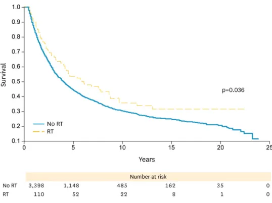
vs. 73%, p=0.021). In stage II patients, there was a trend towards improvement in OS with no RT, while in stage III patients there was higher and statistically significant improvement with RT in OS at 5 years (54% vs. 44%) and 10 years (36% vs. 30%, p=0.036) (Fig. 3). Mean OS for all stages with RT was 14.1 months compared to 15.2 months with no RT.
2. Incidence and OS for clear cell histology by treatment and stage (no RT vs. RT)
A total of 3,467 cases were examined of which 3,399 received no RT and 68 received RT (Table 1). OS was higher in individuals who did not receive RT at 5 years (71% vs. 41%) and 10 years (61% vs. 40%, p<0.0001). For stage I patients, OS was higher and statistically significant in individuals who did not receive RT at 5 years (85% vs. 57%) and 10 years (77% vs. 73%, Fig. 1. Overall survival for stages I–III endometriod, mucinous, and clear cell histologies. RT, radiation therapy.Fig. 2. Cancer specific survival for stages I–III endometriod, mucinous, and clear cell histologies. RT, radiation therapy.
Fig. 3. Overall survival for stage III endometriod, mucinous, and clear cell histologies. RT, radiation therapy.
p=0.002). In stage II patients, OS was higher and statistically significant in individuals who did not receive RT at 5 years (66% vs. 25%) and 10 years (55% vs. 25%, p=0.002). For stage III patients, there was a trend towards improvement with no RT compared to with RT in OS.
3. Incidence and OS for mucinous histology by treatment and stage (no RT vs. RT)
A total of 4,369 cases were examined of which 4,301 received no RT and 68 received RT (Table 1). OS was higher in individuals who did not receive RT at 5 years (76% vs. 70%) and 10 years (65% vs. 49%, p=0.036). For stage I patients, OS was higher and statistically significant in individuals who did not receive RT at 5 years (88% vs. 73%) and 10 years (77% vs. 52%, p=0.001). For stage II patients, there was a trend towards improvement with no RT compared to with RT in OS. In stage III patients there was a statistically significant improvement with RT in OS at 5 years (53% vs. 36%) and 10 years (45% vs. 26%, p=0.052) (Fig. 4).Table 1. Characteristics of total cases according to histology and stage
Histology and stage All stage Stage I Stage II Stage III
Clear cell
No RT 3,399 2,217 418 764
RT 68 35 15 18
Total 3,467 2,252 433 782
Mucinous
No RT 4,301 3,171 337 793
RT 68 38 11 19
Total 4,369 3,209 348 812
Endometrioid
No RT 7,679 4,531 1,307 1,841
RT 314 146 95 73
Total 7,993 4,677 1,402 1,914
RT, radiation therapy.
4. Incidence and OS for endometrioid histology by treatment and stage (no RT vs. RT)
A total of 7,993 cases were examined of which 7,679 received no RT and 314 received RT (Table 1). OS was similar in these patients regardless of their RT status at 5 years (78% vs.
81%) and similar at 10 years (65% vs. 65%) (Fig. 3). Mean OS for all stages with RT (15.3 months) and with no RT (15.5 months).
DISCUSSION
The gynecologic oncologic community has recognized the morbidity and mortality associated with chemotherapy-resistant ovarian malignancies and seeks to identify tumors with certain histologic subtypes that may benefit from adjuvant therapies such as RT and/or immunotherapy earlier to potentially improve outcomes. From an adjuvant RT perspective, previous studies have demonstrated that there may be a benefit with adjuvant RT in ovarian cancer patients with stage III disease and/or patients with serous histologic variants such as clear cell carcinoma, endometriod, and mucinous [5-8]. We examined CSS and OS in the SEER database for all three histologies combined and for each histology with relation to the use of adjuvant RT. Interestingly, the SEER data revealed that both cause specific and OS are higher with no RT in the adjuvant setting for stage (I–III) clear cell, mucinous, and endometriod ovarian cancer. However, for all stage III patients (and specifically with mucinous histology), OS was higher in patients who received adjuvant RT than in those who did not.
Our SEER study revealed that individuals with stage (I–III) clear cell, mucinous, and endometriod ovarian cancer who were treated with adjuvant RT had a lower CSS and OS at 5- and 10-year intervals than those with no RT. We encourage an interpretation of these findings after considering the following elements and aspects of the study. Foremost, over 2004 to 2011, in SEER, only 3% of all clear cell, endometriod, and mucinous ovarian cancer cases are treated with adjuvant RT. Among this small cohort, we are not able to comment on the physician’s reasoning for selecting and offering adjuvant RT, the exact radiation dose, volume of treated disease, fractionation, radiation technique, intent of treatment (palliative, curative, consolidative), treatment compliance, the role of chemotherapy, and the patient characteristics and demographics. Namely, we are not able to capture completely and control for the patient’s performance status, medical comorbidities, ability to tolerate both chemotherapy and consolidative RT, educational, socioeconomic status, and access to care, and the specific referral patterns and presence of multi-disciplinary care in these cases.
It is plausible that patients with adverse clinical features were selected for more aggressive adjuvant therapy such as RT.
Knowing that a majority of women with ovarian cancer are diagnosed with stage III and most will progress after initial therapy there has been an emphasis on better understanding the role of consolidative RT in the stage III setting, especially, since the median progression free survival for advanced ovarian cancer is about 18 months [1]. A study by Sorbe and colleagues [5] examining 98 patients with stage III ovarian cancer after initial cytoreductive surgery followed by chemotherapy who either received consolidative whole abdominal radiotherapy or further chemotherapy alone demonstrated a higher progression-free survival (56% vs.
36%) and OS (69% vs. 57%) in those who received RT than in those who did not. The overall
study subgroup analysis revealed that there was a benefit of RT in terms of OS for all stage III patients. Additionally, like the Sorbe study, there might be a more pronounced benefit with RT in those who received pathologic complete response after initial cytoreductive surgery and platinum-based chemotherapy. Additionally, there may be potential improvements in tolerability, toxicity profiles, and feasibility of consolidative RT given advances in treatment techniques and delivery. Specifically, Rochet and colleagues [11] examined the practicality of whole abdominal intensity modulated RT after surgical cytoreduction and chemotherapy in patients with advanced ovarian cancer. In their study, all patients completed planned treatments with no interruptions [11].
Further characterization of the specifics of stage III patients within the SEER data may depict a more pronounced impact of RT in stage III patients similar to the Chang et al.
[12] and Brown et al. [13] studies. Chang and colleagues [12] examined the patterns of abdomino-pelvic failure in stage III ovarian cancer patients and the potential beneficial role for adjuvant RT. Namely, lymph node positive disease, size of residual disease, and decline in cancer antigen 125 (intermediate risk group) were shown to be significant prognostic indicators for abdominopelvic failures on multivariate analysis [12]. Additionally, there was an abdominopelvic failure free survival benefit in this intermediate risk subgroup using intensity modulated RT [12]. Furthermore, Brown and colleagues [13] demonstrated that there may be a potential role for involved field RT for recurrent nodal and extranodal ovarian malignancy [3,13]. In this study, patients received RT to a total dose of 45 Gy after an initial multi-disciplinary discussion and consideration of the volume of disease, pattern of disease progression, length of disease free intervals, response to prior treatments, resectability of disease, performance status, and potential treatment toxicity [13]. The 5-year rate of in-field disease control after RT was 71% [13]. Therefore, a better understanding of the utility of RT in ovarian cancer will aid in appropriate patient selection.
In rare histologic variants such as clear cell, endometriod, and mucinous ovarian cancer, multiple previous studies have described the absence of survival improvement with standard chemotherapy regimens and conversely the potential role for adjuvant RT [6,7]. In the population-based review by Swenerton et al. [7], among the 351 patients who received RT, patients with clear cell, mucinous, or endometriod histology, there was a 40% reduction in disease-specific mortality and a 43% reduction overall mortality. Additionally, based on a histopathologic review of 1,009 patients with ovarian cancer, it was shown that roughly 90%
of clear cell, endometriod, and mucinous tumors are stage I/II malignancies [8]. Therefore, if these tumors are ultimately defined as potentially platinum-resistant and more sensitive to RT, early, more aggressive adjuvant RT recommendations may procure optimal control of the abdomen and pelvis and potential cure. Interestingly, our SEER study did not demonstrate a benefit with RT in stage I/II disease in clear cell, mucinous, and endometriod histology.
However, there was a specific benefit in stage III mucinous histology patients. A postulation for this potential discrepancy is incomplete definition of patient demographic and clinical risk subgroup, central pathologic review and staging, and treatment delivery, dose, and technique information as mentioned previously. Future studies that better define histopathology and its respective predictive and prognostic nature in relation to adjuvant RT may better elucidate any potential treatment benefit in clear cell, mucinous, and endometriod ovarian cancer. More modern radiotherapy techniques than used in decades past coupled with targeted agents or radiation sensitizers may improve efficacy also. Alternatively, RT may have a deleterious impact on CSS and OS; however, this contradicts some previous reports [5,7].
In conclusion, over 2004 to 2011, only 3% of all clear cell, endometriod, and mucinous ovarian cancer cases were treated with adjuvant RT. Subgroup analysis revealed a benefit of RT in terms of OS for all stage III patients and for stage III patients with mucinous histology.
These findings together with previous studies that demonstrated a potential survival benefit of adjuvant RT for stage I and II patients in these histologies suggest a role of RT. Therefore, further investigation should be performed in the indication for RT, dose and volume treated, RT techniques and delivery, treatment compliance, and the patient’s functional status for non-metastatic clear cell, mucinous, and endometriod ovarian cancer.
REFERENCES
1. Jayson GC, Kohn EC, Kitchener HC, Ledermann JA. Ovarian cancer. Lancet 2014;384:1376-88.
PUBMED | CROSSREF
2. Hoskins PJ, Le N, Gilks B, Tinker A, Santos J, Wong F, et al. Low-stage ovarian clear cell carcinoma:
population-based outcomes in British Columbia, Canada, with evidence for a survival benefit as a result of irradiation. J Clin Oncol 2012;30:1656-62.
PUBMED | CROSSREF
3. Rai B, Bansal A, Patel FD, Sharma SC. Radiotherapy for ovarian cancers - redefining the role. Asian Pac J Cancer Prev 2014;15:4759-63.
PUBMED | CROSSREF
4. Dembo AJ, Bush RS, Beale FA, Bean HA, Pringle JF, Sturgeon J, et al. Ovarian carcinoma: improved survival following abdominopelvic irradiation in patients with a completed pelvic operation. Am J Obstet Gynecol 1979;134:793-800.
PUBMED | CROSSREF
5. Sorbe B, Swedish-Norgewian Ovarian Cancer Study Group. Consolidation treatment of advanced (FIGO stage III) ovarian carcinoma in complete surgical remission after induction chemotherapy: a randomized, controlled, clinical trial comparing whole abdominal radiotherapy, chemotherapy, and no further treatment. Int J Gynecol Cancer 2003;13:278-86.
PUBMED | CROSSREF
6. Sirichaisutdhikorn D, Suprasert P, Khunamornpong S. Clinical outcome of the ovarian clear cell carcinoma compared to other epithelial ovarian cancers when treated with paclitaxel and carboplatin.
Asian Pac J Cancer Prev 2009;10:1041-5.
PUBMED
7. Swenerton KD, Santos JL, Gilks CB, Köbel M, Hoskins PJ, Wong F, et al. Histotype predicts the curative potential of radiotherapy: the example of ovarian cancers. Ann Oncol 2011;22:341-7.
PUBMED | CROSSREF
8. Köbel M, Kalloger SE, Huntsman DG, Santos JL, Swenerton KD, Seidman JD, et al. Cheryl Brown Ovarian Cancer Outcomes Unit of the British Columbia Cancer Agency, Vancouver BC. Differences in tumor type in low-stage versus high-stage ovarian carcinomas. Int J Gynecol Pathol 2010;29:203-11.
PUBMED | CROSSREF
9. Zippin C, Lum D, Hankey BF. Completeness of hospital cancer case reporting from the SEER Program of the National Cancer Institute. Cancer 1995;76:2343-50.
PUBMED | CROSSREF
10. Chino JP, Jones E, Berchuck A, Secord AA, Havrilesky LJ. The influence of radiation modality and lymph node dissection on survival in early-stage endometrial cancer. Int J Radiat Oncol Biol Phys 2012;82:1872-9.
PUBMED | CROSSREF
11. Rochet N, Sterzing F, Jensen AD, Dinkel J, Herfarth KK, Schubert K, et al. Intensity-modulated whole abdominal radiotherapy after surgery and carboplatin/taxane chemotherapy for advanced ovarian cancer:
phase I study. Int J Radiat Oncol Biol Phys 2010;76:1382-9.
PUBMED | CROSSREF
12. Chang JS, Koom WS, Kim SW, Kim S, Kim YB, Kim YT, et al. Risk stratification of abdominopelvic failure for FIGO stage III epithelial ovarian cancer patients: implications for adjuvant radiotherapy. J Gynecol Oncol 2013;24:146-53.
PUBMED | CROSSREF
13. Brown AP, Jhingran A, Klopp AH, Schmeler KM, Ramirez PT, Eifel PJ. Involved-field radiation therapy for locoregionally recurrent ovarian cancer. Gynecol Oncol 2013;130:300-5.