서 론
1. 연구의 필요성
외상 후 스트레스 장애(Post Traumatic Stress Disorder, PTSD)는 자
연재해, 심각한 사고, 테러 행위, 전쟁/전투, 강간 또는 그 밖의 폭력 적인 개인적 폭행과 같은 충격적인 사건을 경험하거나 목격한 사람 들에게 일어날 수 있는 정신 질환이다[1]. 주된 증상으로는 충격적 인 사건의 재경험과 이와 관련된 상황 및 자극에서 회피하려는 행
외상 후 스트레스 장애 난민에 관한 뇌 영상 연구 동향:
주제범위 문헌고찰
윤진수1 ·김민수1 ·추상희1,2
1연세대학교 간호대학, 2김모임간호학연구소
Trends in Brain Imaging Research on Refugees with Post-Traumatic Stress Disorder: A Scoping Review
Yun, Jin Soo
1·Kim, Min Su
1·Chu, Sang Hui
1,21College of Nursing, Yonsei University, Seoul; 2Mo-Im Kim Nursing Research Institute, Seoul, Korea
Purpose: The purpose of this study was to analyze research trends and find whether Post-Traumatic Stress Disorder (PTSD) of refu- gees could affect structural or functional changes of brains of those under MRI, focusing on volumes, functional connectivities, and metabolites. Methods: A literature search was done using PubMed, Embase, RISS, and KMBase to identify studies that matched our research purpose. A total of eight studies were identified using Prisma flow diagram by two reviewers independently. Results: Eight studies were identified. Three studies were on North Korean defectors as subjects. The number of studies that observed structural changes, functional changes, and metabolite changes in brains was 2, 5, and 2, respectively. Although each study observed various parts of the brain, anterior cingulate cortex (ACC) was observed commonly in three studies. The PTSD group showed reduction of ACC volume and N-acetyl-aspartate (NAA) metabolite in ACC compared to the non- PTSD group. When exposed to negative stimuli, the PTSD group showed higher neural activity than the non-PTSD group, but not vice versa. Conclusion: ACC showed significant dif- ference in volume, neural activity, and NAA metabolite between the PTSD and the non-PTSD group, resulting in significant differenc- es in structural changes, functional changes, metabolite changes, respectively. This study showed the need for conducting more re- search using various biomarkers to clarify the relationship between PTSD of refugees and their brain changes.
Key Words: Post-Traumatic Stress Disorders; Magnetic Resonance Imaging; Refugees 국문주요어: 외상 후 스트레스 장애, 자기공명영상, 난민
Corresponding author: Chu, Sang Hui
College of Nursing, Yonsei University•Mo-Im Kim Nursing Research Institute, 50-1 Yonsei-ro, Seodaemun-gu, Seoul 03722, Korea Tel: +82-2-2228-3257 Fax: +82-2-2227-8303 E-mail: [email protected]
* 이 논문은 2019년도 정부(교육부)의 재원으로 한국연구재단의 지원을 받아 수행된 기초연구사업임 (No. 2019R1I1A2A01058746).
* This research was supported by Basic Science Research Program through the National Research Foundation of Korea (NRF) funded by the Ministry of Education (No. 2019R1I1A2A01058746).
Received: June 16, 2021 Revised: August 20, 2021 Accepted: August 26, 2021
This is an Open Access article distributed under the terms of the Creative Commons Attribution Non-Commercial License (https://creativecommons.org/licenses/by-nc/4.0) which permits unrestricted non-commercial use, distribution, and reproduction in any medium, provided the original work is properly cited.
www.bionursingjournal.or.kr https://doi.org/10.7586/jkbns.2021.23.3.159 Journal of Korean Biological Nursing Science 2021;23(3):159-169
동 경향이 있으며, 이후 자신이 겪은 일에 대한 충격으로 주변의 소 리나 자극에 대해 강렬하게 반응하거나 높은 각성상태로 고통을 받기도 한다[2].
국제보건기구(World Health Organisation, WHO)의 ‘난민과 이민 자의 건강증진을 위한 실행 계획, 2019-2023’에 의하면 현재, 전 세계 적으로 2억 5,800만 명의 난민과 이민자가 존재하는 것으로 추정된 다[3]. 난민은 자국의 내전, 분쟁, 또는 박해를 피해 피난을 떠난 사 람들이라는 점에서 더 좋은 일자리 및 교육을 누리기 위해 자발적 으로 이민을 선택하는 이민자와는 구별되는데, 유엔난민기구의 연 례보고서에 따르면 난민의 수는 2012년 4,520만 명에서 2018년 7,080 만 명으로 매년 꾸준히 증가하고 있다[4]. 북미, 오세아니아, 유럽으 로 이주한 난민의 정신건강에 관한 체계적 문헌 고찰 연구에 의하 면 전체 난민 중 9%가 PTSD를, 5%가 주요우울장애를 진단받았는 데, 이러한 난민의 PTSD 유병률은 동일한 연령대의 일반 인구와 비 교 시 약 10배 높은 비율로 PTSD가 난민의 주요 정신 건강 문제임을 시사한다[5]. 또한, 독일의 동독 난민을 대상으로 한 연구에서는 41%
의 대상자가 적응장애를 진단받는 등 사회 적응에도 어려움을 겪 는 것으로 알려져 있다[6].
북한이탈주민은 군사분계선 이북 지역에 주소, 직계가족, 배우 자, 직장 등을 두고 있는 사람으로서 북한을 벗어난 후 외국 국적을 취득하지 아니한 자들이다[7]. 1990년대 이후, 북한의 경제적 고립 및 식량난으로 북한이탈주민이 대거 발생하였고, 1998년에 947명 이었던 남한에 정착한 북한이탈주민의 수는 2019년 12월 기준 약 33,000명으로 증가하였다[8]. 2004년 미국의 북한인권법 제정과 함 께 북한이탈주민의 망명이 허용된 이후 북한이탈주민의 이주현상 이 국제적으로 확산되었고, 현재는 남한, 베트남, 태국, 라오스 등의 국가 또는 난민 지위를 인정하는 미국 및 유럽으로의 이주를 시도 한다는 점과 북한으로 송환 시 박해를 받을 수 있다는 점에서 북한 이탈주민 역시 난민의 범주에 속한다고 할 수 있다[9]. 또한, 북한이 탈 주민 역시 탈북 과정에서 겪는 스트레스와 심리적 외상 경험으 로 인해 남한 사회 적응에 어려움을 겪는 것으로 알려져 있다[10,11].
난민의 경우 거주 지역, 정착 기간에 따라 정신 건강 양상이 다르 게 보고되고 있는데, 난민의 PTSD 유병률은 10-30%로 다양하게 보 고되었고[12], 일부 연구에서는 65%까지 보고되었다[13]. 북한이탈 주민의 PTSD 유병률도 5.9%에서 79.5%로 다양하게 보고되고 있는 데, 이는 남한 국민의 PTSD 유병률로 알려진 0.6-1.6%보다 매우 높 은 수치이다[14]. PTSD 진단 시, 침습 증상, 회피, 부정적 인지 및 감정 의 변화, 각성 등의 증상을 기반으로 진단하는 DSM-5와 ICD-11를 활용하는데[15], 난민의 경우, 본인이 거주했던 문화권을 벗어나서 이주한 특성을 고려하여 PTSD 진단을 내릴 필요성이 있다[16]. 최
근에는 이러한 임상적인 진단 기준과 더불어 뇌 영상 결과와 객관 적인 자료를 활용하여 PTSD를 연구하려는 경향이 증가하고 있고, 특히 난민 대상 PTSD 연구에서 그 효용성이 더 클 것으로 예상된다.
자기 공명 영상(Magnetic Resonance Imaging, MRI) 기술은 신체 부위에 해당하는 타겟에 고주파를 발생시킨 뒤 공명하는 신체의 수소 원자핵 신호 차이를 컴퓨터로 영상화하는 기술이다. 이러한 MRI는 PTSD 환자의 뇌의 구조적(structural)·기능적(functional)인 변화 및 뇌내 대사물질 농도를 연구하는 데에 중요한 역할을 하고 있다. 뇌의 구조적인 변화 연구에서는 MRI, 뇌의 기능적인 변화 연 구에서는 기능적 MRI (functional MRI, fMRI) 가 활용되는데, fMRI 는 뇌의 구조적 변화를 파악할 수 있는 기존 MRI 기능에 뇌의 혈류 및 산소농도 변화를 측정하는 기능이 추가되어 휴식기 뇌 연결망 (resting state functional connectivity, rsFC) 또는 중재 후 뇌의 활성화 정도를 분석하는데 활용되고 있다. PTSD 환자에서는 회백질의 국 소적 위축 또는 분획성 이방성(fractional anisotropy) 감소가 발견되 었고, 백색질, 해마, 편도체, 오른쪽 배측 전전두피질(rostral ventro- medial prefrontal cortex, rvPFC), 등쪽 전방 대상 피질(dorsal anterior cingulate cortex, dACC) 및 미상핵의 부피 감소가 보고되었다[17,18].
이처럼 PTSD 환자의 뇌의 구조적 변화를 연구한 문헌에서 다양한 뇌 부위에서 부피 감소가 보고되었다. 최근에는 뇌의 구조적인 부 피 감소와 동반하여 발생하는 뇌기능의 변화를 측정하기 위해 기 능적 MRI (fMRI), 핵자기공명분광법(magnetic resonance spectrosco- py, MRS), 자기 뇌파 검사(magnetoencephalography), 자화강조영상 (susceptibility-weighted imaging, SWI) 등 새로운 기술을 이용한 종적 인 연구가 제안되고 있다[2].
MRI를 활용하여 PTSD 대상자를 연구한 체계적 문헌고찰 연구 를 살펴보면, PTSD 대상자의 경우 주로 해마 부피감소가 일관성 있 게 나타난 반면, 기능적 측면에 대한 연구는 초기단계로 부위마다 활성화 정도가 다양하게 보고되고 있고, PTSD 난민에 대한 MRI 연 구에 관한 체계적 문헌고찰 연구는 없었다[19,20]. 이처럼 정보가 부 족하거나 제한된 경우, 특정 영역에 대한 근거의 특성 및 범위, 주요 개념을 매핑이 용이한 주제범위 문헌고찰을 통해 향후 연구 방향 을 제시받을 수 있다. 따라서 본 연구에서는 PTSD 난민을 대상으로 MRI를 사용하여 뇌의 구조적·기능적 변화를 측정한 연구에 대한 주제범위 문헌고찰을 실시하여 북한이탈주민을 대상으로 PTSD 진 단과 간호중재 평가에 뇌의 구조적·기능적 평가방법을 활용할 수 있는 근거를 탐색하고자 한다.
2. 연구 목적
본 연구의 목적은 MRI를 활용하여 PTSD 난민을 연구한 문헌을
고찰하여 PTSD와 뇌의 구조적 및 기능적 변화의 관계를 파악하고 자 함이다. 본 연구의 구체적 목표는 다음과 같다.
1) MRI를 활용한 PTSD 난민 연구의 특성을 확인한다.
2) PTSD에 따른 뇌의 구조적·기능적 변화를 확인한다.
연구 방법
1. 연구 설계
본 연구는 PTSD를 갖고 있는 난민을 대상으로 MRI를 이용하여 뇌 구조나 기능을 분석한 국내외 연구 동향을 파악하기 위한 주제 범위 문헌 고찰 연구이다.
2. 연구 대상
PTSD 진단을 받은 난민을 대상으로 MRI 검사를 시행하여 뇌의 구조적 기능적 결과를 포함한 모든 연구를 대상으로 하였다. 연구 대상 문헌의 선정기준은 다음과 같다.
1) 선정기준
선정기준은 1) 19세 이상의 난민을 대상자로 한 연구, 2) PTSD를 갖고 있는 자가 포함된 연구, 3) MRI를 활용한 연구, 4) 영어 또는 한 글로 출판된 연구이다.
2) 제외기준
본 연구에서 review 논문, case report 논문, 학위논문은 제외하였다.
3. 자료수집 1) 자료검색
보건의료분야에서 정신질환 관련 논문이 수록된 3개의 국외 데 이터베이스와 2개의 국내 데이터베이스를 선정하였다. 자료검색은 2021년 1월부터 2월까지 실시하였고, 출판 연도에 제한을 두지 않고 2021년 2월까지 국내외 학술지에 게재된 연구논문을 대상으로 검 색하였다. 국외문헌 검색을 위해 PubMed, CINAHL, psycINFO의 검 색엔진을 이용하였고, 난민에 대한 검색 용어는 (refugee* OR ‘asy- lum seeker*’ OR immigrant* OR migrant* OR ‘displaced person*’ OR
‘displaced people*’), PTSD와 관련된 검색 용어는 (“stress disorders, post-traumatic”[MeSH Terms] OR “post-traumatic stress disorder” OR
“post-traumatic stress disorders” OR “posttraumatic stress disorder” OR
“posttraumatic stress disorders” OR “ptsd”) OR (“stress” AND “disor- ders” AND “post-traumatic”)), MRI와 관련된 검색 용어는 ((“magnetic resonance imaging”) OR (“MR imaging”) OR (MRI))이었다. 국내문헌
검색을 위해 RISS, KMBase 검색엔진을 이용하였고, 난민에 대한 검 색 용어는 (“난민” OR “이민자” OR “북한이탈주민” OR “이주민”), 외 상 후 스트레스장애와 관련된 검색 용어는 (“외상 후 스트레스 장 애” OR “PTSD”), MRI와 관련된 검색 용어는 (“MRI” OR ”magnetic resonance imaging” OR “자기공명 영상장치”)이었다.
2) 자료수집과 선별
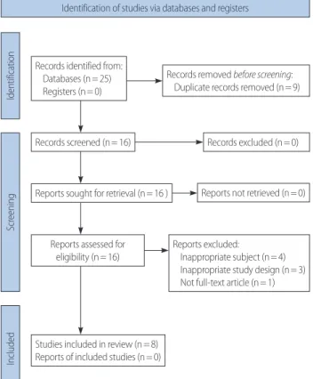
PubMed, CINAHL, psycINFO 검색엔진을 이용하여 검색한 결과 각각 12편, 7편, 6편의 문헌이 도출되었으며, 중복되는 문헌 9편을 제 거하여 총 16편의 논문에 대해 일차적으로 검토를 수행하였다.
RISS, KMBase 검색엔진에서는 0편의 문헌이 검색되었다. 또한 case report 논문 2편, 19세 미만 대상자를 다룬 논문 1편, 대상자가 난민 이 아닌 논문 2편, full-text가 아닌 논문 1편, PTSD 대상자가 아닌 논 문 1편, review 논문 1편을 제외하여 최종적으로 선택된 문헌은 국외 논문 8편이었다(Figure 1). 모든 과정은 2명의 연구자가 독립적으로 수행하여 결과를 비교하였고, 모든 의견이 일치함을 확인하였다.
3) 자료 분석
자료를 추출하기 위해 엑셀 스프레드 시트를 활용하여 자료 기입
Figure 1. Prisma flow diagram.
Identification of studies via databases and registers
IdentificationScreeningIncluded
Records identified from:
Databases (n= 25) Registers (n= 0)
Records screened (n= 16)
Reports sought for retrieval (n= 16 ) Reports not retrieved (n= 0)
Reports excluded:
Inappropriate subject (n= 4) Inappropriate study design (n= 3) Not full-text article (n= 1) Reports assessed for
eligibility (n= 16)
Studies included in review (n= 8) Reports of included studies (n= 0)
Records excluded (n= 0) Records removed before screening:
Duplicate records removed (n= 9)
형식을 자체적으로 개발하여 사용하였다. MRI를 활용한 PTSD 난 민 연구의 특성을 확인하기 위하여 연구유형, 지역, 연구집단, 목적, 측정도구, 주요 결과를 추출하여 기술적 분석을 하였다. 2명의 연구 자가 독립적으로 자료 기입형식을 이용하여, 8편의 개별 논문으로 부터 자료를 반복적으로 추출하면서, 자료 기입형식을 업데이트하 였고, 각자의 자료 추출방식이 연구 질문과 목적에 일치하는지를 모든 연구자가 함께 재검토하고 합의하여 결정하였다.
연구 결과
1. 연구 논문의 특성
총 8편의 논문 중 조사 연구 5편, 실험 연구 3편이 확인되었다. 북 한 이탈주민을 대상으로 한 연구는 3편이었으며, 쿠르드족 난민 및 시리아 난민을 주 대상자로 포함된 연구가 각각 2편이 존재했고, 국 적이 불분명한 난민이 대상자인 연구가 1편이었다. 8편의 연구에서 대상자의 평균 연령은 약 39세 였으며, 연구 대상자로 남성만 포함 된 연구가 5편이었다(Table 1).
2. 뇌의 구조적·기능적 변화
MRI를 통해 뇌의 구조적 변화를 관찰한 연구는 2편, fMRI를 통 해 기능적 변화를 관찰한 연구는 5편, MRS를 통해 뇌의 대사물질 의 변화를 관찰한 연구 2편이 확인되었다. 특히 fMRI를 사용한 연 구는 뇌의 신경학적 변화를 관찰한 실험 연구 3편, 중재 없이 뇌의 휴식기 연결성을 관찰한 연구 2편이 포함되었다(Table 2).
1) 뇌의 구조적 변화
뇌의 구조적 변화를 관찰한 연구는 2편이었는데, 해마와 뇌섬의 부피를 측정한 연구에서는 유의한 부피변화가 나타나지 않았다 [A1]. 반면 대상피질의 다양한 부위(bilateral isthmus of the cingulate, right inferior parietal cortex, bilateral rostral anterior cingulate cortex, left isthmus of the cingulate)의 부피변화를 측정한 연구에서는 건강 한 대상자에 비해 PTSD 증상 유무와 관계없이 트라우마를 경험한 대상자에게는 유의한 부피 감소가 발견되었다[A2].
2) 뇌의 기능적 변화
기능적 변화를 관찰한 연구는 총 5편으로, 중재 없이 뇌의 휴식 기 연결성을 측정한 연구 2편, 중재 시행 후 뇌의 신경활성화 정도를 측정한 연구 3편이었다.
중재 없이 시상과 중심이랑 간 뇌의 휴식기 연결성을 측정한 연 구에서는, 우측 시상과 좌측 중심후이랑 간 기능적 연결성과 좌측
시상과 우측 중심후이랑 간 기능적 연결성이 PTSD 대상자와 건강 한 대상자가 PTSD는 없으나 트라우마에 노출된 대상자보다 더 높 았다. 하지만 좌측 시상과 좌측 중심전이랑 간 기능적 연결성은 건 강한 대상자가, PTSD 대상자와 PTSD는 없으나 트라우마에 노출된 대상자보다 높았다[A3]. 또한, 좌측 편도체와 측두엽 전전두피질 간 기능적 연결성, 그리고 좌측 편도체와 좌측 등 전대상피질 간 기능 적 연결성은 PTSD 발병 비율이 더 큰 북한이탈 주민이 남한 주민보 다 높았다[A4]. PTSD군과 대조군 간 뇌의 기저부위와 피질 간 기능 적 연결성은 어떤 특정부위를 측정했는지에 따라 유의하거나 유의 하지 않았고, 특정 부위에서 PTSD군과 대조군 간 유의한 차이가 발 견된 경우에도 그 부위에 대한 추가연구가 존재하지 않았다.
중재를 포함하여 뇌의 신경활성화 정도를 연구한 3편의 실험연 구에서는 PTSD 증상이 높은 대상자일수록 부정적인 자극이 주어 지면 뇌의 신경활성화 정도가 증가한지만, 보상 중재와 긍정적인 자 극이 주어졌을 때 뇌의 신경활성화 정도가 감소한다는 공통점이 확인되었다. 반면 연구마다 활성화된 뇌 부위는 차이가 존재하였 다. 공포 안면 중재 시행 동안 좌측 해마는 고문을 받은 경험이 있는 집단이 고문을 받은 경험이 없는 집단보다 높은 신경활성화 정도를 보였다[A5]. 긍정적인 사진을 보았을 때 피질 부위에 해당하는 방추 형이랑, 우하측두이랑, 좌중심 후두이랑에서 PTSD 집단이 무증상 집단보다 낮은 신경활성화 정도를 보였다[A6]. 이차성 정신질환은 양극성 장애나 조현병과 같은 일차성 정신질환 없이 환각, 의식 변 화 등 정상적인 현실 인식이 불가능한 상태를 동반하는데[21], 보상 중재가 주어졌을 때, 기저부위인 좌측 선접합 등측 조가비핵과 피 질 부위인 중앙 전전두피질에서 이차성 정신질환이 있는 PTSD환 자가 이차성 정신질환이 없는 PTSD 환자보다 낮은 신경활성화 정 도를 보였다[A7]. 뇌 신경활성도를 파악함으로써 PTSD 증상 유무 뿐만 아니라 PTSD 환자의 이차성 정신질환의 유무를 알아낼 수 있 다는 가능성이 제시되었다.
3) 대사물질 농도 변화
총 2편의 논문에서 대사물질의 농도를 측정하였는데, 측정부위 는 해마와 뇌섬, 전대상피질이다. 해마와 뇌섬에서 PTSD 집단, PTSD가 없지만 트라우마는 있는 집단, 트라우마가 없는 집단 간 유 의한 N-acetyl-aspartate (NAA) 농도차이를 보이지 않았다. 그러나, 전대상피질에서 PTSD 대상자가 non-PTSD 대상자보다 낮은 수치 의 NAA 농도를 보였다[A8]. PTSD 대상자가 모든 뇌 부위에서 NAA 농도의 감소가 발생하는 것이 아님을 알 수 있고, 유의한 변화를 보 인 전대상피질에 대한 추가 연구가 필요함이 확인되었다.
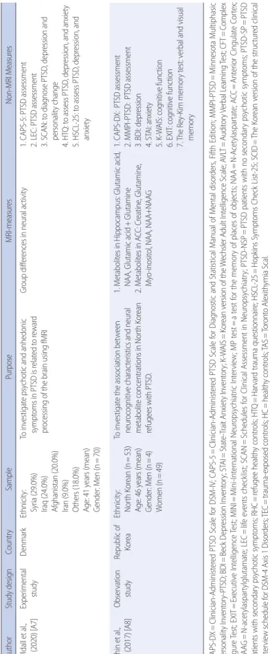
Table 1. Characteristics of the Selected Studies AuthorStudy designCountrySamplePurposeMRI-measuresNon-MRI Measures Eckart et al., (2012) [A1]Observation studyGermanyEthnicity: Albanian (n=1) Serbian (n=1) Romani (n=1) Turkish (n=1) Age: 34 years (mean) Gender: Men (n=47) To investigate the influence of traumatic stress experiences and PTSD on both brain structures and functions in hippocampus and insula 1. Volume of hippocampus and insula 2. NAA and NAA/Creatine ratio in hippocampus and insula
1. CAPS-DX: PTSD assessment 2. MP test: memory test 3. Childhood Trauma Questionnaire: abuse and neglect during childhood 4. M.I.N.I: neuropsychiatric symptoms 5. A shortened version of the Vivo Checklist of War, Detention and Torture Events Eckart et al., (2011) [A2]Observation studyGermanyEthnicity: Kurdish (n=48) Albanian (n=1) Serbian (n=1) Romani (n=1) Turkish (n=1) Age: 33. 5 years (mean) Gender: Men (n=52)
To investigate PTSD-related, structural alterations in cortical regions.Differences in cortical volume (prefrontal, parietal and posterior midline regions)1. CAPS-DX: PTSD assessment 2. M.I.N.I: neuropsychiatric symptoms 3. A shortened version of the Vivo Checklist of War, Detention and Torture Events Jeon et al., (2020) [A3]Observation studyRepublic of KoreaEthnicity: North Korean (n=85) Age: 39 years (mean) Gender: Men (n=20) Women (n=65) To investigate rsFC between the thalamus and cortical brain regions, as well as the relationships of this rsFC with PTSD-related characteristics 1. Differences in resting state functional connectivity in the thalamus 2. Differences in functional connectivity strengths by cortical regions 3. Associations between thalamic rsFC and the clinical features of PTSD
1. CAPS: PTSD assessment 2. BDI: depression 3. Trauma Exposure Check List for North Korean Refugees Kim et al., (2020) [A4]Observation studyRepublic of KoreaEthnicity: North Korean (n=45) South Korean(n=40) Age: 35.5 years (mean) Gender: Men (n=20) Women (n=65)
To investigate in resting state functional connectivity and its relationship with alexithymia in refugees 1. Differences in functional connectivity of the amygdala 2. Correlation between alexithymia and connectivity strength of the amygdala
1. CAPS: PTSD assessment 2. BDI: depression 3. TAS: alexithymia 4. Trauma Exposure checklist for NK Refugees Liddell et al., (2020) [A5]Experimental studyNew ZealandEthnicity: Not Shown Age: 38.8 years (mean) Gender: Men (n=58)
To investigate neurocircuitry associated with threat and reward processing following torture
Group differences in neural activity1. HTQ: trauma assessment 2. DSM-5: to assess PTSD and depression 3. PSSI: PTSD symptom severity 4. HSCL: depression symptom severity Uldall et al., (2020) [A6]Experimental studyDenmarkEthnicity: Syria (29.0%) Iraq (24.0%) Afghanistan (20.0%) Iran (9.0%) Others (18.0%) Age: 42 years (mean) Gender: Men (n=69)
To investigate anhedonia in PTSD using fMRI while presenting positive pictures before and after symptom provocation using personalized Trauma Scripts
Group differences in neural activity1. CAPD-5: to assess current and lifetime PTSD symptoms 2. LEC: PTSD assessment 3. SCAN: to diagnose PTSD, depression and personality change (Continued to the next page)
AuthorStudy designCountrySamplePurposeMRI-measuresNon-MRI Measures Uldall et al., (2020) [A7]Experimental studyDenmarkEthnicity: Syria (29.0%) Iraq (24.0%) Afghanistan (20.0%) Iran (9.0%) Others (18.0%) Age: 41 years (mean) Gender: Men (n=70) To investigate psychotic and anhedonic symptoms in PTSD is related to reward processing of the brain using fMRI
Group differences in neural activity1. CAPS-5: PTSD assessment 2. LEC: PTSD assessment 3. SCAN: to diagnose PTSD, depression and personality change 4. HTQ: to assess PTSD, depression, and anxiety 5. HSCL-25: to assess PTSD, depression, and anxiety Shin et al., (2017) [A8]Observation studyRepublic of KoreaEthnicity: North Korean (n=53) Age: 46 years (mean) Gender: Men (n=4) Women (n=49)
To investigate the association between neurocognitive characteristics and neural metabolite concentrations in North Korean refugees with PTSD.
1. Metabolites in Hippocampus: Glutamic acid, NAA, Glutamic acid+Glutamine 2. Metabolites in ACC: Creatine, Glutamine, Myo-inositol, NAA, NAA+NAAG
1. CAPS-DX: PTSD assessment 2. MMPI-PTSD: PTSD assessment 3 .BDI: depression 4. STAI: anxiety 5. K-WAIS: cognitive function 6. EXIT: cognitive function 7. The Rey-Kim memory test: verbal and visual memory CAPS-DX=Clinician-Administered PTSD Scale for DSM-IV; CAPS-5=Clinician-Administered PTSD Scale for Diagnostic and Statistical Manual of Mental disorders, Fifth Edition; MMPI-PTSD=Minnesota Multiphasic Personality Inventory-PTSD; BDI=Beck Depression Inventory; STAI=State-Trait Anxiety Inventory; K-WAIS=Korean version of the Wechsler Adult Intelligence Scale; AVLT=Auditory Verbal Learning Test; CFT=Complex Figure Test; EXIT=Executive Intelligence Test; MINI=Mini-International Neuropsychiatric Interview; MP test=a test for the memory of places of objects; NAA=N-Acetylaspartate; ACC=Anterior Cingulate Cortex; NAAG=N-acetylaspartylglutamate; LEC=life events checklist; SCAN=Schedules for Clinical Assessment in Neuropsychiatry; PTSD-NSP=PTSD patients with no secondary psychotic symptoms; PTSD-SP=PTSD patients with secondary psychotic symptoms; RHC=refugee healthy controls; HTQ=Harvard trauma questionnaire; HSCL-25=Hopkins Symptoms Check List-25; SCID=The Korean version of the structured clinical interview schedule for DSM-4 Axis 1 Disorders; TEC=trauma-exposed controls; HC=healthy controls; TAS=Toronto Alexithymia Scal.
Table 1. Continued
논 의
본 연구는 PTSD 난민을 대상으로 MRI를 활용한 연구를 고찰함 으로써, 향후 북한이탈주민과 같은 난민 대상 PTSD 진단 및 간호 중재 평가를 위한 객관적인 지표 탐색을 위해 시행된 주제범위 문 헌 고찰 연구이다. PTSD 난민에게 사용할 수 있는 뇌 바이오마커 탐색을 위해 MRI 활용 연구에 관한 주제 범위 문헌 고찰을 시행한 결과, 아직 연구가 8편으로 그 수가 제한적임을 확인하였다.
최근 정신건강 분야 연구에서 MRI를 PTSD 진단과 중재의 평가 에 활용하는 연구가 증가하고 있는데[22-24], 대부분 전쟁을 경험한 군인, 성폭력을 경험한 여성, 아동기 학대 경험이 있는 청소년 등을 대상으로 연구가 활발히 진행되고 있다[25-28]. PTSD난민을 대상으 로 한 연구에서 MRI를 활용한 연구가 8편으로 제한된 것은 난민이 가지고 있는 사회문화적 환경과 관련이 있다. 특정 지역 또는 캠프 에 거주하는 난민을 연구에 참여시키기 위해서는 관계기관의 협조 가 필요하고, 캠프 방문이 가능해지더라도 난민의 어려운 사회경제 Table 2. Outcomes of the Selected Studies
Author Characteristic of participants MRI outcome
Eckart et al., PTSD: Traumatized non-PTSD: Healthy subject (n)= 20:16:11 Volume
(2012) [A1] CAPS DX-score Hippocampus: No significant difference among 3 group.
PTSD: Traumatized= 68.90:13.05 (p< .001) Insula: No significant difference among 3 group.
NAA concentration
Left and right hippocampus: No significant difference among 3 group.
Insula: No significant difference among 3 group Eckart et al., PTSD: Traumatized non-PTSD: Healthy subject (n)= 20:19:13 Volume
(2011) [A2] CAPS DX-score
PTSD: Traumatized= 68.90:13.05 (p< .001)
Bilateral isthmus of the cingulate: Traumatized< nontraumatized (p= .017), PTSD< nontraumatized (p= .013)
Right inferior parietal cortex: Traumatized< nontraumatized (p= .049), PTSD< nontraumatized (p= .001)
Bilateral rostal anterior cingulate cortex: Traumatized< nontraumatized (p= 0.026), PTSD< nontraumatized (p= .004)
Left isthmus of the cingulate: Traumatized< nontraumatized (p= .02), PTSD< nontraumatized (p= .002)
Jeon et al., PTSD: Traumatized non-PTSD: Healthy subject (n)= 23:22:40 Resting state functional connectivity (rsFC)
(2020) [A3] CAPS total, current (mean) Right thalamus and left postcentral gyrus: PTSD,HC> TEC (p= .001) PTSD: Traumatized non-PTSD= 41.22:5.55 (p< .001) Left thalamus and left precentral gyrus: HC> PTSD, TEC (p= .018) CAPS total, lifetime (mean) Left thalamus and right postcentral gyrus: PTSD, HC> TEC (p= .002) PTSD: Traumatized non-PTSD= 68.96:12.00 (p< .001)
Kim et al., NK with PTSD: NK without PTSD (n)= 10:35 Functional connectivity (2020) [A4] Current CAPS total of NK (mean)= 23.78
Lifetime CAPS total of NK (mean)= 41.11
Left amygdala and bilateral dorsolateral prefrontal cortex (dlPFC): NK> SK (p= .047: left, p= .003: right)
Left amygdala and left dorsal anterior cingulate cortex (dACC): NK> SK (p= .001) Liddell et al., Torture survivor group: Non-torture survivor group (n)= 31:27 Neural activity in happy face processing
(2020) [A5] PTSD diagnosis (DSM-5) Right ventral striatum: TS< NTS (p< .001) TS group: NTS group= 14:10 ( p= .531) Neural activity in fear face processing
Left hippocampus: TS> NTS (p< .001)
Uldall SW et al., PTSD: Healthy subject (n)= 38:31 Neural activity when viewing positive pictures (2020) [A6] CAPS-5 score (PTSD participants) Fusiform gyrus: RHC> PTSD (p< .001)
Arousal symptoms (mean) Right inferior temporal gyrus : RHC> PTSD (p< .001) PTSD: Non-PTSD= 17.5:8.3 (p< .001) Left middle occipital gyrus : RHC> PTSD (p< .001) Avoidance symptoms (mean)
PTSD: Non-PTSD= 7.8:3.6 (p= .002)
Correlation between neural activity and anhedonia when viewing positive pictures
Dissociation symptoms (mean) Neural activity & anhedonia: Negative (p= .003) PTSD: Non-PTSD= 7.8:3.5 (p= .003)
Uldall SW et al., PTSD-NSP: PTSD-SP: RHC (n)= 18:21:31 Neural activity with monetary reward task
(2020) [A7] Harvard Trauma Questionnaire (mean) Left precommissural dorsal putamen (pre-DPU): PTSD-SP< PTSD-NSP (p< .050) PTSD-NSP: PTSD-SP: RHC= 2.8:3.1:1.4 (p= .010) Medial prefrontal cortex (mPFC): PTSD< RHC (p= .007)
Shin et al., PTSD: non-PTSD (n)= 30:23 NAA concentration
(2017) [A8] CAPS-DX-score Anterior Cingulate Cortex (ACC): PTSD< Non-PTSD (p< .019)
PTSD: Non-PTSD= 51.7:13.5 (p< .001)
적 여건은 연구 참여의 제한이 되는 것으로 보고되고 있다[29].
본 연구에서 확인된 PTSD 난민을 대상으로 MRI를 사용하여 뇌 의 구조적·기능적 변화를 측정한 연구는 독일, 덴마크, 뉴질랜드에 서 시행된 연구가 5편, 남한에서 북한이탈주민을 대상으로 한 연구 가 3편이었다. 해외에서 연구된 문헌 5편은 모두 남성만을 연구 대 상자로 선정한 반면, 국내에서 연구된 문헌 3편의 연구 대상자는 여 성이 남성보다 더 많이 포함되었다. 이는 북한이탈주민 중 여성이 71%라는 사실과 관련이 있다[30]. 일반적으로 여성은 남성보다 PTSD에 취약한 것으로 알려져 있는데[31], 이러한 성별의 차이는 고 정된 성역할이나 유전, 그리고 호르몬이 PTSD에 영향을 주기 때문 이다[32]. 또한 성별에 따라 경험한 트라우마의 종류와 빈도도 다른 것으로 알려져 있는데, 남성은 주로 강제추방이나 수감, 반면에 여 성은 성폭력이나 납치에 주로 노출되어 트라우마를 경험하는 것으 로 알려져 있다[33]. 따라서 향후 PTSD 대상자를 선정하여 연구를 진행할 때에는 이러한 성별의 차이를 고려할 필요가 있다.
뇌의 구조적 변화를 측정한 문헌 2편에서는 해마와 뇌섬, 또는 전 대뇌피질 부위의 부피를 측정하였는데, 해마는 대상을 인식하고 맥 락을 파악하는 등 현실적인 상황 파악에 관여하고[34], 뇌섬은 공포 상황에서 뇌의 가교 역할 및 감정의 상기를 담당하는 것으로 알려 져 있다[35]. 사건의 재경험과 자극으로부터의 회피가 PTSD의 주된 증상이므로, 해마와 뇌섬의 기능은 PTSD증상과 관련이 높아 연구 의 주된 관심부위이다[36]. 참전군인을 대상으로 PTSD 심각도와 해 마의 부피 간 상관관계를 연구한 결과, PTSD 심각도가 높을수록 해마의 부피가 감소하였고[37], 뇌섬 또한 PTSD 대상자에서 부피가 감소됨이 보고된 바 있다[38]. 그러나 본 연구에서 확인된 PTSD 난 민을 대상으로 해마와 뇌섬의 부피변화를 관찰한 문헌은 1편뿐으 로, 해마와 뇌섬의 부피 변화가 PTSD 난민에게 유의하게 나타나는 지 알아보기 위해서는 추가적인 연구가 필요할 것이다.
전대상피질은 해마와 편도체 등의 감정조절 변연계와 연결되어 있어 자극과 반응에 주요한 역할을 수행하며, 특히 공포반응에 대 한 선택적 집중과 소멸을 담당한다는 점에서 공포에 대한 과활성화 가 발생하는 PTSD증상과 연관성이 깊다[39]. 142명의 참전 군인을 대상으로 전대상피질의 부피와 PTSD 증상의 상관관계를 연구한 결과, 전대상피질의 부피가 더 작은 대상자일수록 상황에서 더 높 은 신경흥분상태를 보인다는 선행연구 결과는 이를 뒷받침한다 [40]. PTSD난민에서 전대상피질의 위축은 자극의 습관화를 방해하 여 위협적 자극에 대한 과반응을 유도하는 것으로 보인다.
PTSD대상자의 뇌의 기능적 변화를 알아보기 위해 중재 없이 시 상과 중심이랑 간 뇌의 휴식기 연결성을 측정한 2편의 연구결과에 서, 좌측 시상과 좌측 중심전이랑 간 기능적 연결성은 PTSD대상자
가 건강한 대상자보다 낮은 반면[A3], 좌측 편도체와 측두엽 전전두 피질 간 기능적 연결성, 그리고 좌측 편도체와 좌측 등 전대상피질 간 기능적 연결성은 PTSD대상자가 유의하게 높게 나타나[A4], 뇌의 휴식기 연결성을 난민 PTSD의 진단 바이오마커로 활용하기 위해 서는 추가적인 연구가 필요함을 확인하였다.
특정 뇌 부위에서 PTSD대상자와 건강한 대상자 간의 신경활성 도 차이를 보기 위해 긍정안면중재 및 공포안면중재와 보상중재를 사용하기도 하였는데, PTSD 대상자가 대조군에 비해 긍정 안면 중 재 및 보상중재에는 더 낮은 신경활성화정도를, 공포안면중재에는 더 높은 신경활성화정도를 보였다[A5,A6,A7]. 이는 편도체-청반-전 대상 회로에서, PTSD 대상자에게 발견되는 원심성 비아드레날린성 투사가 청반으로부터 이루어지면 전대상의 공포 조건화 기능이 저 하되어 공포 반응에 대한 과활성화가 발생하는 것으로 여겨진다 [41]. 또한 PTSD 대상자에게서 발견되는 편도체와 전대상피질 사이 의 유의한 휴식기 연결성은 기억의 재구성을 통해 단편적인 공포자 극만 등장해도 과거의 트라우마에서 벗어나지 못하고 괴로워하는 데 기여하는 것으로 유추된다[A3]. 즉, 트라우마 경험 이후 공포자 극을 담당하는 편도체와 전대상피질 간 휴식기 연결성이 높아지 고, 편도체-청반-전대상 회로의 비아드레날린성 투사로 인한 공포 자극 과활성화가 복합적으로 나타나 PTSD 증상 중 하나인 공포자 극에 대한 과활성화 반응을 일으키는 것으로 사료된다. 따라서, 긍 정적 자극에는 신경이 둔해지지만, 부정적 자극에는 신경이 과활성 화되는 PTSD 대상자의 뇌의 기전을 이해하고, 이들을 위한 간호중 재 개발 및 평가에 활용할 필요가 있다.
뇌의 대사물질 변화를 연구한 문헌은 2편으로, 전대상피질의 NAA수치는 PTSD대상자가 건강한 대상자에 비해 낮게 측정된 반 면[A8], 해마와 뇌섬에서는 대사물질 농도의 집단 간 유의미한 차이 를 보이지 않았다[A1]. NAA는 단백질 안정성을 유지하는 뇌에 존재 하는 유기화합물로, 단백질 접힘 오류로 유발되는 알츠하이머병, 파킨슨병, 헌팅턴 무도병 등의 신경퇴행성 질환의 바이오마커로 가 장 널리 활용되고 있다[42]. 과거에 동물실험에서 해마 NAA 수치의 감소가 PTSD증상을 예견하는 지표로 쓰일 가능성이 제시되었으 며[43], 최근 47명의 성인 대상자를 대상으로 진행된 연구에서, PTSD대상자에게도 해마의 NAA수치의 감소가 발견되었다[44]. 전 대상피질이 해마와 편도체의 신경활동을 제어하는 특성을 고려할 때[45], PTSD 대상자가 건강한 대상자보다 전대상피질에서 NAA 농 도가 낮게 발견된 연구 결과는 전대상피질이 해마보다 더 선제적인 PTSD예측지표로서의 활용 가능성을 제시하고 있다.
최근 기술의 발달로 인하여, 간호중재를 개발하고 평가하는 데 있어서 환자보고 증상 이외에도 객관적인 데이터를 활용할 필요성
이 대두되고 있다. 특히, 임상전문가에 의한 임상적 진단에 의존하 는 PTSD 관련 분야에서 MRI를 활용한 진단 및 간호중재 평가가 가 능해진다면 문화권이 다른 국가나 사회에서 이주한 난민을 대상으 로 PTSD에 대한 조기 스크리닝과 적극적인 간호중재 도입이 가능 해질 것으로 예상된다. 향후 fMRI 데이터를 기반으로 대상자의 PTSD상태와 심각성, 증상 등을 예측하고 평가할 수 있다면 PTSD 를 가진 대상자들에게 보다 질 높은 간호를 조기에 제공할 수 있을 것으로 기대된다. 또한 이러한 연구는 북한이탈주민을 대상으로 PTSD를 조기 스크리닝하고 필요한 간호중재를 개발하고 평가하는 간호연구를 활성화하는데도 기여할 것으로 예측된다.
결 론
PTSD 난민을 대상으로 뇌영상기법을 사용한 연구에 관해 주제범 위 문헌 고찰을 한 결과 최종 선택된 논문은 8편으로, 아직 연구가 제 한적으로 이루어짐이 확인되었다. 제한된 논문 편수에도 불구하고 MRI를 통해 PTSD가 구조적·기능적 변화를 일으키는 뇌 부위는 다 양하게 탐색되고 있어 아직 연구가 초기단계임이 확인되었다. PTSD 와 뇌의 구조적 또는 기능적 변화와의 관계를 파악하는 체계적인 연 구는 향후 간호사에 의한 데이터 기반 조기 스크리닝과 비침습적 간 호중재의 도입과 평가에서 유용한 도구로 활용될 것으로 기대되므 로 관련 연구가 간호학 분야에서 활발히 연구될 필요가 있다.
CONFLICT OF INTEREST The authors declared no conflict of interest.
AUTHOSHIPS
CSH contributed to the conception and design of this study; YJS and KMS collected data; YJS performed the analysis and interpretation; YJS and KMS drafted the manuscript; YJS and CSH critically revised the manuscript; CSH supervised the whole study process. All authors read and approved the final manuscript.
REFERENCES
1. American Psychiatric Association. Diagnostic and statistical manual of mental disorders. 5th ed. Washington, DC: American Psychiatric Association.; 2013.
p.271-280. https://doi.org/10.1176/appi.books.9780890425596
2. Bisson JI. Post-traumatic stress disorder. British Medical Journal Publishing
Group. 2007;334(7597):789-793. https://doi.org/10.1136/bmj.39162.538553.80 3. Pant S, Eder B, Vračar A, Mosca D, Orcutt M. WHO’s global action plan to pro- mote the health of refugees and migrants. British Medical Journal Publishing Group. 2019;366:l4806. https://doi.org/10.1136/bmj.l4806.
5. Fazel M, Wheeler J, Danesh J. Prevalence of serious mental disorder in 7000 ref- ugees resettled in western countries: a systematic review. The Lancet. 2005;
365(9467):1309-1314. https://doi.org/10.1016/S0140-6736(05)61027-6 6. Bauer M, Priebe S. Psychopathology and long-term adjustment after crises in
refugees from East Germany. International journal of social psychiatry. 1994;
40(3):165-176. https://doi.org/10.1177/002076409404000302
7. Kang JW. Human rights and refugee status of the North Korean Diaspora.
North Korean Review. 2013;9(2):4-17.
8. Song YH. Current status and issues of North Korean defector migration abroad.
JPI Policy Forum. 2012;107(0):1-24.
9. Noland M, Haggard S, Chang Y, Kurlantzick J, Lankov A, Mason J. The North Korean refugee crisis: human rights and international response. Washington, DC: U.S. Committee for Human Rights in North Korea; 2006. p. 23-30.
10. Kim YH, Cho YA. A study on the prevalence and the influencing factors of the mental health problems among recent migrant North Koreans: a focus on 2007 entrants. Unification Policy Studies. 2010;19(2):141-174.
11. Han NY, Lee SH, Yoo SY, Kim SJ, Jun JY, Won SD, et al. Predictors of PTSD among North Korean defectors visited psychiatric department of North Korean defectors treatment center. Journal of Korean Neuropsychiatric Association.
2015;54(1):105-111. https://doi.org/10.4306/jknpa.2015.54.1.105
12. Steel Z, Chey T, Silove D, Marnane C, Bryant RA, van Ommeren M. Associa- tion of torture and other potentially traumatic events with mental health out- comes among populations exposed to mass conflict and displacement: a sys- tematic review and meta-analysis. The Journal of the American Medical Asso- ciation. 2009;302(5):537-549. https://doi.org/10.1001/jama.2009.1132.
13. Marshall EA, Butler K, Roche T, Cumming J, Taknint JT. Refugee youth: a re- view of mental health counselling issues and practices. Canadian Psychology.
2016;57(4):308-319. https://doi.org/10.1037/cap0000068.
14. Ministry of Health and Welfare. Epidemiological survey of psychiatric illnesses in Korea[Internet]. Seoul: Statistics Korea; 2017 [cited 2020 Mar 22]. Available from: http://kosis.kr/statHtml/statHtml.do?orgId=117&tblId=TX_117_2009_
HB121&conn_path=I2
15. Miao XR, Chen QB, Wei K, Tao KM, Lu ZJ. Posttraumatic stress disorder: from diagnosis to prevention. Military Medical Research. 2018;5(1):32. https://doi.
org/10.1186/s40779-018-0179-0.
16. Clarke SK, Jaffe J, Mutch R. Overcoming communication barriers in refugee health care. Pediatric Clinics of North America. 2019;66(3):669-686. Epub 2019/05/01. https://doi.org/10.1016/j.pcl.2019.02.012
17. Bolzenius JD, Velez CS, Lewis JD, Bigler ED, Wade BS, Cooper DB, et al. Diffu- sion imaging findings in US service members with mild traumatic brain injury and posttraumatic stress disorder. The Journal of Head Trauma Rehabilitation.
2018;33(6):393-402. https://doi.org/10.1097/htr.0000000000000378 18. Logue MW, van Rooij SJ, Dennis EL, Davis SL, Hayes JP, Stevens JS, et al. Small-
er hippocampal volume in posttraumatic stress disorder: a multisite ENIGMA- PGC study: subcortical volumetry results from posttraumatic stress disorder consortia. Biological psychiatry. 2018;83(3):244-253. https://doi.org/10.1016/
j.biopsych.2017.09.006
19. O’Doherty DC, Chitty KM, Saddiqui S, Bennett MR, Lagopoulos J. A system- atic review and meta-analysis of magnetic resonance imaging measurement of
structural volumes in posttraumatic stress disorder. Psychiatry Research. 2015;
232(1):1-33. https://doi.org/10.1016/j.pscychresns.2015.01.002
20. Milani AC, Hoffmann EV, Fossaluza V, Jackowski AP, Mello MF. Does pediatric post-traumatic stress disorder alter the brain? Systematic review and meta-anal- ysis of structural and functional magnetic resonance imaging studies. Psychia- try and Clinical Neurosciences. 2017;71(3):154-169. https://doi.org/10.1111/
pcn.12473
21. Keshavan MS, Kaneko Y. Secondary psychoses: an update. World Psychiatry.
2013;12(1):4-15. https://doi.org/10.1002/wps.20001
22. Begemann MJ, Schutte MJ, van Dellen E, Abramovic L, Boks MP, van Haren NE, et al. Childhood trauma is associated with reduced frontal gray matter vol- ume: a large transdiagnostic structural MRI study. Psychological Medicine.
2021:1-9. https://doi.org/10.1017/s0033291721002087
23. Matthijssen X, Wouters F, Sidhu N, van der Helm-van Mil A. Value of imaging detected joint inflammation in explaining fatigue in RA at diagnosis and during the disease course: a large MRI study. RMD Open. 2021;7(2):e001599. https://
doi.org/10.1136/rmdopen-2021-001599
24. Ranti D, Murrough J, Balchandani P, Morris L. Trauma Exposure and Perivas- cular Spaces in Depression: a 7-Tesla MRI study. Biological Psychiatry. 2021;
89(9):S188-S189. https://doi.org/10.1016/j.biopsych.2021.02.478
25. Fu S, Ma X, Li C, Wang T, Li C, Bai Z, et al. Aberrant regional homogeneity in post-traumatic stress disorder after traffic accident: a resting-state functional MRI study. NeuroImage: Clinical. 2019;24:101951. https://doi.org/10.1016/j.
nicl.2019.101951
26. Lebois LAM, Li M, Baker JT, Wolff JD, Wang D, Lambros AM, et al. Large-scale functional brain network architecture changes associated with trauma-related dissociation. The American Journal of Psychiatry. 2021;178(2):165-173. https://
doi.org/10.1176/appi.ajp.2020.19060647
27. Berman Z, Assaf Y, Tarrasch R, Joel D. Assault-related self-blame and its associa- tion with PTSD in sexually assaulted women: an MRI inquiry. Social Cognitive and Affective Neuroscience. 2018;13(7):775-784. https://doi.org/10.1093/scan/
nsy044
28. Lindauer RJ, Vlieger EJ, Jalink M, Olff M, Carlier IV, Majoie CB, et al. Effects of psychotherapy on hippocampal volume in out-patients with post-traumatic stress disorder: a MRI investigation. Psychological Medicine. 2005;35(10):1421- 1431. https://doi.org/10.1017/s0033291705005246
29. Halabi JO. Nursing research with refugee clients: a call for more qualitative ap- proaches. International Nursing Review. 2005;52(4):270-275. https://doi.org/
10.1111/j.1466-7657.2005.00440.x
30. Kim E, Yun M, Jun JY, Park WS. Pre-migration trauma, repatriation experienc- es, and PTSD among North Korean refugees. Journal of Immigrant and Minor- ity Health. 2019;21(3):466-472. https://doi.org/10.1007/s10903-018-0742-5 31. Ainamani HE, Elbert T, Olema DK, Hecker T. Gender differences in response
to war-related trauma and posttraumatic stress disorder–a study among the Congolese refugees in Uganda. BMC Psychiatry. 2020;20(1):17. https://doi.org/
10.1186/s12888-019-2420-0
32. Christiansen DM, Berke ET. Gender-and sex-based contributors to sex differ- ences in PTSD. Current Psychiatry Reports. 2020;22(4):19. https://doi.org/10.
1007/s11920-020-1140-y
33. Rodolico A, Vaccino N, Riso MC, Concerto C, Aguglia E, Signorelli MS. Preva- lence of post-traumatic stress disorder among asylum seekers in Italy: a popula- tion-based survey in sicily. Journal of Immigrant Minor Health. 2020;22(3):
634-638. https://doi.org/10.1007/s10903-019-00948-9
34. Milani ACC, Hoffmann EV, Fossaluza V, Jackowski AP, Mello MF. Does pediat- ric post-traumatic stress disorder alter the brain? Systematic review and meta- analysis of structural and functional magnetic resonance imaging studies. Psy- chiatry and Clinical Neurosciences. 2017;71(3):154-169. https://doi.org/10.
1111/pcn.12473
35. Craig AD. How do you feel--now? The anterior insula and human awareness.
Nature Reviews Neuroscience. 2009;10(1). https://doi.org/10.1038/nrn2555 36. Lambert HK, McLaughlin KA. Impaired hippocampus-dependent associative
learning as a mechanism underlying PTSD: a meta-analysis. Neuroscience &
Biobehavioral Reviews. 2019;107:729-749. https://doi.org/10.1016/j.neubior- ev.2019.09.024
37. Gilbertson MW, Shenton ME, Ciszewski A, Kasai K, Lasko NB, Orr SP, et al.
Smaller hippocampal volume predicts pathologic vulnerability to psychological trauma. Nature Neuroscience. 2002;5(11):1242-1247. https://doi.org/10.1038/
nn958
38. Chen S, Li L, Xu B, Liu J. Insular cortex involvement in declarative memory def- icits in patients with post-traumatic stress disorder. BMC Psychiatry. 2009;9:39.
https://doi.org/10.1186/1471-244x-9-39
39. Rauch SL, Shin LM, Phelps EA. Neurocircuitry models of posttraumatic stress disorder and extinction: human neuroimaging research—past, present, and fu- ture. Biological Psychiatry. 2006;60(4):376-382. https://doi.org/10.1016/j.bio- psych.2006.06.004
40. Young DA, Chao L, Neylan TC, O’Donovan A, Metzler TJ, Inslicht SS. Associa- tion among anterior cingulate cortex volume, psychophysiological response, and PTSD diagnosis in a Veteran sample. Neurobiology of Learning and Mem- ory. 2018;155:189-196. https://doi.org/10.1016/j.nlm.2018.08.006
41. Hamner MB, Lorberbaum JP, George MS. Potential role of the anterior cingu- late cortex in PTSD: review and hypothesis. Depress Anxiety. 1999;9(1):1-14.
42. Warepam M, Ahmad K, Rahman S, Rahaman H, Kumari K, Singh LR. N- Acetylaspartate is an important brain osmolyte. Biomolecules. 2020;10(2):286.
https://doi.org/10.3390/biom10020286
43. Siegmund A, Kaltwasser SF, Holsboer F, Czisch M, Wotjak CT. Hippocampal N-acetylaspartate levels before trauma predict the development of long-lasting posttraumatic stress disorder-like symptoms in mice. Biological Psychiatry.
2009;65(3):258-262. https://doi.org/10.1016/j.biopsych.2008.08.023 44. Rosso IM, Crowley DJ, Silveri MM, Rauch SL, Jensen JE. Hippocampus gluta-
mate and N-acetyl aspartate markers of excitotoxic neuronal compromise in posttraumatic stress disorder. Neuropsychopharmacology. 2017;42(8):1698- 1705. https://doi.org/10.1038/npp.2017.32
45. Hamner MB, Lorberbaum JP, George MS. Potential role of the anterior cingu- late cortex in PTSD: review and hypothesis. Depression and anxiety. 1999;
9(1):1-14.
Appendix: List of studies reviewed
A1. Eckart C, Kaufmann J, Kanowski M, Tempelmann C, Hinrichs H, Elbert T, et al. Magnetic resonance volumetry and spectroscopy of hippocampus and in- sula in relation to severe exposure of traumatic stress. Psychophysiology. 2012;
49(2):261-270. https://doi.org/10.1111/j.1469-8986.2011.01303.x.
A2. Eckart C, Stoppel C, Kaufmann J, Tempelmann C, Hinrichs H, Elbert T, et al.
Structural alterations in lateral prefrontal, parietal and posterior midline re- gions of men with chronic posttraumatic stress disorder. Journal Psychiatry and Neurosciences. 2011;36(3):176-86. Epub 2010/12/02. https://doi.org/10.
1503/jpn.100010
A3. Jeon S, Lee YJ, Park I, Kim N, Kim S, Jun JY, et al. Resting State Functional Con- nectivity of the Thalamus in North Korean Refugees with and without Post- traumatic Stress Disorder. Scientific Reports. 2020;10(1):3194. https://doi.
org/10.1038/s41598-020-59815-5
A4. Kim N, Park I, Lee YJ, Jeon S, Kim S, Lee KH, et al. Alexithymia and frontal- amygdala functional connectivity in North Korean refugees. Psychological medicine. 2020;50(2):334-41. https://doi.org/10.1017/s0033291719000175.
A5. Liddell B, Malhi GS, Felmingham KL, Cheung J, Outhred T, Das P, et al. The impact of torture on interpersonal threat and reward neurocircuitry. The Aus- tralian and New Zealand Journal of Psychiatry. 2021;55(2):153-166, https://doi.
org/10.1177/0004867420950819
A6. Uldall SW, Madsen KH, Siebner HR, Lanius R, Frewen P, Fischer E, et al. Pro- cessing of Positive Visual Stimuli Before and After Symptoms Provocation in Posttraumatic Stress Disorder-A Functional Magnetic Resonance Imaging Study of Trauma-Affected Male Refugees. Chronic Stress (Thousand Oaks).
2020;4:2470547020917623. https://doi.org/10.1177/2470547020917623 A7. Uldall SW, Nielsen M, Carlsson J, Glenthøj B, Siebner HR, Madsen KH, et al.
Associations of neural processing of reward with posttraumatic stress disorder and secondary psychotic symptoms in trauma-affected refugees. European Journal of Psychotraumatology. 2020;11(1):1730091. https://doi.org/10.1080/2 0008198.2020.1730091
A8. Shin JE, Choi CH, Lee JM, Kwon JS, Lee SH, Kim HC, et al. Association be- tween memory impairment and brain metabolite concentrations in North Ko- rean refugees with posttraumatic stress disorder. PLoS One. 2017;12(12):
e0188953. https://doi.org/10.1371/journal.pone.0188953.
![Table 1. Characteristics of the Selected Studies AuthorStudy designCountrySamplePurposeMRI-measuresNon-MRI Measures Eckart et al., (2012) [A1]Observation studyGermanyEthnicity:Albanian (n=1) Serbian (n=1) Romani (n=1) Turkish (n=1) Age: 34 years (mean) Ge](https://thumb-ap.123doks.com/thumbv2/123dokinfo/4853094.284797/5.892.116.797.131.1102/characteristics-selected-authorstudy-designcountrysamplepurposemri-measuresnon-measures-observation-studygermanyethnicity.webp)