Electromyographic Analysis of Thoracic and Lumbar Erector Spinae Activity Using the Abdominal Drawing-in Maneuver and Chin Tuck During Prone Thoracic
Extension Exercises
Ki-song Kim
1, MPH, PT, Gyu-wan Lee
l, MSc, PT, Dong-joon Choi
2, BHSc, PT, Heon-seock Cynn
3, PhD, PT, Oh-yun Kwon
3, PhD, PT
1Dept. of Physical Therapy, Gangnam Severance Hospital Yonsei University, College of Medicine,
2Dept. of Ergonomic Therapy, The Graduate School of Health Science, Yonsei University,
3Dept. of Physical Therapy, College of Health Science, Yonsei University, Dept. of Ergonomic Therapy, The Graduate School of Health Science, Yonsei University
Abstract 1)
This present study investigated the effects of the abdominal drawing-in maneuver (ADIM) and chin tuck (CT) on middle thoracic erector spinae, lower thoracic erector spinae, and lumbar erector spinae muscle activity during three prone thoracic extension (PTE) exercises. Twelve healthy subjects performed preferred PTE, ADIM PTE, and ADIM-CT PTE. Surface electromyography was used to collect data on the muscle activity of dominant middle and lower thoracic erector spinae muscles and the lumbar erector spinae. Middle and lower thoracic erector spinae muscle activity significantly increased when ADIM and CT was performed (p<.05). However, lumbar erector spinae muscle activity significantly decreased in ADIM PTE compared to preferred PTE (p=.017) and significantly increased in ADIM-CT PTE compared to ADIM PTE (p=.004). In conclusion, ADIM-CT PTE effectively increased middle and lower thoracic erector spinae muscle activity, and ADIM PTE decreased lumbar erector spinae muscle activity. Hence, ADIM PTE could be a recommended exercise maneuver to strengthen thoracic erector spinae without over activation of lumbar erector spinae.
[Ki-song Kim, Gyu-wan Lee, Dong-joon Choi, Heon-seock Cynn, Oh-yun Kwon. Electromyographic Analysis of Thoracic and Lumbar Erector Spinae Activity Using the Abdominal Drawing-in Maneuver and Chin Tuck During Prone Thoracic Extension Exercises. Phys Ther Kor. 2012;19(4):1-7.]
Key Words: Abdominal drawing-in maneuver; Chin tuck; Prone thoracic extension; Surface electromyography.
Introduction
The weakness or fatigue of trunk muscles can in- crease the risk of neuromuscular deficits, which, ac- cording to Panjabi's theory of the spinal stabilizing system, causes uncontrolled intervertebral movements (Granata and Gottipati, 2008; Panjabi, 1992). Thoracic extension exercise is performed to activate or strengthen weak and fatigued thoracic erector spinae
(TES). Various thoracic extension exercises are ap- plied in the clinical setting and previous studies have reported positive results (Pfeifer et al, 2004; Sinaki, 1982; Sinaki and Lynn, 2002; Sinaki et al, 2005).
However, prone thoracic extension (PTE) can in-
duce an undesirable compensatory deviation. Previous
studies showed that lumbar extension can increase
with exaggerated anterior pelvic tilt and/or cervical
lordosis, leading to microtrauma and pain during
Corresponding author: Oh-yun Kwon [email protected]
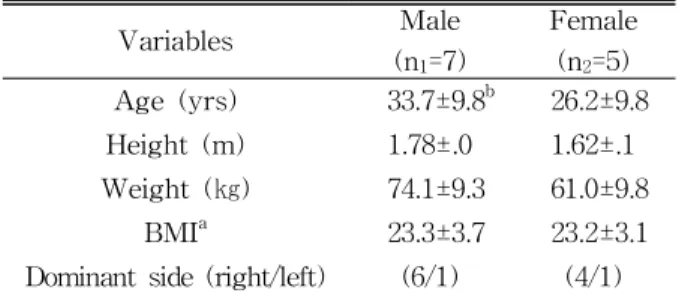
Variables Male (n
1=7)
Female (n
2=5) Age (yrs) 33.7±9.8
b26.2±9.8 Height (m) 1.78±.0 1.62±.1 Weight (㎏) 74.1±9.3 61.0±9.8
BMI
a23.3±3.7 23.2±3.1
Dominant side (right/left) (6/1) (4/1)
a
body mass index,
bmean±standard deviation.
Table 1. General characteristics for this subjects (N=12) PTE (Sinaki et al, 1996). Moreover, excessive use of
lumbar erector spinae (LES) is often associated with chronic low back pain (Holmström et al, 1992).
Furthermore, repetitive lumbar extension and anterior pelvic tilt may contribute to over activation of LES and less activation of TES. Hence, prescribing PTE alone may also increase cervical and/or lumbar lor- dosis (Kuramoto et al, 2011), which is not desirable for individuals with lumbar extension syndrome.
Several modes of exercise have been developed for improving the muscular function of the back (Menacho et al, 2010). Hongo et al (2007) found that modified PTE is required for preventing unwanted effects in order to increase the strength of TES and the quality of life of patients with osteoporosis (Hongo et al, 2007). Another previous study advo- cated internal or external stabilization for the appli- cation of TES without increasing lumbar lordosis (Kisner and Colby, 2007).
Abdominal drawing-in maneuver (ADIM) was ad- vocated to stabilize lumbopelvis and to reduce un- wanted compensation in various previous studies (Cynn et al, 2006; Oh et al, 2007; Park et al, 2011).
The pressure biofeedback unit was utilized in differ- ent body position to educate and confirm the lumbo- pelvic stabilization (Richardson et al, 2004).
The chin tuck (CT) exercise has been recom- mended to provide craniocvercial stability and to ac- tivate deep cervical stabilizers, such as rectus capitis anterior, rectus capitis lateralis, longus capitis, and longus colli. In particular, weakness of deep cervical stabilizers and over activation of global muscles (e.g., sternocleidomastoid and scalenus muscles) were no- ticed in patients with cervical pain. CT has been practiced in the clinical interventions for patients with chronic cervical pain to correct the forward head posture. In the previous study, there was no significant differences in improvement of craniocer- vical flexor performance in the fifty female patients with chronic mild neck pain (O'Leary et al, 2007).
However, no previous study was performed to in- vestigate whether ADIM can alter the muscle activ-
ity of TES and LES during PTE. In particular, the role of CT exercises in craniocervical stability has not been evaluated in the literature. Thus, this study aimed at determining the effect of ADIM and CT on the muscle activity of the middle and lower TES and LES during three different PTEs in healthy subjects.
The research hypothesis is that during PTE, ADIM and CT increase the muscle activity of middle and lower TES and decrease the muscle activity of LES.
Methods
Subjects
A power analysis was performed to calculate the sample size in this study. From the data of a pilot study of 5 subjects, the necessary sample size of 12 subjects was to achieve the effect size of .4 (calculated by partial η
2of .14) with an alpha level of .05, and the power of .8. Twelve healthy subjects were recruited to accommodate the calculated sample size. The characteristics of the subjects are shown in Table 1. The inclusion criteria for the subjects were as follows: 1) no current pain at neck, thoracic, and lumbar pain. 2) not participating in any regular flex- ibility or strengthening exercise programs. 3) no his- tory of surgery at neck, thoracic, or lumbar spine.
The exclusion criteria for the subjects were was fol-
lows: 1) previous pain at neck, thoracic, or lumbar
spine within six months of entering this study; 2)
taking any muscle relaxant or steroid injection re-
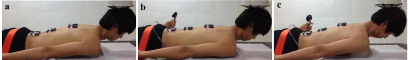
Figure 1. The prone thoracic extension exercises (a: preferred prone thoracic extension (PTE), b:
abdominal drawing-in maneuver (ADIM) PTE, c: ADIM-CT (chin tuck) PTE).
lated to pain in neck, thoracic, or lumbar spine. This study was approved by the Human Studies Committee of Yonsei University Wonju Campus. All participants provided written informed consent before the data collection began.
Instruments
2)Surface EMG
1)was used to collect data on the muscle activity of middle and lower TES and LES.
EMG data were collected at the sampling rate of 1024 ㎐ and analyzed with MYOLAB (BTS Company, Italy) software. A bandpass filter (20-450
㎐) and notch filter (60 ㎐) were used, and the data were processed to produce the root mean square (RMS) (Cynn et al, 2006). Data were collected from the dominant side of the trunk. Electrode placement sites were shaved and then swabbed with alcohol to decrease skin resistance. Disposable Ag/AgCl surface electrodes were placed over each erector spinae mus- cle on the belly, approximately 2 ㎝ lateral to the spinous process of T7 or T8 for middle thoracic erector spinae muscle, T12 for lower thoracic erector spinae, and L3 for lumbar erector spinae (Cram et al, 1998). The raw data were processed to produce RMS with a window of 50 milliseconds. For normalization, the mean RMS of three trials of 3-second maximal voluntary contraction was calculated for the three erector spinae. Maximal voluntary isometric con- tractions (MVIC) were collected during prone back extension with maximal resistance for 5 seconds.
The manual muscle testing position was used, as described by Kendall et al (2005). The muscle activ- ity was expressed as a percentage of the calculated
root mean square of MVIC (%MVIC).
Procedures
The primary investigator demonstrated PTE and instructed each subject with consistent verbal and tactile cues. All subjects were pain free and comfort- able after the familiarization period. The target bar for PTE was placed at a height of 10 ㎝ from the occiput of each prone subject. Figure 1 shows each prone thoracic extension exercise.
For familiarization with ADIM, the pressure bio- feedback unit was placed between the pad on the physical treatment table and the subject's lower ab- domen and was then inflated to 70 ㎜Hg. The sub- ject was asked to perform ADIM and decrease the pressure from 70 ㎜Hg to 60 ㎜Hg. The subject was given verbal feedback from a secondary investigator to maintain around 60 ㎜Hg during PTE (Oh et al, 2007).
For familiarization with the CT exercise, the pri- mary examiner taught the subjects the correct meth- od of performing the CT exercise in the sitting posture. To standardize the CT, the subjects were positioned in the sitting posture with the forehead and chin positioned vertically. An imaginary line parallel to the plinth extended from the tragus of the ear, bisecting the neck longitudinally. This CT meth- od was modified from the test position of craniocer- vical flexion (Kelly et al, 2012). Subjects performed the CT and received verbal and tactile feedback from the secondary examiner during the familiarization session.
For familiarization for ADIM PTE, subjects per- formed thoracic extension to contact occiput with the
1) BTS FREEEMG 300, BTS Company, MI, Italy.
target bar and ADIM, consecutively. Subjects re- ceived information from the secondary examiner about the pressure biofeedback unit during ADIM PTE for 5 seconds. This familiarization period was finished after the pressure had been stabilized in three consecutive trials.
For familiarization with ADIM PTE with chin tuck, subjects performed thoracic extension to attain contact of the occiput with the target bar and ADIM-CT, consecutively. Subjects received in- formation from the secondary examiner about the pressure biofeedback unit during ADIM PTE for 5 seconds. This familiarization period was finished af- ter the pressure had been stabilized in three consec- utive trials.
Data collection
Data on the muscle activity of the TES and LES was collected in the preferred PTE. Following the preferred PTE, ADIM with the pressure biofeedback unit was familiarized. Subjects performed ADIM PTE followed by a 5-second isometric contraction. A metronome was used to control the time and speed of movement. The starting signal was provided by a beeper sound cue in the MYOLAB software. The data collection was repeated in ADIM-PTE. Finally, ADIM with chin tuck PTE was performed in ADIM-CT PTE. Subjects were instructed to perform ADIM PTE and then were asked to tuck their chins gently. The principal investigator placed the tip of the index finger to the subject’s chin to maintain and confirm the chin tuck maneuver. The same procedure for data collection procedure was repeated in ADIM-CT PTE. PTE was performed at a comfort- able speed and consisted of a concentric phase, iso- metric phase, and eccentric phase. EMG data were collected within 5 seconds in the isometric phase.
Subjects performed three trials of each PTE con- dition with 1 minute rests. The mean value was used for the data analysis.
Statistical Analysis
SPSS ver. 12.0 software was used for the stat- istical analyses. One-way analysis of variance (ANOVA) with repeated measures was employed for comparing the three different exercise conditions (preferred, ADIM, ADIM-CT). In the case of sig- nificant differences between exercise conditions, Bonferroni’s post hoc test was performed. In all analyses, the level of statistical significance was set at α=.05.
Results
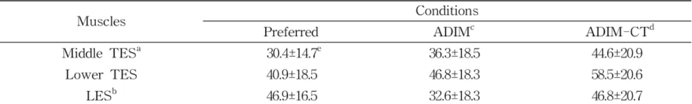
For middle TES muscle activity, there was a sig- nificant difference in three conditions (F=21.848, p<.001). The post hoc test revealed significant dif- ferences in all comparisons (preferred PTE vs.
ADIM PTE, p=.031; preferred PTE vs. ADIM-CT PTE, p=.001; ADIM PTE vs. ADIM-CT PTE, p=.002). The means of %MVIC in three conditions increased as follows: preferred PTE (30.4±14.7) <
ADIM PTE (36.3±18.5) < ADIM-CT PTE (44.6±20.9). For lower TES muscle activity, there was a significant difference in three conditions (F=12.836, p=.002). The post hoc test revealed sig- nificant differences in all comparisons (preferred PTE vs. ADIM PTE, p=.005; preferred PTE vs.
ADIM-CT PTE, p=.001; ADIM PTE vs. ADIM-CT
PTE, p=.003). The means of %MVIC in three con-
ditions were increased as follows: preferred PTE
(40.9±18.5) < ADIM PTE (46.8±18.3) < ADIM-CT
PTE (58.5±20.6). For LES, there was a significant
difference in three conditions (F=9.149, p=.001). The
post hoc test demonstrated significantly lower dif-
ferences in the ADIM PTE condition compared to
the preferred PTE (p=.017) and ADIM-CT PTE
conditions (p=.004). However, no significant differ-
ence was observed between preferred PTE and
ADIM-CT PTE (p=1.000). The means of %MVIC in
the three conditions were as follows: preferred
PTE=46.9±16.5, ADIM PTE=32.6±18.3, and
ADIM-CT PTE=46.8±20.7.
Muscles Conditions
Preferred ADIM
cADIM-CT
dMiddle TES
a30.4±14.7
e36.3±18.5 44.6±20.9
Lower TES 40.9±18.5 46.8±18.3 58.5±20.6
LES
b46.9±16.5 32.6±18.3 46.8±20.7
a