415
© Copyright The Korean Academy of Asthma, Allergy and Clinical Immunology • The Korean Academy of Pediatric Allergy and Respiratory Disease http://e-aair.org INTRODUCTION
Drug-Induced Hypersensitivity Syndrome (DIHS) or Drug Reaction with Eosinophilia and Systemic Symptoms (DRESS) is a severe systemic reaction typically occurring 2-6 weeks after exposure to certain drugs. DIHS is characterized by fever, skin rash, internal organ dysfunction, and hematologic abnormali- ties either eosinophilia or atypical lymphocytosis.1
There is no gold standard for DIHS diagnosis other than the proposed diagnostic criteria.2,3 Sulfasalazine-induced hyper- sensitivity syndrome is rare but well-recognized.4 Nevertheless, when clinical presentation is not straightforward, other causes must be excluded. The available diagnostic criteria are used for the validation of suspected cases, but are not designed for early diagnosis or distinguishing drug hypersensitivity from other diseases that give similar reactions.
The enzyme-linked immunospot (ELISPOT) assay is a sensi- tive method capable of detecting a small number of antigen- specific cytokine-producing cells. Recently, this technique has been introduced to confirm the diagnosis of delayed-type drug hypersensitivity reactions and may be useful in patients with a remote history of drug allergy.5 We report herein a case of DIHS that presented with infectious mononucleosis-like reaction, complicated by acalculous cholecystitis and hypotension. Sul- fasalazine hypersensitivity was proven by interferon-gamma
A Case of Sulfasalazine-Induced Hypersensitivity Syndrome Confirmed by Enzyme-Linked Immunospot Assay
Parkpoom Phatharacharukul,
1Jettanong Klaewsongkram
2*
1Faculty of Medicine, Chulalongkorn University, Bangkok, Thailand
2Division of Allergy and Clinical Immunology, Department of Medicine, Faculty of Medicine, Allergy and Clinical Immunolgy Research Group, Chulalongkorn University, Bangkok, Thailand
(IFN-γ) ELISPOT assay.
CASE REPORT
A 24-year-old Thai male presented with high fever and ab- dominal pain for 4 days. The patient first noticed his fever ac- companied with fatigue and a bitemporal throbbing pain with- out organ-specific symptoms, 2 weeks prior to admission when attending an outpatient clinic. Viral infection was the presump- tive diagnosis. His low-grade fever remained for several weeks until 4 days prior to admission, when high fever and a progres- sive maculopapular rash developed. He also experienced epi- gastric pain, which brought him to our hospital again seeking medical attention.
The patient was diagnosed with bilateral chronic uveitis 2 years ago, when topical steroids had been prescribed. Low-dose oral prednisolone (15 mg/day) was subsequently added before ta-
Case Report
Allergy Asthma Immunol Res. 2013 November;5(6):415-417.
http://dx.doi.org/10.4168/aair.2013.5.6.415 pISSN 2092-7355 • eISSN 2092-7363
A 24-year-old male with a history of spondyloarthropathy presented with high fever, cervical lymphadenopathy and generalized maculopapular rash.
He was treated with prednisolone for chronic uveitis before being switched to sulfasalazine 3 weeks prior to admission. Laboratory findings revealed marked leukocytosis with frequent atypical lymphocytes. Sulfasalazine was discontinued and the etiology of mononucleosis syndrome explored.
During admission, he developed acalculous cholecystitis and hypotension. All symptoms quickly improved following administration of systemic cor- ticosteroids. The investigation for infectious mononucleosis yielded negative results and a diagnosis of sulfasalazine-induced hypersensitivity syn- drome was confirmed using enzyme-linked immunospot assays.
Key Words: Drug hypersensitivity; enzyme-linked immunospot assay; sulfasalazine
This is an Open Access article distributed under the terms of the Creative Commons Attribution Non-Commercial License (http://creativecommons.org/licenses/by-nc/3.0/) which permits unrestricted non-commercial use, distribution, and reproduction in any medium, provided the original work is properly cited.
Correspondence to: Jettanong Klaewsongkram, MD, Division of Allergy and Clinical Immunology, Department of Medicine, Faculty of Medicine,
Allergy and Clinical Immunolgy Research Group, Chulalongkorn University, Bangkok 10330, Thailand.
Tel: +662-2564152; Fax: +662-2542323; E-mail: [email protected] Received: October 31, 2012; Accepted: December 26, 2012
•There are no financial or other issues that might lead to conflict of interest.
Phatharacharukul et al.
Allergy Asthma Immunol Res. 2013 November;5(6):415-417. http://dx.doi.org/10.4168/aair.2013.5.6.415 Volume 5, Number 6, November 2013
416 http://e-aair.org
pering off within 11 months. Two months later, a diagnosis of spondyloarthropathy was suspected by the rheumatologist, when Achilles tenosynovitis, plantar fasciitis, and tenderness over the sacroiliac joints developed. Sulfasalazine was then pre- scribed starting at 1 g/day, gradually increasing to 2 g/day for 2 weeks prior to the development of fever.
Physical examination revealed high fever (38.5°C), cervical lymphadenopathy, pharyngitis with whitish patches on soft palate and buccal mucosa. The patient had a scattered maculo- papular rash over his trunk and extremities and abdominal ex- amination revealed mild epigastrium tenderness. A complete blood count revealed marked leukocytosis with a total white blood cell count of 23,740/µL, 50% neutrophils, 16% lympho- cytes, 31% monocytes, and 3% eosinophils. Atypical lympho- cytes were detected in peripheral blood smears. Blood culture and anti-viral antibody profiles were investigated; sulfasalazine was promptly discontinued.
On the third day of admission, the patient developed severe abdominal pain and mild icteric sclera was noted. Liver func- tion tests showed direct hyperbilirubinemia with total bilirubin 3.34 mg/dL and direct bilirubin 2.94 mg/dL. ASL and ALT lev- els were 536 and 734 U/mL, respectively, and serum alkaline phosphatase was 301 IU/mL. Upper abdominal ultrasonogra- phy revealed gallbladder wall thickening with pericholecystic fluid collection and positive sonographic Murphy’s sign. Intra- venous ceftriaxone was then administered. On the ninth day of admission, he developed hypotension (blood pressure of 70/40 mmHg) before being rescued with 1,500-mL intravenous fluid.
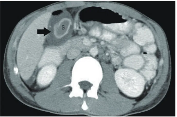
Abdominal computed tomography scans showed a collapsed gallbladder with a moderate amount of pericholecystic fluid, but no gallstones could be demonstrated (Fig. 1); hepatospleno- megaly and minimal ascites were also noticed. Dexamethasone (5 mg) was administered every 12 h intravenously for six doses, resulting in cessation of the fever and abdominal pain, after the third dose.
The viral studies yielded negative results for IgM and IgG
against Epstein-Barr virus, cytomegalovirus, and Dengue virus.
Blood culture results showed no bacterial growth. Anti-HIV and anti-human herpes virus 6 (HHV-6) IgM antibodies were also negative, but anti-HHV-6 IgG was positive (17.05 units with a cut-off value of 11 units). Two days after the initiation of steroids, the numbers of IFN-γ–releasing cells in the peripheral blood were measured by ELISPOT assay (Mabtech, Stockholm, Swe- den) upon stimulation with four drugs as described previous- ly.6 Significant numbers of IFN-γ–secreting cells were demon- strated (1,048 spots forming cells/106 PBMCs) upon incubation with 100 µg/mL sulfasalazine, but not with other drugs admin- istered concurrently (ceftriaxone), previously (amoxicillin), or never (ceftazidime) (Fig. 2). Dexamethasone was then replaced with prednisolone 1 mg/kg/day before being tapered off over 3 weeks. His liver function tests returned to normal levels in 1 month; no complications were noted after 2 years of follow-up.
His symptoms are now well controlled with NSAIDs and meth- otrexate.
DISCUSSION
The diagnosis of DIHS or DRESS can be challenging. Several diseases including acute infectious mononucleosis, hemato- logic malignancies, and collagen vascular disorders are known to share similar clinical presentations.7 Management of DIHS frequently requires systemic corticosteroids and may lead to deleterious effects in immunocompromised patients. Acalcu- lous cholecystitis has been reported in drug-induced hypersen- sitivity syndrome, but is uncommon.8 Although this patient ful- filled the diagnostic criteria for DIHS, atypical presentation of acalculous cholecystitis and the lack of peripheral blood eosin- ophilia deferred systemic corticosteroid administration while bacterial and disseminated viral infections were being excluded.
Despite the pathogenesis of DIHS not being fully understood, a complex interaction between drug-specific immune respons- es accompanied with viral reactivation is a possible mecha- nism.9 The evidence of human herpes virus 6 reactivation, sup- ported by the elevated anti-HHV-6 IgG levels, confirms the as- sociation between the development of DIHS and human her- pes virus 6 infection, as reported previously.10 The lymphocyte transformation test (LTT) is a useful proliferation-based assay to assess T cell responses to drugs, but the technique is time- consuming.11 ELISPOT assays are more sensitive in the detection of low-frequency antigen-specific T cells, and IFN-γ ELISPOT as-
Fig. 1. Acalculous cholecystitis with pericholecystic fluid collection as demon- strated by abdominal computed tomography scans. G, gallbladder.
G
Fig. 2. Sulfasalazine-specific interferon-gamma responses as demonstrated by enzyme-linked immunospot assay.
Negative
control Amoxicillin Ceftriaxone Ceftazidime Sulfasalazine Positive control IFN-γ
Sulfasalazine Allergy Confirmed by ELISPOT Assay
Allergy Asthma Immunol Res. 2013 November;5(6):415-417. http://dx.doi.org/10.4168/aair.2013.5.6.415 AAIR
417 http://e-aair.org says are effective for the diagnosis of drug-induced maculopap-
ular rash.12
We report a case of sulfasalazine-induced hypersensitivity syndrome complicated with acalculous cholecystitis and hypo- tension, the diagnosis and treatment of which was delayed due to its atypical features. Our findings suggest the potential role of IFN-γ ELISPOT assays in confirming the diagnosis of DIHS in patients in whom the diagnosis is still doubtful, or identifying the culprit drug in patients with a history of multiple drug use.
ACKNOWLEDGMENTS
We would like to thank Supranee Buranapraditkun, Ph.D. for her assistance with the ELISPOT assays. This study was sup- ported by the Special Task Force for Activating Research (STAR), Chulalongkorn University.
REFERENCES
1. Kano Y, Shiohara T. The variable clinical picture of drug-induced hypersensitivity syndrome/drug rash with eosinophilia and sys- temic symptoms in relation to the eliciting drug. Immunol Allergy Clin North Am 2009;29:481-501.
2. Kardaun SH, Sidoroff A, Valeyrie-Allanore L, Halevy S, Davidovici BB, Mockenhaupt M, Roujeau JC. Variability in the clinical pattern of cutaneous side-effects of drugs with systemic symptoms: does a DRESS syndrome really exist? Br J Dermatol 2007;156:609-11.
3. Shiohara T, Iijima M, Ikezawa Z, Hashimoto K. The diagnosis of a DRESS syndrome has been sufficiently established on the basis of typical clinical features and viral reactivations. Br J Dermatol 2007;
156:1083-4.
4. Kunisaki Y, Goto H, Kitagawa K, Nagano M. Salazosulfapyridine in- duced hypersensitivity syndrome associated with reactivation of human herpes virus 6. Intern Med 2003;42:203-7.
5. Beeler A, Engler O, Gerber BO, Pichler WJ. Long-lasting reactivity and high frequency of drug-specific T cells after severe systemic drug hypersensitivity reactions. J Allergy Clin Immunol 2006;117:
455-62.
6. Tanvarasethee B, Buranapraditkun S, Klaewsongkram J. The po- tential of using enzyme-linked immunospot to diagnose cephalo- sporin-induced maculopapular exanthems. Acta Derm Venereol 2013;93:66-9.
7. Knowles SR, Shapiro LE, Shear NH. Anticonvulsant hypersensitivi- ty syndrome: incidence, prevention and management. Drug Saf 1999;21:489-501.
8. Cacoub P, Musette P, Descamps V, Meyer O, Speirs C, Finzi L, Rou- jeau JC. The DRESS syndrome: a literature review. Am J Med 2011;
124:588-97.
9. Shiohara T, Inaoka M, Kano Y. Drug-induced hypersensitivity syn- drome (DIHS): a reaction induced by a complex interplay among herpesviruses and antiviral and antidrug immune responses. Al- lergol Int 2006;55:1-8.
10. Descamps V, Valance A, Edlinger C, Fillet AM, Grossin M, Lebrun- Vignes B, Belaich S, Crickx B. Association of human herpesvirus 6 infection with drug reaction with eosinophilia and systemic symp- toms. Arch Dermatol 2001;137:301-4.
11. Pichler WJ, Tilch J. The lymphocyte transformation test in the diag- nosis of drug hypersensitivity. Allergy 2004;59:809-20.
12. Rozieres A, Hennino A, Rodet K, Gutowski MC, Gunera-Saad N, Berard F, Cozon G, Bienvenu J, Nicolas JF. Detection and quantifi- cation of drug-specific T cells in penicillin allergy. Allergy 2009;64:
534-42.