situations likes stress fractures, previous surgical interventions, metabolic bone disease can also be possible reasons5,6). Altered mechanical axis of the lower limb and anatomic landmarks inter- fere with correct implantation of TKA components, and this can be a reason of malalignment that can lead to patellar maltracking, early loosening, higher rates of polyethylene wear1-5,7). Retained hardware and canal sclerosis, which are frequently seen in pa- tients with extra-articular deformity, can also be an obstacle in TKA. Under such abnormal situations, usage of conventional in- strumentations of TKA including intramedullary femoral guide and intra- and extramedullary tibial guide is highly restricted.
TKA in knees with extra-articular deformity can be done through staged or one-stage operation. The lower incidences of anesthesia, operation and complication are the relative advan- tages of one-stage operation over staged operation. Surgical navi- gation system can be a good option that facilitates simultaneous treatment of osteoarthritis and extra-articular deformity of the knee. The purpose of this study was to report good clinical and radiological results of navigation-assisted TKA for osteoarthritic knees with extra-articular deformity.
Navigation-Assisted Total Knee Arthroplasty for Patients with Extra-Articular Deformity
Seung Joon Rhee, MD, Chang Hyo Seo, MD, and Jeung Tak Suh, MD
Department of Orthopedic Surgery, Pusan National University Hospital, Busan, Korea
Purpose: Since the existence of an extra-articular deformity seriously alters the normal geometry and kinetics around the knee joint, difficulties are often encountered in total knee arthroplasty (TKA) using a standard surgical technique. The purpose of this study was to evaluate the usefulness of surgical navigation system as a treatment option for osteoarthritic knees with extra-articular deformity.
Materials and Methods: The authors retrospectively reviewed medical records of the patients who underwent primary TKA between 2007 and 2012.
Knees with preoperative radiography showing an angular deformity within the region from the middle third of the femur to the middle third of the tibia in the ipsilateral limb of the arthritic knees were considered as cases having extra-articular deformity. Thirteen knees of the 13 patients were found to have undergone TKA using a navigation system for osteoarthritis with ipsilateral extra-articular deformity. The hip-knee-ankle angle, Knee Society score (KSS), and range of motion were measured before and after the operation to evaluate the improvement.
Results: The mean hip-knee-ankle angle in the coronal plane was improved to 0.2o±4.5o in valgus alignment postoperatively. The KSS was improved to 89.6±4.6 points postoperatively at the last follow-up, with over 90% of good and excellent results. The range of motion was improved to 118.5o±10.5o postoperatively.
Conclusions: Navigation-assisted TKA is a good treatment option of osteoarthritic knees with extra-articular deformity.
Keywords: Knee, Extraarticular, Deformity, Arthroplasty, Navigation pISSN 2234-0726 · eISSN 2234-2451
Knee Surgery & Related Research
Received August 5, 2013; Revised (1st) October 7, 2013;
(2nd) October 30, 2013; Accepted November 11, 2013 Correspondence to: Jeung Tak Suh, MD
Department of Orthopedic Surgery, Pusan National University Hospital, 179 Gudeok-ro, Seo-gu, Busan 602-739, Korea
Tel: +82-51-240-7248, Fax: +82-51-247-8395 E-mail: [email protected]
Introduction
Recovery of normal mechanical alignment with adequate soft tissue balancing is a key factor and a goal to successful total knee arthroplasty (TKA)1-4). However, in the presence of extra- articular deformities of either the femur or the tibia, such a goal is much more difficult to achieve since the deformity aggravates the distortion of the 3-dimensional geometry of the arthritic knee.
While the extra-articular deformity of the knee is commonly caused by malunion after a previous traumatic event, other
194
This is an Open Access article distributed under the terms of the Creative Commons Attribution Non-Commercial License (http://creativecommons.org/licenses/by-nc/3.0/) which permits unrestricted non-commercial use, distribution, and reproduction in any medium, provided the original work is properly cited.
Copyright © 2013 KOREAN KNEE SOCIETY www.jksrr.org
Materials and Methods
Authors retrospectively reviewed medical records of the pa- tients who underwent primary TKA between 2007 and 2012 to enroll the cases with extra-articular deformity. Knees with pre- operative radiography showing angular deformity in the middle or distal third of the femur or in the proximal or middle third of the tibia with or without involvement of the articular surface in the ipsilateral limb of the arthritic knees were considered as having extra-articular deformity6). Thirteen knees of 13 patients were found to have had ipsilateral extra-articular deformity as- sociated with arthritic knees. All of the patients underwent TKA and intra-articular correction of the extra-articular deformity us- ing navigation system. There were 4 male and 9 female patients, and their average age at operation was 69 years (range, 52 to 83
years). Extra-articular deformities were located in the femur in 9 patients, and tibia in 4 patients. There were 3 uniplanar and 10 biplanar deformities. The mean follow-up period was 3.1 years.
All deformities were caused by malunion of fracture. Three pa- tients had retained hardware due to prior operation for trauma.
The average interval between the initial trauma and operation was 26 years (range, 10 to 42 years) (Table 1).
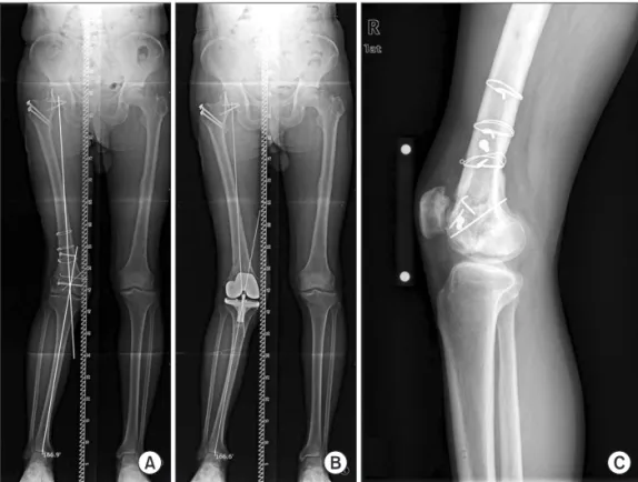
Preoperative and postoperative orthoroentgenogram (an- teroposterior full-length weight-bearing radiography) of all the patients were checked. The hip-knee-ankle axis and angulation in both coronal and sagittal planes of the femur or tibia were measured (Fig. 1). For femoral deformities, on the preoperative orthoroentgenogram, the proposed distal femoral cut was drawn perpendicular to the mechanical axis of the femur. If the defor- mity was more than 20o or if the plane of the distal cut compro-
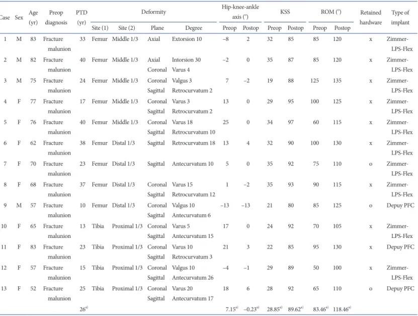
Table 1. Data on the Extra-Articular Deformity and Pre- (Preop) and Postoperative (Postop) Hip-Knee-Ankle Axis, Knee Society Score (KSS), Range of Motion (ROM) of the Knee
Case Sex Age (yr)
Preop diagnosis
PTD (yr)
Deformity Hip-knee-ankle
axis (o) KSS ROM (o) Retained
hardware
Type of implant Site (1) Site (2) Plane Degree Preop Postop Preop Postop Preop Postop
1 M 83 Fracture malunion
33 Femur Middle 1/3 Axial Extorsion 10 –8 2 32 85 85 120 x Zimmer-
LPS-Flex 2 M 82 Fracture
malunion
40 Femur Middle 1/3 Axial Coronal
Intorsion 30 Varus 4
–2 0 35 87 85 120 x Zimmer-
LPS-Flex 3 M 75 Fracture
malunion
24 Femur Middle 1/3 Coronal Sagittal
Valgus 3 Retrocurvatum 2
7 –2 19 88 125 135 x Zimmer-
LPS-Flex 4 F 77 Fracture
malunion
17 Femur Middle 1/3 Coronal Sagittal
Varus 3 Retrocurvatum 2
13 0 29 95 100 125 x Zimmer-
LPS-Flex 5 F 76 Fracture
malunion
40 Femur Middle 1/3 Coronal Sagittal
Varus 18 Retrocurvatum 10
25 0 34 97 60 115 x Zimmer-
LPS-Flex 6 F 62 Fracture
malunion
38 Femur Distal 1/3 Sagittal Retrocurvatum 18 13 4 32 90 100 130 x Zimmer-
LPS-Flex 7 F 70 Fracture
malunion
23 Femur Distal 1/3 Sagittal Antecurvatum 10 5 0 35 92 75 110 o Zimmer-
LPS-Flex 8 F 68 Fracture
malunion
37 Femur Distal 1/3 Coronal Sagittal
Varus 15 Retrocurvatum 12
1 –2 35 93 90 115 x Zimmer-
LPS-Flex 9 M 57 Fracture
malunion
10 Femur Distal 1/3 Coronal Sagittal
Valgus 10 Antecurvatum 6
–13 –13 21 80 85 125 o Depuy PFC
10 F 65 Fracture malunion
13 Tibia Proximal 1/3 Coronal Sagittal
Varus 5 Antecurvatum 15
17 0 24 92 70 105 x Zimmer-
LPS-Flex 11 F 83 Fracture
malunion
23 Tibia Proximal 1/3 Coronal Sagittal
Varus 10 Retrocurvatum 3
21 3 22 85 95 130 x Depuy PFC
12 F 57 Fracture malunion
15 Tibia Proximal 1/3 Coronal Sagittal
Valgus 10 Antecurvatum 26
–4 –1 29 89 50 100 x Zimmer-
LPS-Flex 13 F 52 Fracture
malunion
25 Tibia Proximal 1/3 Coronal Sagittal
Varus 20 Antecurvatum 17
18 6 28 92 65 110 o Depuy PFC
26a) 7.15a) –0.23a) 28.85a) 89.62a) 83.46a) 118.46a)
PTD: posttraumatic duration, Hip-knee-ankle axis: +, varus; –, valgus.
a)Values are presented as mean.
mised the collateral ligament attachment, other surgical options, such as corrective osteotomy, was considered. For tibial deformi- ties, the proposed proximal tibial cut was drawn perpendicular to the mechanical axis of the tibia. If the distal tibial axis did not pass through the tibial plateau or if the deformity was more than
30o, other surgical options, such as corrective osteotomy, was considered.
Preoperative orthoroentgenogram and anteroposterior and lateral radiographs of the knee were exported from the PACS, Marosis m-view 5.4 (Marotech Inc., Seoul, Korea) program as a
Fig. 1. (A) Measurement of the preoperative hip-knee-ankle axis using orthoroentgeno- gram (anteroposterior full-length weight- bearing radiography), which is showing 25o of varus. (B) Measurement of the femoral anatomical axis deformity using anteropos- terior and lateral knee radiography, which is showing 18o of varus and 10o of retrocurva- tum.
Fig. 2. Preoperative templating using Or- thoview (Meridian Technique Limited) program and imported JPG images: (A) Resizing using a 10 cm ruler, (B) Measuring of the femoral alignment, (C) Measuring of the size of the femur and tibia in the coro- nal plane, (D) Measuring of the size of the femur and tibia in the sagittal plane.
JPG file. Then, templating was done using Orthoview (Meridian Technique Limited, Southampton, UK) program and exported JPG images. After re-sizing the images to match the size of the 10 cm ruler as a first step, leg alignment was measured using the alignment tool. Then, the size and shape of the femur and tibia in the coronal and sagittal planes was measured (Fig. 2). As a final step, template of the femur and tibia was overlapped in the coronal and sagittal plane to approximate the proper size of the implants and to look for any deformity that would be an obstacle to component placement (Fig. 3). Assessment to determine the need for augmentation using bone grafts, wedges, or stem exten- sions was done. In this series, no bone graft was required in the real operation, but metal block augmentation was required in 3 cases in the femoral side and tibial stem extension was used in 1 case. Knee Society score (KSS) was assessed before and after the operation.
All the operations were performed by a single surgeon. Two dif- ferent TKA designs were implanted, NexGen LPS-Flex (Zimmer Inc., Warsaw, IN, USA) in 10 patients and P.F.C. Sigma (DePuy Orthopaedics Inc., Warsaw, IN, USA) in 3 patients. In all cases, TKAs were performed using an image free Medtronic Electro- magnetic knee navigation system (Zimmer Inc.).
Medial parapatellar arthrotomy was performed using an anteri- or midline approach in 11 patients, whereas, in patients who had a previous knee surgery, the procedure was carried out through the original incision site to avoid skin necrosis or additional scar- ring. Then, navigation tracker fixation was done by inserting two pins in the distal femur and proximal tibia. The location of the hip joint center was identified as the average center of a large and three-planar femur-to-pelvis rotation. Since Medtronic Elec- tromagnetic knee navigation system (Zimmer Inc.) is an image free navigation system that uses a surface mapping guide for the placement of the cutting guides, registration of anatomical land- marks and axes including femur anteroposterior axis and the me- dial and lateral epicondyles with instrumented pointer was per-
formed to define the anatomical reference frames for the femur and tibia, which provided target orientations for all relevant bone cuts and rotational alignment. Bone cutting was planned for the distal femur and proximal tibia separately with regard to the me- chanical axis of each bone. The distal femoral bone cut was at 0o in the coronal plane and at 3o of flexion in the sagittal plane, and the proximal tibial cut was at 0o in the frontal plane and 5o poste- rior slope in the sagittal plane. For rotational alignment, the epi- condylar axis and the Whiteside line were taken for the femur8). Tibial trial was left free to rotate during cycles of knee flexion and extension, and the orientation in full extension was chosen for its optimal rotational alignment.
Following those bone cutting, the extension gap or flexion gap was seen in trapezoid rather than rectangular shape. But, both collateral ligament insertions were all intact. Soft tissue balancing was performed during the trial component placement and after the final component placement under the guidance of the naviga- tion system.
Orthoroentgenogram was obtained immediately after surgery, 1 month postoperatively, and consecutively at follow-up. The fol- low-up period was 3.1 years on average. The femoral mechanical axis was defined as a line drawn from the hip center to the deep- est part of the femoral intercondylar notch, and tibial mechani- cal axis as a line drawn from the midpoint of the tibial spine to the midpoint of the talar dome. The hip-knee-ankle angles were measured using those axes (Fig. 1A). The range of motion and KSS were also recorded for clinical evaluation.
Results
The mean preoperative hip-knee-ankle angle in the coronal plane was 7.2o±11.8o in varus alignment (range, 25o varus to 13o valgus). Of the 9 femoral extra-articular deformities, 5 were in the middle third and 4 were in the distal third of the femur. The mean femoral angular deformity was varus 3.4o in the coronal Fig. 3. Determination of the implant size based on the measurement results on the coronal plane (A) and sagittal plane (B).
plane and 4.0o recurvatum in the sagittal plane. All 4 tibial extra- articular deformities were in the proximal third of the tibia.
The mean angular deformity of the tibia was 6.3o varus in the coronal plane and 13.8o antecurvatum in the sagittal plane. At the postoperative follow-up, it was 0.2o±4.5o in valgus alignment (range, 6o varus to 13o valgus) (Fig. 4). The KSS was improved from 28.8±5.7 points preoperatively (range, 19 to 35 points) to 89.6±4.6 points at the last follow-up (range, 80 to 97 points), with over 90% of good and excellent results. The range of motion was improved from a mean of 83.5o±19.8o preoperatively (range, 50o to 125o) to 118.5o±10.5o postoperatively (range, 100o to 135o) (Table 1).
Discussion
Currently, operation assisted by a navigation system is often performed in various surgical fields. Navigation helps surgeons identify what they are doing and how they are doing by visualiz- ing the surgical field with reconstructed images or representative numbers and guides throughout the operation. It is helpful for avoiding serious mistakes during surgery and ultimately reducing postoperative complication rates. This is not an exception in the field of orthopaedics, and TKA is a renowned example of naviga- tion applicable orthopaedic surgery. The advantages with respect to component alignment and soft tissue balancing are already well established in several studies9-17). The importance of this study lies in that authors applied the navigation system for TKA in patients with extra-articular deformity. Since the existence of
an extra-articular deformity seriously alters the normal geometry and kinetics around the knee joint, difficulties are often encoun- ter in operations using standard surgical techniques. So, surgical strategies, such as simultaneous femoral osteotomy with TKA and staged TKA following corrective osteotomy, were adapted to overcome such an unfavorable condition and achieve successful TKA in patients with extra-articular deformity. There are reports showing good clinical and radiological results of those traditional staged surgery, but staged surgery is inevitably affected by com- plications including infections, arthrofibrosis, and nonunion of the osteotomy site. Since Wang and Wang6) first reported intra- articular bone resection could be a new alternative surgical strat- egy in correcting extra-articular deformity, despite the risk asso- ciated with collateral ligament injury and grossly asymmetric gap, this technique has become the common choice of surgery. Less incision, lower incidence of complications, and earlier rehabilita- tion are good reasons to prefer intra-articular bone resection in the correction of extra-articular deformity. Moreover, combined use of a navigation system made this surgical option more prefer- able by effectively overcoming the difficulties. Navigation systems provide information on accurate bone cutting, proper orientation of prosthesis component, and soft tissue balancing under the sit- uation that operators cannot recognize normal anatomical land- marks or insert intramedullary guide due to the extra-articular deformity of the femur or tibia.
There were 9 femoral extra-articular deformities and 4 tibial extra-articular deformities in our study. Although their causative deformities were located either in the femur or the tibia alone, Fig. 4. Postoperative radiographs showing the results of navigation-assisted total knee arthroplasty in case No. 5: 0o hip-knee- ankle axis (A), 0o femoral mechanical axis in the sagittal plane (B), and anteroposterior and lateral knee radiographs (C).
the deformity affected the whole lower limb and changed the limb alignment significantly. So, the treatment and evaluation of surgical results were focused on the correction of the hip-knee- ankle angle. We designed our operative goal to neutralize the hip- knee-ankle angle to 0o. In the operative field, we confirmed that the hip-knee-ankle angle was corrected to 0o in all cases, but the postoperative hip-knee-ankle angle was not the same as the in- traoperative measurement on the postoperative orthoroentgeno- gram. In the weight loaded standing orthoroentgenogram, we identified 0o of hip-knee-ankle angle in 5 cases and less than 2o of deviation from 0o in 4 cases, but the rest 4 cases showed more than 2o of deviation from 0o. We consider that the less than 2o of hip-knee-ankle angle deviation might be due to the characteristic errors of electromagnetic navigation and somewhat inevitable.
Since the electromagnetic navigation system makes calculations based on the data transmitted from the trackers, the erroneous fixation of trackers on the wrong position or inadvertent move- ment of trackers can distort the resultant presentation. For the 4 cases with over 2o of hip-knee-ankle angle deviation, we suspect ligament balancing was a major problem. Due to the severe soft tissue contractures associated with poor arc of knee motion and compromised, scarred soft tissue, patients required extensive soft tissue and ligamentous release and even collateral ligament reconstruction. So, we suspect poor preoperative soft tissue con- dition and unsatisfactory ligamentous balancing could have been the reasons for unsatisfactory hip-knee-ankle angle correction.
Especially, case No. 9 had shown severe valgus instability gait due to the numerous and long standing series of operations for his trauma and posttraumatic arthritis, muscle weakness around the knee joint was severe, and the laxity of medial collateral ligament was disastrous, and even after our operation, the limb alignment returned to valgus position again (Fig. 5). In our opinion, the operations on cases with hip-knee-ankle angle deformities ex- ceeding 15o in any direction are more difficult. However, it needs to be proven statistically with detailed parameters later based on accumulated cases.
There were 2 cases with rotational deformities in the femur.
Case No. 1 had 10o externally rotated femur as a result of trau- matic deformity. But, in this case, we didn’t concern much about the rotational alignment itself because the patient had fully adapted to his rotational problem with subclinical gait abnormal- ity. For case No. 2, a detailed study of rotational alignment was done before the operation because of apparent rotational defor- mity in the orthoroentgenogram. Preoperative computed tomog- raphy revealed that his femoral anteversion was about 46o, which was about 30o internally rotated than normal range. However, rotational deformity itself couldn’t be corrected by intra-articular correction. Even 10o correction would injure the collateral liga- ment insertion and possibly cause seriously imbalanced posterior condylar resection. Soft tissue release could not achieve the bal- ance, either. So, in this case we decided to correct the hip-knee- ankle angle only (Fig. 6).
Fig. 5. Radiographic images of case No. 9:
13o of valgus hip-knee-ankle axis in the pre- operative orthoroentgenogram (A), 13o of valgus hip-knee-ankle axis in the 1-month postoperative orthoroentgenogram (B), and preoperative lateral knee radiograph showing the retained hardware in the distal femur (C).
There were 3 cases with remaining hardware of past trauma operation. Case No. 7 had a cerclage wire in the femur and Case No. 13 had 2 cerclage wires in the proximal tibia. But they didn’t disturb any procedure related to the navigation-assisted TKA.
Case No. 9 had multiple cerclages wires, Kirshner wires and screws in the distal femur that would probably be an obstacle if it were a conventional TKA using intramedullary cutting guide (Fig.
5). However, by using the navigation, TKA could be performed relatively with ease and necessitated minimum removal of the remaining hardware. But, the remaining hardware was removed to lower the risk of infection and patient discomfort.
As for complication, partial patellar tendon detachment oc- curred in 2 cases. Primary repair using Ethibond suture was done in the operative field. Their knees were immobilized for 6 weeks postoperatively with subsequent uncomplicated healing and full restoration of extensor mechanism function.
The advantages of navigation-assisted TKA in knees with extra- articular deformity lie in the planning and assessment of the most adequate planes for the bone cuts, guidance of the surgeon to avoid compromising the epicondyles and the consequent imbalance of the ligaments. In addition, the use of a navigation system instead of intramedullary rods reduces blood loss and the potential risk of fat emboli since the intramedullary canal re- mains intact18,19). Prolonged operative time and cost is a problem that needs to be overcome when using a navigation system in
TKA. Finally, after removal of the hardware, the remaining holes can be sites for stress with an associated increase in the rate of the periprosthetic fractures7,20,21).
The limitation of our study is that the number of patients was too small due to the rarity of TKA with extra-articular deformity.
High standard of fracture treatment left not so many cases of residual deformity, which the authors tried to look for. For these reasons, further meaningful statistical analysis couldn’t be done.
In addition, comparison between navigation-assisted and con- ventional TKAs couldn’t be carried out, because the authors only included the cases with TKA using a navigation system.
Conclusions
Navigation-assisted TKA could enable mechanical axis correc- tion of the lower extremity without separate corrective osteotomy in the selected patients with osteoarthritic knees and extra-artic- ular deformities. Since the navigation helped us idenfity the ana- tomical parameters, we could overcome the limitations caused by the extra-articular deformity and achieve near normal alignment with relatively few difficulties. The postoperative improvement in the hip-knee-ankle angle, KSS, range of motion proved the effec- tiveness of a navigation system in TKA. We think that navigation- assisted TKA is a good treatment option for osteoarthritic knees with extra-articular deformity.
Fig. 6. (A) Preoperative orthoroentgeno- gram showing 2o of valgus hip-knee-ankle axis with large, externally rotated lesser trochanter implying internally rotated extra articular deformity. (B) Axial computed tomography showing 47.3o of anterversion of the femur. (C) Reconstructed image of the femur showing 46o of anterversion. (D) Postoperative orthoroentgenogram show- ing 0o of hip-knee-ankle axis with the knee joint still internally rotated.
Conflict of Interest
No potential conflict of interest relevant to this article was re- ported.
Acknowledgments
This work was supported by a 2-Year Research Grant of Pusan National University.
References
1. Berger RA, Rubash HE, Seel MJ, Thompson WH, Crossett LS. Determining the rotational alignment of the femoral component in total knee arthroplasty using the epicondylar axis. Clin Orthop Relat Res. 1993;(286):40-7.
2. Oswald MH, Jakob RP, Schneider E, Hoogewoud HM.
Radiological analysis of normal axial alignment of femur and tibia in view of total knee arthroplasty. J Arthroplasty.
1993;8:419-26.
3. Wasielewski RC, Galante JO, Leighty RM, Natarajan RN, Rosenberg AG. Wear patterns on retrieved polyethylene tibial inserts and their relationship to technical consider- ations during total knee arthroplasty. Clin Orthop Relat Res.
1994;(299):31-43.
4. Garg A, Walker PS. Prediction of total knee motion using a three-dimensional computer-graphics model. J Biomech.
1990;23:45-58.
5. Lonner JH, Siliski JM, Lotke PA. Simultaneous femoral os- teotomy and total knee arthroplasty for treatment of osteo- arthritis associated with severe extra-articular deformity. J Bone Joint Surg Am. 2000;82:342-8.
6. Wang JW, Wang CJ. Total knee arthroplasty for arthritis of the knee with extra-articular deformity. J Bone Joint Surg Am. 2002;84:1769-74.
7. Bottros J, Klika AK, Lee HH, Polousky J, Barsoum WK. The use of navigation in total knee arthroplasty for patients with extra-articular deformity. J Arthroplasty. 2008;23:74-8.
8. Whiteside LA, Arima J. The anteroposterior axis for femoral rotational alignment in valgus total knee arthroplasty. Clin Orthop Relat Res. 1995;(321):168-72.
9. Bathis H, Perlick L, Tingart M, Luring C, Zurakowski D, Grifka J. Alignment in total knee arthroplasty: a comparison of computer-assisted surgery with the conventional tech- nique. J Bone Joint Surg Br. 2004;86:682-7.
10. Bathis H, Shafizadeh S, Paffrath T, Simanski C, Grifka J, Lur-
ing C. Are computer assisted total knee replacements more accurately placed? A meta-analysis of comparative studies.
Orthopade. 2006;35:1056-65.
11. Bolognesi M, Hofmann A. Computer navigation versus standard instrumentation for TKA: a single-surgeon experi- ence. Clin Orthop Relat Res. 2005;440:162-9.
12. Chauhan SK, Scott RG, Breidahl W, Beaver RJ. Computer- assisted knee arthroplasty versus a conventional jig-based technique: a randomised, prospective trial. J Bone Joint Surg Br. 2004;86:372-7.
13. Ensini A, Catani F, Leardini A, Romagnoli M, Giannini S. Alignments and clinical results in conventional and navigated total knee arthroplasty. Clin Orthop Relat Res.
2007;457:156-62.
14. Hart R, Janecek M, Chaker A, Bucek P. Total knee arthro- plasty implanted with and without kinematic navigation. Int Orthop. 2003;27:366-9.
15. Jenny JY, Clemens U, Kohler S, Kiefer H, Konermann W, Miehlke RK. Consistency of implantation of a total knee arthroplasty with a non-image-based navigation system: a case-control study of 235 cases compared with 235 conven- tionally implanted prostheses. J Arthroplasty. 2005;20:832-9.
16. Luring C, Oczipka F, Perlick L, Tingart M, Grifka J, Bathis H. Two year follow-up comparing computer assisted ver- sus freehand TKR on joint stability, muscular function and patients satisfaction. Knee Surg Sports Traumatol Arthrosc.
2009;17:228-32.
17. Sparmann M, Wolke B, Czupalla H, Banzer D, Zink A. Po- sitioning of total knee arthroplasty with and without naviga- tion support: a prospective, randomised study. J Bone Joint Surg Br. 2003;85:830-5.
18. Kalairajah Y, Cossey AJ, Verrall GM, Ludbrook G, Spriggins AJ. Are systemic emboli reduced in computer-assisted knee surgery?: a prospective, randomised, clinical trial. J Bone Joint Surg Br. 2006;88:198-202.
19. Jeon SH, Kim JH, Lee JM, Seo ES. Efficacy of extramedul- lary femoral component alignment guide system for blood saving after total knee arthroplasty. Knee Surg Relat Res.
2012;24:99-103.
20. Fehring TK, Mason JB, Moskal J, Pollock DC, Mann J, Wil- liams VJ. When computer-assisted knee replacement is the best alternative. Clin Orthop Relat Res. 2006;452:132-6.
21. Klein GR, Austin MS, Smith EB, Hozack WJ. Total knee arthroplasty using computer-assisted navigation in patients with deformities of the femur and tibia. J Arthroplasty.
2006;21:284-8.