149
∙Received: March 28, 2014. Accepted: April 21, 2014.
∙Corresponding author : Ki Choon Moon
Department of Nuclear Medicine, Seoul National University Bundang Hospital,82 Gumi-ro 173beon-gil, Bundang-gu, Seongnam 463-707, Korea
Tel: +82-31-787-2952, Fax: +82-31-787-4018 E-mail: [email protected]
Original Article 핵의학 영상검사 후 시행된 핵의학 검체검사에서의 영향
분당서울대학교병원 핵의학과
문기춘⋅권원현⋅김정인⋅이인원The Effect on The Result, in Case of the In-vitro Test Performance after an Imaging Test
Ki Choon Moon, Won Hyun Kwon, Jung In Kim and In Won Lee
Department of Nuclear Medicine, Seoul National University Bundang Hospital, Seongnam, Korea
Purpose: At our hospital blood is collected from a patient before an imaging test, with the concern of any effect possible when a nuclear medicine imaging test and an in-vitro test are carried out at the same time. However, occasionally, the blood collection is performed after an imaging test, with the reasons that the patient is not properly guided or the patient doesn't follow the guide correctly. In that case, we prefer to gather blood again after a few days. The purpose of this study is not only to see whether there is any effect of an imaging test on the result of the in-vitro test performed with the blood collected after the imaging test, but also to study how many days waiting after each test is appropriate to take a blood sample, if the effect exists. Materials and Methods:
From September to October 2013, blood were collected from 13 patients in our hospital regardless of age and sex each time before and after the injection of the radioactive isotope from the tests : PET-CT, Gated Myocardial SPECT, and DTPA GFR Scan. Considering a half-life, AFP, CA19-9, CEA, TSH, and T3 were carried out right after the blood collection. In case of an iodine therapy, blood were taken each time before and after taking radioactive iodine, and, after AFP, CA19-9, and CEA, the difference between them in consistency and in cpm were compared. Results: With 10 patients after the imaging tests and 3 patients after the iodine therapy, their serum cpm was over 10,000. Over time, the cpm decreased in accordance with the half-life (
18F 110minutes,
99m
Tc 6hours,
201Tl 72hours,
131I 7days). Between the two cases, one before and the other after the injection of the radioactive isotope, the cpm and the results of AFP, CA19-9, CEA, TSH, and T3 from three patients each test, PET-CT, Gated Myocardial SPECT, and DTPA GFR Scan, were very similar. In addition, in case of an iodine therapy, there was also not a meaningful difference in the cpm and the results of AFP, CA19-9, and CEA, from three patients in an iodine therapy, between the two cases, one before and the other after taking the radioactive iodine. Conclusion: In case a blood collection was performed after the imaging test which required a radioactive isotope injection, the cpm increased, differently according to the kind of the radioactive isotope. However, the results of the in-vitro tests like AFP, CA19-9, CEA, TSH, T3, etc were nearly not affected. As the result, it's considered that there will not be any significant effect also from other tests, as the result from the performed seven tests. (Korean J Nucl Med Technol 2014;18(1):149-152)
Key Words : Radioactive isotope (
18F,
99mTc ,
201Tl,
131I), Half-life, CPM
서 론
본원은 핵의학과 영상검사와 검체검사가 동시에 있을 경
우 검사결과에 조금의 영향이라도 미칠 것을 우려해 영상 검사 시행 전 채혈을 하도록 환자들에게 안내하고 있는데 간혹 안내가 제대로 이루어지지 않거나 환자가 지시를 잘 따르지 않아서 영상검사를 먼저하고 채혈을 하는 경우가 있다. 이러한 경우 며칠 후 다시 채혈을 하도록 권유하고 있는데 과연 영상검사 시행 후 채혈 시 검체검사에 미치는 영향은 있는지, 영향이 있다면 며칠 경과 후 채혈을 하는 것이 적당한지 각 검사별로 차이를 알아보는 데 중점을 두었다.
J Nucl Med Technol Vol. 18, No. 1, May 2014
핵의학기술 제18권 제1호 2014
150
사용되는 동위원소: 18F (FDG: fluorodeoxyglucose), 반감기: 110분 Fig. 1. PET-CT.사용되는 동위원소: 99mTc. 201Tl, 반감기: 6시간, 72시간 Fig. 2. Gated Myocardial SPECT.
Table 1. PET-CT
주사 - 2:10, 8:45, 9:15 채혈 - 2:50, 9:15, 10:00
AFP CA 19-9 CEA TSH T3
DOSE CPM DOSE CPM DOSE CPM DOSE CPM DOSE CPM
1 5.2 1046 <5 831 <1.0 186 0.49 243 67 13497
5.6 1103 <5 761 <1.0 158 0.69 301 72 13405
2 1.2 521 7 1057 1.2 355 3.29 1016 127 7929
1.3 546 5 984 1.1 334 2.81 882 120 8258
3 2.2 840 21 1706 1.8 458 2.88 902 122 8204
2.0 787 19 1626 1.7 439 2.39 761 117 8646
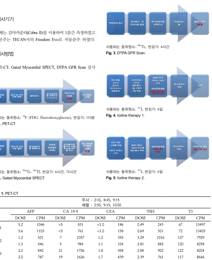
사용되는 동위원소: 99mTc, 반감기: 6시간 Fig. 3. DTPA GFR Scan.
사용되는 동위원소: 131I, 반감기: 6일 Fig. 4. Iodine therapy 1.
사용되는 동위원소: 131I, 반감기: 6일 Fig. 5. Iodine therapy 2.
대상 및 방법 1. 실험대상
2013년 9월부터 10월까지 본원 내원 환자 13명의 환자 검체.
2. 검사기기
검체는 감마카운터(Cobra II)를 이용하여 1분간 측정하였고 검체분주는 TECAN사의 Freedom Evo로 자동분주 하였다.
3. 검사방법
PET-CT, Gated Myocardial SPECT, DTPA GFR Scan 검사
시 방사성동위원소 투입 전 ․ 투입 후 각각 채혈하여 AFP, CA19-9, CEA, TSH, T3 검사를 실시하고, 옥소 치료 시 방 사성 옥소 복용 전․ 복용 후 각각 채혈하여 AFP, CA19-9, CEA, TG, TG Ab 검사를 실시하였다.
문기춘 외 3인. 핵의학 영상검사 후 시행된 핵의학 검체검사에서의 영향
151
Table 2. Gated myocardial SPECT주사 - 9:00 9:45, 9:35 10:10, 10:00,10:45 채혈 - 9:50, 10:13, 10:48
AFP CA 19-9 CEA TSH T3
DOSE CPM DOSE CPM DOSE CPM DOSE CPM DOSE CPM
1 0.8 284 34 2152 1.0 272 2.51 995 60 13812
1.0 324 31 2032 1.0 258 2.98 1165 52 14399
2 1.6 411 <5 811 <1.0 175 1.92 846 86 13615
1.5 387 <5 763 <1.0 166 1.85 818 70 11401
3 1.9 759 16 1398 4.9 787 0.21 58 103 9813
1.6 640 17 1313 4.4 696 0.28 64 90 10901
Table 3. DTPA GFR Scan
주사 - 2:25, 2:00, 2:35 채혈 - 2:30, 2:05, 2:40
AFP CA 19-9 CEA TSH T3
DOSE CPM DOSE CPM DOSE CPM DOSE CPM DOSE CPM
1 2.5 835 30 1728 <1.0 265 1.16 404 113 6830
2.4 810 31 1811 <1.0 308 1.45 499 109 7043
2 2.4 1070 12 1520 <1.0 271 1.05 258 138 5587
2.6 1165 10 1435 1.1 288 0.81 203 126 6007
3 3.6 1579 5 1180 1.2 308 2.04 511 138 5562
3.2 1398 5 1215 1.0 272 1.85 462 124 6102
Table 4. 옥소치료
채혈1-10/12,10/19,10/30 채혈2–10/14,10/21,11/1
TG TG Ab AFP CA 19-9 CEA
DOSE CPM DOSE CPM DOSE CPM DOSE CPM DOSE CPM
1 <0.2 140 6840 1042 1.4 594 5 916 <1.0 201
<0.2 119 8310 895 1.2 519 7 1031 <1.0 188
2 408 18211 31 6105
686 31243 54 5528
3 38.28 19249 <20 5511
48.92 21381 20 5018
결 과
결론 및 고찰
PET-CT, Gated Myocardial SPECT, DTPA GFR Scan을 시 행한 환자혈액 혈청 cpm은 10,000 이상을 넘었으며 시간이 지남에 따라 반감기에 맞게 (18F 110분, 99mTc 6시간, 201Tl 72 시간, 131I 7일) cpm이 감소하는 것을 확인하였다. 그러나 영
상검사 시행 후 Tumor marker, Hormone 검사를 시행했을 때 전, 후 결과값은 유의한 차이를 보이지 않았으며(전. 후 Difference: 10% 내외 다만, TSH 등의 저농도 값에서 전. 후 20% 이상 차이를 나타내는 경우가 있었다.) 방사성 옥소 복 용 후 TG와 TG Ab 결과값에 미치는 영향 또한 없었다. TG 값의 차이가 40% 이상 차이나는 경우가 있었지만 이는 rH TSH를 주사하고 72시간 후가 가장 높게 나타나고 시간이 지나면서 TG값이 낮게나오는 현상 때문이다.
핵의학기술 제18권 제1호 2014
152
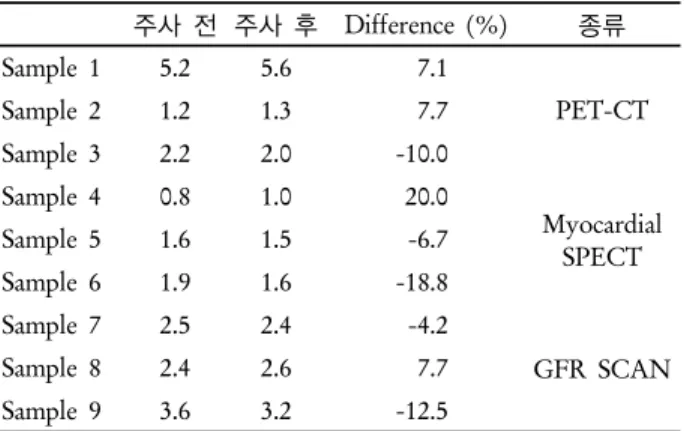
Table 9. Difference (TG)주사 전 주사 후 Difference(%) 종류
Sample 1 0.2 0.2 0.0
TG
Sample 2 408 686 40.5
Sample 3 38.28 48.92 21.7
Table 5. Difference (AFP)
주사 전 주사 후 Difference (%) 종류
Sample 1 5.2 5.6 7.1
PET-CT
Sample 2 1.2 1.3 7.7
Sample 3 2.2 2.0 -10.0
Sample 4 0.8 1.0 20.0
Myocardial SPECT
Sample 5 1.6 1.5 -6.7
Sample 6 1.9 1.6 -18.8
Sample 7 2.5 2.4 -4.2
GFR SCAN
Sample 8 2.4 2.6 7.7
Sample 9 3.6 3.2 -12.5
Table 6. Difference (CEA)
주사 전 주사 후 Difference (%) 종류
Sample 1 1.0 1.0 0.0
PET-CT
Sample 2 1.2 1.1 -9.1
Sample 3 1.8 1.7 -5.9
Sample 4 1.0 1.0 0.0
Myocardial SPECT
Sample 5 1.0 1.0 0.0
Sample 6 4.9 4.4 -11.4
Sample 7 1.0 1.0 0.0
GFR SCAN
Sample 8 1.0 1.1 9.1
Sample 9 1.2 1.0 -20.0
Table 7. Difference (CA 19-9)
주사 전 주사 후 Difference (%) 종류
Sample 1 5 5 0.0
PET-CT
Sample 2 7 5 -40.0
Sample 3 21 19 -10.5
Sample 4 34 31 -9.7
Myocardial SPECT
Sample 5 5 5 0.0
Sample 6 16 17 5.9
Sample 7 30 31 3.2
GFR SCAN
Sample 8 12 10 -20.0
Sample 9 5 5 0.0
Table 8. Difference (TSH)
주사 전 주사 후 Difference(%) 종류
Sample 1 0.49 0.69 29.0
PET-CT Sample 2 3.29 2.81 -17.1
Sample 3 2.88 2.39 -20.5
Sample 4 2.51 2.98 15.8 Myocardial SPECT
Sample 5 1.92 1.85 -3.8
Sample 6 0.21 0.28 25.0
GFR SCAN
Sample 7 1.16 1.45 20.0
Sample 8 1.05 0.81 -29.6 Sample 9 2.04 1.85 -10.3
요 약
핵의학과 영상검사 후에 검체검사를 시행할 경우 결과값에 미치는 영향을 알아보기 위해 PET-CT, Gated Myocardial SPECT, DTPA GFR Scan을 시행하기 전․ 후에 채혈하여 Tumor marker (AFP,CEA,CA19-9), Hormone (TSH,T3,TG,TG Ab)검 사를 시행하여 Difference를 구하였다.
대부분의 결과가 10% 이내의 차이를 나타냈지만 Table 7 의 sample 2와 Table 8의 sample 1, sample 6, sample 8의 저 농도 값에서 20%를 넘는 차이를 나타내는 경우가 있었다.
그렇지만 cpm값은 Table 7의 sample 2는 984(전), 1057(후) Table 8의 sample 1은 243(전), 301(후) sample 6은 58(전),
64(후) sample 8은 258(전), 203(후)으로 매우 흡사한 값을 나타냈다. 이 같은 값을 바탕으로 볼 때 영상검사를 시행한 후에 검체검사를 시행하더라도 결과값에 미치는 영향은 없 는 것으로 판단된다.
REFERENCES
1. Haugen BR, Pacini F, Reiners C, Schlumberger M, Ladenson PW, Sherman SI, et al. A comparison of recombinant human thyrotropin and thyroid hormone withdrawal for the detection of thyroid remnant or cancer.cer. J Clin Endocrinol Metab 1999;84:3877-3880.