Quantitative Expression Analysis of Functional Genes in Four Dog Breeds
Jeong-An Gim
1,2†, Sang-Hoon Kim
1†, Hee-Eun Lee
1,2, Hoim Jeong
1,2, Gyu-Hwi Nam
1,2, Min Kyu Kim
3, Jae-Won Huh
4, Bong-Hwan Choi
5and Heui-Soo Kim
1,2*
1Department of Biological Sciences, College of Natural Sciences, Pusan National University, Busan 46241, Korea
2Genetic Engineering Institute, Pusan National University, Busan 46241, Korea
3Laboratory of Animal Reproduction and Physiology, Department of Animal Science and Biotechnology, College of Agriculture and Life Science, Chungnam National University, Daejeon 34134, Korea
4National Primate Research Center, Korea Research Institute of Bioscience and Biotechnology, Ochang, Chungbuk 28116, Korea
5Division of Animal Genomics and Bioinformatics, National Institute of Animal Science, Rural Development Administration, Jeonju, Jeongbuk 54875, Korea
Received July 16, 2015 /Revised August 21, 2015 /Accepted August 25, 2015
One of the domesticated species; the dog has been selectively bred for various aims by human. The dog has many breeds, which are artificially selected for specific behaviors and morphologies. Dogs contribute their life to human as working dogs for guide, rescue, detection or etc. Working dogs re- quires good personality, such as gentleness, robustness and patience for performing their special duty.
Many studies have concentrated on finding genetic marker for selecting the high-quality working dog.
In this study, we confirmed quantitative expression patterns of eight genes (ABAT; 4-Aminobutyrate Aminotransferase, PLCB1; Phospholipase C, Beta 1, SLC10A4; Solute Carrier Family 10, Member 4,
WNT1; Wingless-Type MMTV Integration Site Family, Member 1, BARX2; BarH-Like Homeobox 2, NEUROD6; Neuronal Differentiation 6, SEPT9; Septin 9 and TBR1; T-Box, Brain, 1) among brains tis-sues from four dog breeds (Beagle, Sapsaree, Shepherd and Jindo), because these genes were ex- pressed and have functions in brain mostly. Specially, BARX2, SEPT9, SLC10A4, TBR1 and WNT1 genes were highly expressed in Beagle and Jindo, and Sapsaree and German Shepherd were vice versa. The biological significance of total genes was estimated by database for annotation, visual- ization and integrated discovery (DAVID) to determine a different gene ontology (GO) class. In these analyses, we suppose to these eight genes could provide influential information for brain develop- ment, and intelligence of organisms. Taken together, these results could provide clues to discover bio- marker related to functional traits in brain, and beneficial for selecting superior working dogs.
Key words :
Brain tissue, dog breeds, functional genes, gene expression profile, qRT-PCR
†Authors contributed equally.
*Corresponding author
*Tel : +82-51-510-2259, Fax : +82-51-581-2962
*E-mail : [email protected]
This is an Open-Access article distributed under the terms of the Creative Commons Attribution Non-Commercial License (http://creativecommons.org/licenses/by-nc/3.0) which permits unrestricted non-commercial use, distribution, and reproduction in any medium, provided the original work is properly cited.
Journal of Life Science 2015 Vol. 25. No. 8. 861~869 DOI : http://dx.doi.org/10.5352/JLS.2015.25.8.861
Introduction
The dog (Canis lupus familiaris) had been domesticated from wild gray wolves (Canis lupus). Based on archeological data, the dogs had had first artificial selection that from 100,000 to 15,000 years ago in multiple locations, including Europe, the Middle East and East Asia [48, 49]. Nowadays, the dog population is separated into more than 400 breeds exist in worldwide [36]. The dogs evolved through a mu-
tually valuable relationship with human beings, and their abilities have been developed to perform an outstanding va- riety of working or special roles. These roles include military watch, security guards, shepherds, guides, rescue and pets [45]. Working dogs are required to good personality of gen- tleness, robustness and patience for performing their special duty. Several animal personality are influenced by the activ- ity of specific genes [17, 42]. Specially, the brain specific genes controls behavior, personality, or aggression, therefore it is needed to study for confirming the gene expression pat- terns in brain. Microarray studies have been performed to assess changes in behavior with gene expression patterns in the brain [46]. We tried to identify such expression changes of eight genes in brain of four domesticated dog breeds (Beagle, German Shepherd, Sapsaree and Jindo), then compared to their expression patterns.
A major goal in the brain and behavioral sciences is to
identify genes that influence social behavior and understand how their gene products influence the structure and function of the nervous system [17, 42, 46]. Thus, the aim of this study was to predict the relationships among personality traits (calmness, trainability, dog sociability, boldness, and etc.) of dogs. Gene expression profiles may reflect the complete per- sonality of regulatory pathways; therefore we describe the gene expression patterns of eight genes (ABAT, BARX2,
NEUROD6, SEPT9, SLC10A4, PLCB1, TBR1, WNT1) in dogbreeds.
The domestic dogs display an extraordinary level of phe- notypic diversity in personality and behavior, because dog breeding was introduced as various methods by human dur- ing the nineteenth century [48, 50]. As the results of artificial selection, each dog breeds have fixed phenotypic traits [12, 47]. In this point, it is important to select the suitable traits of special dog from many dog breeds. In this study, we pre- sented each gene expression patterns in four dog breeds, which provides the clue to study of each dog breeds of spe- cific traits.
The Beagle is one breed of small to medium-sized dog, and developed primarily for tracking wild animals as de- tection dogs. Their great sense of smell and tracking ability is used to detect food items, drug, or explosive detection [6]. Due to their strength, intelligence, trainability and obedi- ence, German Shepherds are generally used in search-and- rescue, police, or military dogs around the world. German Shepherds have abilities of tracking, patrolling, or detection by training [39]. Therefore, the German Shepherd is the widely used breeds for military, police or scent-work roles.
As the Korean traditional breeds, Sapsaree is known as dauntlessness and loyalty aspects. Jindo is also one of the Korean traditional breeds, and well known for its unwaver- ing loyalty and gentle nature. Because the Sapsaree and Jindo are an active and intelligent dog, it requires frequent interaction with people or another dog in the family [14, 28]. Although they are frequently used as pet dog or guide dog, they are not frequently used as special dogs. In order to use these breeds as special dogs, gene expression patterns in brain could be good clues.
Materials and Methods
Tissue samples, RNA isolation and synthesis of cDNA
One post-mortem brain tissue sample was extracted from
four dog breeds (Beagle, German Shepherd, Sapsaree, and Jindo). Beagle and Sapsaree brain tissues were obtained by Chungnam National University with approval by the Animal Ethics Committee (CNU-00199). German Shepherd and Jindo brain tissues by Rural Development Administra- tion (Jeonju, Korea), and the animals received care in accord- ance with the standard guidelines for the Care and Use of Laboratory Animals provided by the National Institute of Animal Science Animal Care Committee, and the experiment was executed with approval from the animal ethics commit- tee under the operation rule of animal experiment ethics at the National Institute of Animal Science (approval number:
2014-085).
Cellular RNA of dog brain tissues was isolated by TRIzol® Reagent (Invitrogen, Carlsbad, CA, USA) to purify total RNA according to the manufacturer’s guidelines. After RNA isolation, the quality and quantity of the resulting sin- gle-stranded RNAs were assessed using a ND-1000 spec- trophotometer (NanoDrop, Wilmington, DE, USA). Total RNA was treated and reverse-transcribed using a Prime- Script RT reagent kit with genomic DNA Eraser (Takara Bio, Shiga, Japan) according to the instructions of the manu- facturer. Eight pairs of primer were designed to detect the mRNA of each gene according to the open reading frames (ORFs) in the whole genome (Table 1).
Quantitative real time RT-PCR amplification Quantitative real-time RT-PCR was performed with the Rotor-Gene Q system (QIAGEN, Hilden, Germany) with a gene-specific primer set (Table 1). Each of amplification re- action mixture (20 μl) contained 7 μl of H
2O, 10 μl of QuantiTech SYBR Green PCR Master Mix (QIAGEN, Hilden, NW, Germany), 1 μl each of forward and reverse primers at 10 nmol/ μl, and 1 μl of cDNA template. In addition, to confirm non-specific background amplification, we ampli- fied a template control without cDNA. Real-time RT-PCR amplifications for target genes and housekeeping genes were conducted as follows: 30 cycles each of 95°C for 15 s, anneal- ing temperature for 15 s, and 72°C for 15 s. Annealing tem- perature range is set from 54 to 57°C depends on genes.
Melting curve analysis was performed for 30 s at 65-99°C.
To guarantee reproducibility, we amplified all samples in
triplicate and the results were averaged. As a standard con-
trol, we used GAPDH (Glyceraldehyde-3-phosphate de-
hydrogenase) in gene expression, for normalization of re-
al-time RT-PCR amplification.
Table 1. Gene description and the information of primers used in this study for real-time RT-PCR analysis Gene
name Description Location
(canFam3) Accession No. Primer Sequences (5′-3′) Annealing temp. (°C)
Product size (bp) ABAT 4-aminobutyrate
aminotransferase
chr6:33,388, 930-33, 425,870
ENSCAFT000000 30230
F:
R:
TGGAAGAGGTGGAGGATCTGA CCTGGAGATGTCTCTCAGCTT
56 143
BARX2 BARX homeobox
2 chr5:5,238,
743-5, 310,648
ENSCAFT000000 45519,
ENSCAFT000000 16227
F:
R:
CTGCAAGTGAAGACTTGGTATC GTTCATCTTCTCCTCAGCCT
57 153
PLCB1 Phospholipase C,
beta 1 chr24:13,337,
234-13, 587,167
ENSCAFT000000 46106,
ENSCAFT000000 09472
F:
R:
ACCCACTGGAATCTGGAGTT ATGAGGGCTCAAACATGCTG
56 171
NEUROD6 Neuronal differentiation 6
chr14:43,914, 149-43, 915,162
ENSCAFT000000 05020
F:
R:
CTGAGAATCGGCAAGAGACC GCTGTGGTAGGGTGGGTAGA
54 192
SEPT9 Septin 9
chr9:3,497, 801-3,626, 550
ENSCAFT000000 08383,
ENSCAFT000000 47802
F:
R:
CCTCAGAAGGAGTTTGACGA CCTCTTCCCATTCACTTGGT
56 117
SLC10A4 Solute carrier family 10, member 4
chr13:44,293, 981-44, 299,528
ENSCAFT000000 03107
F:
R:
AAGGTTTCCCTGTGGTCTCTG TCCAGGCAGACAGTCCTCTT
56 206
WNT1 Wingless-type MMTV integration site family, member 1
chr27:5,589, 117-5, 592,060
ENSCAFT000000 13918,
ENSCAFT000000 45709
F:
R:
ACGACGGTGTTCTCCGAGA GCGGTTGCCATAGAGGAC
56 162
TBR1 T-box, brain, 1 chr36:6,989, 703-6, 997,934
ENSCAFT000000 15885
F:
R:
GTCCTTGCACAAGTACCAGC ATTGTGTAATGTCGGTGTTCTG
57 155
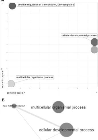
Gene ontology analysis and functional annotations Functional annotation of eight genes was analyzed by the DAVID (Database for Annotation, Visualization and in- tegrated Discovery) [16]. DAVID calculates P-values to dem- onstrate GO terms enrichment, where P-values less than 0.05 are considered to be strongly enriched in the annotation cat- egory after Benjamini multiple test correction. We then per- formed gene ontology (GO) term analyses and each genes were grouped as GO terms. The results of significant GO terms were queried to the REViGO program in order to con- struct a scatterplot and interactive graph [44], and then we summarized GO terms on the 2D semantic space by seman- tic similarities. P-values were originated from the Benjamini and Hochberg false discovery rate (FDR), and the color of circles indicates enriched GO terms with FDR <0.05.
Results and Discussion
Gene expression patterns in four breeds
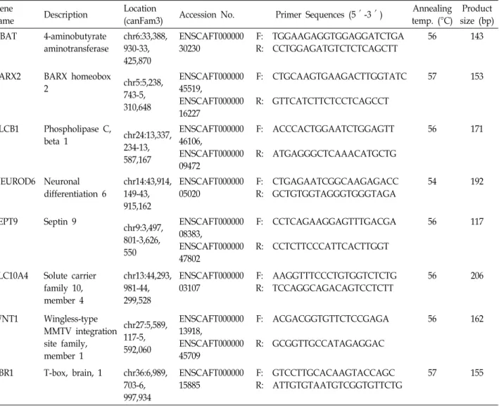
Our gene expression data indicate that similar expression patterns were presented in four breeds of dog. Generally, Beagle and Jindo are dominant expressed patterns, whereas German shepherd and Sapsaree are lower expressed than Beagle and Jindo (Fig. 1).
ABAT has several psychiatry roles, and regulate specially
gamma-aminobutyric acid (GABA) in neuronal cells by cata-
lyzing the degradation of GABA. Thus low-expressed ABAT
gene induces to enhanced amount of GABA in the synaptic
junctions, then increases GABA-mediated signaling by
means of the GABA receptors [26]. Specially, GABA has no
means to penetrate the blood-brain barrier, so it must be
synthesized in the brain. Therefore to elucidate ABAT ex-
pression patterns in the brain is important for neuronal proc-
Fig. 1. Quantitative real-time RT-PCR analysis is performed for the comparison of eight genes (ABAT, BARX2, NEUROD6, SEPT9, SLC10A4, PLCB1, TBR1, WNT1) of expression levels in four dog breeds. The tanscript copy number of eight genes were normalized to GAPDH housekeeping gene copy number in each sample. The values and their error bars indicate means, and standard deviation (n=3).
esses and brain development [51]. In the brain of four dog breeds, ABAT is high-expressed in Beagle, and low-ex- pressed in Shepherd. The two breeds of Sapsaree and Jindo, originated from Korea, have similar expression patterns.
BARX2, SLC10A4, TBR1 and WNT1 genes have same ex-
pression patterns. BARX2 gene is known as transcription fac- tor of cell adhesion [33]. The mechanisms of cell adhesion is very important for brain morphology, and functions such as learning, signal transduction, and memory [40]. When the early development of the nervous system, neurons maintain synapses by formation of cell-cell adhesions. Differently ex- pressed patterns could provide the formation of brain cell, and the features of personality or intelligence. TBR1 has a role in glutamatergic projection in neuron differentiation.
Glutamatergic neurons express receptors for the excitatory neurotransmitter glutamate as opposed to receptors for the inhibitory neurotransmitter GABA [23]. Solute carriers (SLCs) have a role for the transmembrane transport of various ma- terials, such as amino acids, sugars, or inorganic ions.
SLC10A4, one of the solute carrier family, has a role of carry- ing solution in blood or body fluid. SLC10A1 genes were involved in blood-brain barrier [19], and SLC10A4 also has
possible important roles in brain. Therefore, the problems of SLC in the brain could induce mental illness such as ADHD, depression or psychiatric disorders [38]. The study of this gene in the dog could help to elucidate the canine mental illness or behavior disorders.
Proto-oncogene protein WNT1, has been originally con- sidered as a candidate gene for Joubert syndrome, an autoso- mal recessive disorder with cerebellar hypoplasia as a lead- ing feature. Joubert syndrome is a rare genetic disorder that affects the cerebellum, an area of the brain that controls bal- ance and coordination [31, 32]. Wnt signaling pathway con- trolled by WNT1, is stimulated by BDNF (Brain-derived neu- rotrophic factor), then induce the proliferation and differ- entiation of neural stem cells [13]. These genes same ex- pression patterns have crucial roles in brain, and each gene pathway studies will be performed as further studies.
Three genes, NEUROD6, SEPT9 and PLCB1, were similar expressed patterns and highest expressed in Beagle.
NEUROD6 is transcription factors, and associated to differ-
entiation or development of the nervous system. It also regu-
lates fasciculation and targeted axogenesis in the neocortex
[8]. SEPT9 (septin 9) is involved in cytokinesis and cell cycle
Table 2. The function of each genes used in this study
Gene Function References
ABAT
∙ 4-aminobutyrate transaminase activity
∙ Transaminase activity
∙ Pyridoxal phosphate binding
∙ Succinate-semialdehyde dehydrogenase binding
∙ Protein homodimerization activity
[5, 24, 35]
BARX2
∙ BMP signaling and chondrogenesis
∙ Transcription regulatory region sequence-specific DNA binding
∙ Sequence-specific DNA binding transcription factor activity
[25, 33, 41]
PLCB1
∙ Phosphatidylinositol phospholipase C activity
∙ Signal transducer activity
∙ GTPase activator activity
∙ Calcium ion binding
∙ Protein binding
[4, 11, 37]
NEUROD6 ∙ Transcription regulatory region sequence-specific DNA binding [2, 3, 8, 27]
SEPT9 ∙ Filament-forming cytoskeletal GTPase activity
∙ Play a role in cytokinesis [20, 34]
SLC10A4 ∙ Bile acid:sodium symporter activity [1, 7, 15]
TBR1 ∙ RNA polymerase II core promoter sequence-specific DNA binding
∙ Sequence-specific DNA binding transcription factor activity [10, 22, 23]
WNT1
∙ Ligand for members of the frizzled family of seven transmembrane receptors.
∙ Ligand for the coreceptor RYK, thus triggering Wnt signaling.
∙ Signaling molecule important in CNS development.
[9, 18]
Table 3. The function of eight genes based on GO term analysis
GO ID GO terms Count P-value Genes
GO:0030154 GO:0048869 GO:0032501 GO:0009891 GO:0031328 GO:0010628 GO:0010557 GO:0051173 GO:0045935 GO:0051254 GO:0045941 GO:0045893
cell differentiation
cellular developmental process multicellular organismal process
positive regulation of biosynthetic process positive regulation of cellular biosynthetic process positive regulation of gene expression
positive regulation of macromolecule biosynthetic process
positive regulation of nitrogen compound metabolic process
positive regulation of nucleobase, nucleoside, nucleotide and nucleic acid metabolic process
positive regulation of RNA metabolic process positive regulation of transcription
positive regulation of transcription, DNA-dependent
4 4 6 3 3 3 3 3 3 3 3 3
0.038 0.042 0.030 0.043 0.042 0.031 0.039 0.037 0.035 0.022 0.029 0.021
SEPT9, TBR1, WNT1, BARX2 SEPT9, TBR1, WNT1, BARX2 ABAT, SEPT9, NEUROD6, TBR1, WNT1, BARX2 TBR1, WNT1, BARX2 TBR1, WNT1, BARX2 TBR1, WNT1, BARX2 TBR1, WNT1, BARX2 TBR1, WNT1, BARX2 TBR1, WNT1, BARX2 TBR1, WNT1, BARX2 TBR1, WNT1, BARX2 TBR1, WNT1, BARX2
control, and known as a candidate for the ovarian tumor
suppressor gene [20]. Mutations in this gene cause heredi- tary neuralgic amyotrophy [30], and a chromosomal trans- location involving this gene on chromosome 17 and the MLL gene on chromosome 11 results in acute myelomonocytic leukemia [29]. SEPT9 is highest expressed in Beagle, but we
could not detect to specific expression patterns. SEPT9 is re- lated to not only neural diseases but also diseases such as leukemia or tumor, therefore the specific expression patterns is need to be specified.
PLCB1 catalyzes the formation of inositol 1,4,5-tri-
sphosphate and diacylglycerol from phosphatidylinositol
A
B
Fig. 2. Molecular functions of eight genes in this study. After DAVID analysis, GO terms were obtained (P-values
<0.05). Each GO terms of eight genes were queried to the REViGO program. (A) Scatter plots of GO terms from eight genes are viewered, and circles indicated by color depict significantly enriched GO terms with FDR
<0.05. (B) Integrated graph of biological process from eight genes is presented by integrating nodes. Each GO terms (FDR <0.05) of each biological process were de- picted in the 2D semantic space as default options from a REViGO. The circle size of each GO terms is relative to statistical significance, and edge thickness depicts be- tween two nodes.
4,5-bisphosphate by using calcium as a cofactor [21]. This reaction plays an important role in the intracellular trans- duction of many extracellular signals.
The general features of total eight genes in the organisms were listed, and these genes have crucial roles for cellular metabolisms, DNA binding, or cell signaling (Table 2). The variable gene expression patterns in four breeds of dog brain can provide clues for the studies of breed-specific traits.
Function prediction of eight genes
A GO analysis was performed in order to confirm the biological function of the eight genes. The gene set was con- firmed as cellular GO terms, such as cell differentiation, mul- ticellular organismal process, and cellular developmental process. The full list of statistically significant GO terms is enumerated (Table 3). Total 12 functions were enriched, and the most significant function is cell differentiation (P-value
= 0.038) with four genes. Multicellular organismal process includes six of eight genes, except SLC10A4 and PLCB1 genes. BARX2, TBR1, and WNT1 genes were enriched in positive regulation of various biological processes, such as DNA-dependent transcription, RNA metabolic process, and nucleic acid metabolic process. Interestingly, these two genes were high-expressed in Beagle and Jindo, whereas low-ex- pressed in Shepherd and Sapsaree. According to these facts, we summarized that positive regulation of biological process is enhanced in the brain of two breeds, Beagle and Jindo.
We used the REViGO program to get the functional rela- tionship of GO terms in the network structure. The scatter plots and integrated graph show the cell metabolism-related GO terms, including cell differentiation, multicellular organ- ismal process, cellular developmental process, and positive regulation of transcription, DNA-template (Fig. 2). WNT1 gene is included in all GO terms, and crucial factor of each network structure. WNT1 regulate the wnt signaling path- way, then induce neural stem cell differentiation [13].
Therefore, WNT1 gene can provide a clue for the study of wnt signaling brain cell, and their mental features. SEPT9 gene is also included in the scatter plots and integrated graph. SEPT9 gene is related to cell division, such as cell cytokinesis and cell cycle [20]. SEPT9 is also high-expressed in human lymphoid and malignant brain tumors [43]. We predict to these genes can provide crucial information for brain development, and intelligence of organisms.
In conclusion, we selected eight genes related to brain functions, then confirmed their expression patterns, func-
tions, and network. Total genes have a similar expression patterns, and their function is related to cell differentiation, cellular developmental process and multicellular organismal process. This data provides the fundamental clues for the studies of brain functions.
Acknowledgement
This research was supported by awards from the
AGENDA project (Project No. PJ009254) in the National
Institute of Animal Science, Rural Development Administra- tion (RDA).
References
1. Abe, T., Kanemitu, Y., Nakasone, M., Kawahata, I., Yamak- uni, T., Nakajima, A., Suzuki, N., Nishikawa, M., Hishinu- ma, T. and Tomioka, Y. 2013. SLC10A4 is a protease-acti- vated transporter that transports bile acids. J. Biochem.
mvt031.
2. Bartholomä, A. and Nave, K. A. 1994. NEX-1: a novel brain-specific helix-loop-helix protein with autoregulation and sustained expression in mature cortical neurons. Mech.
Dev. 48, 217-228.
3. Baxter, K. K., Uittenbogaard, M., Yoon, J. and Chiaramello, A. 2009. The neurogenic basic helix-loop-helix transcription factor NeuroD6 concomitantly increases mitochondrial mass and regulates cytoskeletal organization in the early stages of neuronal differentiation. ASN Neuro. 1, 195-211.
4. Berstein, G., Blank, J. L., Jhon, D. Y., Exton, J. H., Rhee, S.
G. and Ross, E. M. 1992. Phospholipase C-β1 is a GTPase-ac- tivating protein for G q/11, its physiologic regulator. Cell 70, 411-418.
5. Biase, D., Barra, D., Simmaco, M., John, R. A. and Bossa, F.
1995. Primary Structure and Tissue Distribution of Human 4‐Aminobutyrate Aminotransferase. Eur. J. Biochem. 227, 476-480.
6. Bielfelt, S., Redman, H. and McClellan, R. 1971. Sire-and sex-related differences in rates of epileptiform seizures in a purebred beagle dog colony. Am. J. Vet. Res. 32, 2039-2048.
7. Borges, K. 2013. Slc10A4―what do we know about the func- tion of this “secret ligand carrier” protein? Exp. Neurol. 248, 258-261.
8. Bormuth, I., Yan, K., Yonemasu, T., Gummert, M., Zhang, M., Wichert, S., Grishina, O., Pieper, A., Zhang, W., Goeb- bels, S., Tarabykin, V., Nave, K. A. and Schwab, M. H. 2013.
Neuronal basic helix-loop-helix proteins Neurod2/6 regu- late cortical commissure formation before midline inter- actions. J. Neurosci. 33, 641-651.
9. Brault, V., Moore, R., Kutsch, S., Ishibashi, M., Rowitch, D.
H., McMahon, A. P., Sommer, L., Boussadia, O. and Kemler, R. 2001. Inactivation of the (β)-catenin gene by Wnt1-Cre- mediated deletion results in dramatic brain malformation and failure of craniofacial development. Development 128, 1253-1264.
10. Bulfone, A., Smiga, S. M., Shimamura, K., Peterson, A., Puelles, L. and Rubenstein, J. L. 1995. T-brain-1: a homolog of Brachyury whose expression defines molecularly distinct domains within the cerebral cortex. Neuron 15, 63-78.
11. Caricasole, A., Sala, C., Roncarati, R., Formenti, E. and Terstappen, G. C. 2000. Cloning and characterization of the human phosphoinositide-specific phospholipase C-beta 1 (PLCβ1). Biochim. Biophys. Acta 1517, 63-72.
12. Chase, K., Jones, P., Martin, A., Ostrander, E. A. and Lark, K. G. 2009. Genetic mapping of fixed phenotypes: disease
frequency as a breed characteristic. J. Hered. 100, S37-S41.
13. Chen, B. Y., Wang, X., Wang, Z. Y., Wang, Y. Z., Chen, L.
W. and Luo, Z. J. 2013. Brain‐derived neurotrophic factor stimulates proliferation and differentiation of neural stem cells, possibly by triggering the Wnt/β‐catenin signaling pathway. J. Neurosci. Res. 91, 30-41.
14. Cho, G. 2005. Microsatellite polymorphism and genetic rela- tionship in dog breeds in Korea. Asian Australas. J. Anim.
Sci. 18, 1071-1074.
15. da Silva, T. C., Polli, J. E. and Swaan, P. W. 2013. The solute carrier family 10 (SLC10): beyond bile acid transport. Mol.
Aspects Med. 34, 252-269.
16. Dennis Jr, G., Sherman, B. T., Hosack, D. A., Yang, J., Gao, W., Lane, H. C. and Lempicki, R. A. 2003. DAVID: database for annotation, visualization, and integrated discovery.
Genome Biol. 4, P3.
17. Dingemanse, N. J., Kazem, A. J., Réale, D. and Wright, J.
2010. Behavioural reaction norms: animal personality meets individual plasticity. Trends Ecol. Evol. 25, 81-89.
18. Fortress, A. M., Schram, S. L., Tuscher, J. J. and Frick, K.
M. 2013. Canonical Wnt signaling is necessary for object rec- ognition memory consolidation. J. Neurosci. 33, 12619-12626.
19. Geier, E. G., Chen, E. C., Webb, A., Papp, A. C., Yee, S.
W., Sadee, W. and Giacomini, K. M. 2013. Profiling Solute Carrier Transporters in the Human Blood–Brain Barrier.
Clin. Pharmacol. Ther. 94, 636-639.
20. Hall, P. A., Jung, K., Hillan, K. J. and Russell, S. 2005.
Expression profiling the human septin gene family. J. Pathol.
206, 269-278.
21. Han, J. Y., Shin, E. S., Lee, Y., Ghang, H., Kim, S., Hwang, J., Kim, J. and Lee, J. 2013. A genome-wide association study for irinotecan-related severe toxicities in patients with ad- vanced non-small-cell lung cancer. Pharmacogenomics J. 13, 417-422.
22. Hevner, R. F., Miyashita-Lin, E. and Rubenstein, J. L. 2002.
Cortical and thalamic axon pathfinding defects in Tbr1, Gbx2, and Pax6 mutant mice: evidence that cortical and tha- lamic axons interact and guide each other. J. Comp. Neurol.
447, 8-17.
23. Hevner, R. F., Shi, L., Justice, N., Hsueh, Y., Sheng, M., Smiga, S., Bulfone, A., Goffinet, A. M., Campagnoni, A. T.
and Rubenstein, J. L. 2001. Tbr1 regulates differentiation of the preplate and layer 6. Neuron 29, 353-366.
24. Jirholt, J., Asling, B., Hammond, P., Davidson, G., Knutsson, M., Walentinsson, A., Jensen, J. M., Lehmann, A., Agreus, L. and Lagerstrom-Fermer, M. 2011. 4-aminobutyrate ami- notransferase (ABAT): genetic and pharmacological evi- dence for an involvement in gastro esophageal reflux disease. PLoS One 6, e19095.
25. Jones, P. L. 2003. Homeobox genes in pulmonary vascular development and disease. Trends Cardiovasc. Med. 13, 336- 345.
26. Jung, M., Lippert, B., Metcalf, B., Böhlen, P. and Schechter, P. 1977. γ-Vinyl GABA (4-amino-hex-5-enoic acid), a new selective irreversible inhibitor of GABA-T: Effects on brain GABA metabolism in mice. J. Neurochem. 29, 797-802.
27. Kay, J. N., Voinescu, P. E., Chu, M. W. and Sanes, J. R.
2011. Neurod6 expression defines new retinal amacrine cell subtypes and regulates their fate. Nat. Neurosci. 14, 965-972.
28. Kim, K., Tanabe, Y., Park, C. and Ha, J. 2001. Genetic varia- bility in East Asian dogs using microsatellite loci analysis.
J. Hered. 92, 398-403.
29. Kreuziger, L. M. B., Porcher, J. C., Ketterling, R. P. and Steensma, D. P. 2007. An MLL-SEPT9 fusion and t (11;
17)(q23; q25) associated with de novo myelodysplastic syndrome. Leuk. Res. 31, 1145-1148.
30. Kuhlenbäumer, G., Hannibal, M. C., Nelis, E., Schirmacher, A., Verpoorten, N., Meuleman, J., Watts, G. D., De Vriendt, E., Young, P. and Stögbauer, F. 2005. Mutations in SEPT9 cause hereditary neuralgic amyotrophy. Nat. Genet. 37, 1044- 1046.
31. Lancaster, M. A., Gopal, D. J., Kim, J., Saleem, S. N., Silhavy, J. L., Louie, C. M., Thacker, B. E., Williams, Y., Zaki, M.
S. and Gleeson, J. G. 2011. Defective Wnt-dependent cer- ebellar midline fusion in a mouse model of Joubert syndrome. Nat. Med. 17, 726-731.
32. Louie, C. M. and Gleeson, J. G. 2005. Genetic basis of Joubert syndrome and related disorders of cerebellar development.
Hum. Mol. Genet. 14, R235-R242.
33. Meech, R., Edelman, D. B., Jones, F. S. and Makarenkova, H. P. 2005. The homeobox transcription factor Barx2 regu- lates chondrogenesis during limb development. Development 132, 2135-2146.
34. Nagata, K. I., Asano, T., Nozawa, Y. and Inagaki, M. 2004.
Biochemical and cell biological analyses of a mammalian septin complex, Sept7/9b/11. J. Biol. Chem. 279, 55895-55904.
35. Osei, Y. D. and Churchich, J. E. 1995. Screening and se- quence determination of a cDNA encoding the human brain 4-aminobutyrate aminotransferase. Gene 155, 185-187.
36. Parker, H. G., Kim, L. V., Sutter, N. B., Carlson, S., Lorentzen, T. D., Malek, T. B., Johnson, G. S., DeFrance, H. B., Ostran- der, E. A. and Kruglyak, L. 2004. Genetic structure of the purebred domestic dog. Science 304, 1160-1164.
37. Peruzzi, D., Aluigi, M., Manzoli, L., Billi, A. M., Di Giorgio, F. P., Morleo, M., Martelli, A. M. and Cocco, L. 2002.
Molecular characterization of the human PLC beta1 gene.
Biochim. Biophys. Acta 1584, 46-54.
38. Rask-Andersen, M., Masuram, S., Fredriksson, R. and Schiöth, H. B. 2013. Solute carriers as drug targets: current use, clinical trials and prospective. Mol. Aspects Med. 34, 702-710.
39. Ruefenacht, S., Gebhardt-Henrich, S., Miyake, T. and
Gaillard, C. 2002. A behaviour test on German Shepherd dogs: heritability of seven different traits. Appl. Anim. Behav.
Sci. 79, 113-132.
40. Sakisaka, T. and Takai, Y. 2005. Cell adhesion molecules in the CNS. J. Cell Sci. 118, 5407-5410.
41. Sander, G., Bawden, C. S., Hynd, P. I., Nesci, A., Rogers, G. and Powell, B. C. 2000. Expression of the homeobox gene, Barx2, in wool follicle development. J. Invest. Dermatol. 115, 753-756.
42. Stamps, J. and Groothuis, T. G. 2010. The development of animal personality: relevance, concepts and perspectives.
Biol. Rev. 85, 301-325.
43. Storlazzi, C., Brekke, H., Mandahl, N., Brosjö, O., Smeland, S., Lothe, R. and Mertens, F. 2006. Identification of a novel amplicon at distal 17q containing the BIRC5/SURVIVIN gene in malignant peripheral nerve sheath tumours. J.
Pathol. 209, 492-500.
44. Supek, F., Bošnjak, M., Škunca, N. and Šmuc, T. 2011.
REVIGO summarizes and visualizes long lists of gene ontol- ogy terms. PLoS One 6, e21800.
45. Svartberg, K. 2006. Breed-typical behaviour in dogs—
Historical remnants or recent constructs? Appl. Anim. Behav.
Sci. 96, 293-313.
46. Toth, A. L., Varala, K., Newman, T. C., Miguez, F. E., Hutchison, S. K., Willoughby, D. A., Simons, J. F., Egholm, M., Hunt, J. H. and Hudson, M. E. 2007. Wasp gene ex- pression supports an evolutionary link between maternal behavior and eusociality. Science 318, 441-444.
47. Vaysse, A., Ratnakumar, A., Derrien, T., Axelsson, E., Rosengren Pielberg, G., Sigurdsson, S., Fall, T., Seppala, E.
H., Hansen, M. and Lawley, C. T. 2011. Identification of ge- nomic regions associated with phenotypic variation between dog breeds using selection mapping. PLoS Genet. 7, e1002316.
48. Vila, C., Savolainen, P., Maldonado, J. E., Amorim, I. R., Rice, J. E., Honeycutt, R. L., Crandall, K. A., Lundeberg, J. and Wayne, R. K. 1997. Multiple and ancient origins of the domestic dog. Science 276, 1687-1689.
49. Wayne, R. K., Geffen, E., Girman, D. J., Koepfli, K. P., Lau, L. M. and Marshall, C. R. 1997. Molecular systematics of the Canidae. Syst. Biol. 46, 622-653.
50. Wayne, R. K. and Ostrander, E. A. 1999. Origin, genetic di- versity, and genome structure of the domestic dog. Bioessays 21, 247-257.
51. Yoon, B. E., Woo, J. and Lee, C. J. 2012. Astrocytes as GABA- ergic and GABA-ceptive cells. Neurochem. Res. 37, 2474-2479.
초록:개의 네 품종에서 기능 유전자들에 대한 정량적 발현 분석
김정안
1,2†․김상훈
1†․이희은
1,2․정호임
1,2․남규휘
1,2․김민규
3․허재원
4․최봉환
5․김희수
1,2*
(1부산대학교 자연과학대학 생명과학과, 2부산대학교 유전공학연구소, 3충남대학교 농업생명과학대학 동물자원
과학부, 4한국생명공학연구원 국가영장류센터, 5농촌진흥청 국립축산과학원)