Dependency on p53 in Expression Changes of ATF3 and NAG-1 Induced by EGCG, Genistein, and Resveratrol
Min-Jeong Kim, Hyun-Ji Kim, Yu-Mi Seo, Eun-Joo Lee and Jong-Sik Kim*
Department of Biological Sciences, Andong National University, Andong 36729, Korea Received November 8, 2017 /Revised May 3, 2018 /Accepted May 14, 2018
Epigallocatechin-3-gallate (EGCG), one of catechins of green tea, has been known to possess anti-oxi- dation, anti-inflammation, and anti-cancer effects. The present study analyzed global gene expression changes in EGCG-treated HCT116 cells and p53-null HCT116 cells by oligo DNA microarray analysis.
Among the differentially expressed genes in EGCG-treated HCT116 cells, four were selected that are known as tumor suppressor genes (activating transcription factor 3 [ATF3], cyclin dependent kinase inhibitor 1A [CDKN1A], DNA damage-inducible transcript 3 [DDIT3] and non-steroidal anti-in- flammatory drug activated gene [NAG-1]) and their expression was compared to the expression of genes in p53-null HCT116 cells. We found that the expression of these genes was not dependent on their p53 status except for NAG-1, which was only up-regulated in HCT116. The results of RT-PCR and Western blot analysis showed that ATF3 up-regulation by EGCG was not affected by the presence of p53, whereas NAG-1 expression was not induced in p53-null HCT116 cells. We also detected ATF3 and NAG-1 expression changes through genistein and resveratrol treatment. Interestingly, genistein could not up-regulate ATF3 regardless of p53 status, but genistein could induce NAG-1 only in HCT116 cells. Resveratrol could significantly induce NAG-1 as well as ATF3 independent of p53 presence. These results indicate that EGCG, genistein and resveratrol may have different anti-cancer effects. Overall, the results of this study may help to increase our understandings of molecular mecha- nisms on anti-cancer activities mediated by EGCG, genistein and resveratrol in human colorectal can- cer cells.
Key words : ATF3, HCT116, NAG-1, phytochemicals, p53
*Corresponding author
*Tel : +82-54-820-5798, Fax : +82-54-820-7705
*E-mail : [email protected]
This is an Open-Access article distributed under the terms of the Creative Commons Attribution Non-Commercial License (http://creativecommons.org/licenses/by-nc/3.0) which permits unrestricted non-commercial use, distribution, and reproduction in any medium, provided the original work is properly cited.
Journal of Life Science 2018 Vol. 28. No. 5. 615~620 DOI : https://doi.org/10.5352/JLS.2018.28.5.615
서 론
녹차는 세계적으로 물 다음으로 많이 소비되는 음료로서 다량의 flavonoid를 함유하고 있으며[8], 녹차에 함유된 다양 한 생리활성 성분 중 주성분으로 알려진 카테킨(catechin)은 polyphenol의 화합물로서 녹차 잎 건조 중량의 30%를 차지한 다. 녹차에 함유된 주요 카테킨은 EC (epicatechin), ECG (epicatechin gallate), EGC (epigallocatechin), 그리고 EGCG (epigallocatechin gallate) 등이 있으며, 이 중 EGCG가 가장 효과적인 암 예방 활성이 있다고 보고되었다[4]. EGCG에 의 한 항암 활성은 다양한 암 세포에서 다양한 기전에 의해 일어 날 수 있음이 보고되었는데, Shin 등[17]에 의하면 두경부암 세포모델에서 EGCG는 β-catenin의 전사활성의 억제와 분해 를 증가 시킴으로써 항암활성을 가지는 것으로 보고하였다.
최근에는 EGCG가 식도암 세포주와 유방암 세포주 MCF-7과 T40D에서 다양한 기전에 의해 apoptosis를 촉진함이 보고되 었다[6, 11, 14].
대표적인 암 억제유전자인 p53은 인간 암에서 돌연변이가 50% 이상 일어나는 것으로 알려져 있으며[16], p53은 전사조 절인자로서 세포주기 중지, apoptosis, DNA 복구, 노화 등에 관련된 하위 유전자의 발현을 조절함으로써 작용한다[9, 10].
또한, 전사조절인자의 역할과는 관계없이 세포사멸 신호에 의 해 미토콘드리아로 이동하여 세포사멸을 유도하는 것으로도 보고되었다[20]. 따라서, 항암제나 항암 활성을 가진 천연물의 활성과 작용기전에 있어서 암 세포의 p53의 상태는 매우 중요 하다고 판단된다.
파이토케미칼은 식물 유래의 비 영양성분을 지칭하는 것으 로써, 다양한 작용기전에 의해 암을 예방하는 암 화학예방 (cancer chemoprevention) 활성을 가지고 있는 것들이 많이 알려져 있다[12, 18]. 암 예방법의 기전으로는 파이토케미칼에 의해 유도되는 암세포사멸을 들 수 있는데, 이 경우 암 억제단 백질 p53 의존 경로를 통하는 경우가 많이 보고되었다[13].
본 연구에 사용한 녹차 유래의 카테킨 성분 중 하나인 EGCG, 대두 유래의 genistein, 그리고 포도껍질에 풍부한 resveratrol 도 각각 p53 의존성으로 암세포사멸을 유도하는 기전이 보고 된 바 있다[15, 19, 23].
- Note -
Table 1. Sequences of oligonucleotide primers used for RT-PCR
Gene name GenBank Acc. No. Primer sequences
ATF3 NM_004024 Forward: 5'-TGGTGTTTGAGGATTTTGCT-3'
Reverse: 5'-ATTTCTTTCTCGTCGCCTCT-3'
CDKN1A NM_078467 Forward: 5’-CGATGGAACTTCGACTTTGT-3’
Reverse: 5’-GTCCACATGGTCTTCCTCTG-3’
DDIT3 NM_004083 Forward: 5’-CATTGCCTTTCTCCTTCGGG-3’
Reverse: 5’-TGCTGGTTCTGGCTCCTCCT-3’
NAG-1 NM_004864 Forward: 5'-CTCTCAGATGCTCCTGGTGT-3'
Reverse: 5'-GAATCTTCCCAGCTCTGGTT-3'
p53 NM_022767 Forward: 5’-CTCACCATCATCACACTCGA-3’
Reverse: 5’-GAGAGGAGCTGGTGTTGTTG-3’
GAPDH NM_002046 Forward: 5'-CTGACCTGCCGTCTAGAAAA-3'
Reverse: 5'-GAGCTTGACAAAGTGGTCGT-3' 본 연구에서는 대장암 세포주 HCT116와 p53-null HCT116
에 EGCG를 처리한 후, EGCG에 의한 항암기전을 유전자 발현 수준에서 이해하고자 oligo DNA microarray 실험을 수행하 여 각 세포에서 차별적으로 발현되는 유전자를 선별하였다.
그리고, EGCG에 의해 발현이 증가된 암 억제유전자 중 ATF3 (activating transcription factor 3)와 NAG-1 (non-steroidal anti-inflammatory drug activated gene) 을 선정하여 이들 유 전자의 발현에 대한 p53 의존성을 분석하였다. 또한 파이토케 미칼 genistein과 resveratrol에 의한 ATF3와 NAG-1 단백질 발현의 p53 의존성을 규명하였다. 이러한 연구결과는 파이토 케미칼 EGCG, genistein, resveratrol에 의한 항암 활성 및 작 용기전을 이해하는데 도움을 줄 것으로 사료된다.
재료 및 방법
세포주 및 파이토케미칼
대장암 세포주 HCT116는 American Type Culture Collec- tion (ATCC, Fredrick, MD, USA)에서 구입하였고, p53-null HCT116 세포주는 Johns Hopkins 의과대학의 Bert Vogelstein 박사로부터 분양 받았다. 두 종류의 세포주 배양은 Dulbecco's Modified Eagle Medium (DMEM, Gibco, Grand Island, NY, USA)에 10% FBS (Fetal Bovine Serum, Gibco)을 첨가해 사용 하였다. 파이토케미칼인 EGCG, genistein, resveratrol과 대조 구와 용매로 사용한 DMSO (Dimethyl Sulfoxide)는 Sigma사 (St. Louis, MO, USA)로부터 구입하였다.
Oligo DNA microarray 실험
Oligo DNA microarray 실험은 지노믹트리사(Daejeon, Korea)에 위탁하여 수행하였고, Agilent Human whole ge- nome 4 X 44K arrays (Agilent Techonlogies, Palo Alto, CA, USA)를 사용하여 분석하였다. 즉, Cy3와 Cy5로 각각 표지된 cDNAs는 Agilent in situ hybridization kit을 이용하여 mix
되고 분절화 되었다. Hybridization은 교반되는 hybridization oven에서 16시간 수행하였으며, hybridization과 일련의 세척 을 마친 microarray는 Agilent scanner로 스캔하여 이미지를 얻었다. Agilent Feature Extraction Software (version 7.5)를 통하여 얻어진 data는 모든 매개 변수에 대한 기본 값을 사용 하였다. Error값과 p-value값을 포함한 이미지와 GEML 파일 은 Agilent Feature Extraction Software로부터 얻어서 Rosetta Resolver (version 3.2, build 3.2.2.0.33) (Rosetta Bio software, Kirkland, WA, USA)를 사용하여 나타내었다. 각 실험으로부 터 intensity plot이 만들어졌고, p value 값이 0.001 미만인 유전자를 선정하였다. 데이터 분석은 Gene Spring 7.3 pro- gram (Agilent)을 이용하여 fluorescence 발현 정도를 측정하 여 분석하였다.
RT-PCR
수확한 세포주로부터 추출한 total RNA 3.0 μg을 주형으로 PrimeScriptTM RT-PCR Kit (TaKaRa, Japan)을 이용하여 제조 사의 메뉴얼에 따라 cDNA를 제조하였다. 먼저 Total RNA를 3.0 μg이 되도록 sample을 준비하고 oligo dT (2.5 mM)와 dNTP mixture (10 mM)를 1 μl씩 첨가한 뒤 증류수를 이용하 여 최종 부피를 10 μl로 한 다음 65℃에서 10분간 반응시켰다.
이후 4℃에 방치하고 5X PrimeScriptTM buffer 4 μl, RNase Inhibitor (40 units/μl) 0.5 μl, 그리고 RNase free DW 5 μl를 첨가하여 45℃에서 30분 동안 cDNA를 합성하였다. 합성된 cDNA를 주형으로 하여 유전자 특이적인 oligo primer를 이용 하여 PCR (polymerase chain reaction)을 수행하였다. PCR에 사용된 primer는 Table 1과 같으며, Bioneer사(Daejeon, Ko- rea)로부터 주문 제작하여 사용하였다.
Western blot analysis
인간 대장암 세포주 HCT116 세포주와 p53-null HCT116 를 60 mm dish에 각각 접종 후 약 24시간 후에 파이토케미칼
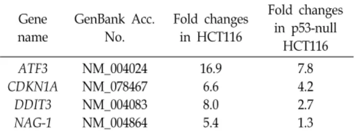
Table 2. Several commonly up-regulated genes more than 2- folds by EGCG in HCT116 cells and/or p53-null HCT 116 cells
Gene name
GenBank Acc.
No.
Fold changes in HCT116
Fold changes in p53-null
HCT116 ATF3
CDKN1A DDIT3 NAG-1
NM_004024 NM_078467 NM_004083 NM_004864
16.9 6.6 8.0 5.4
7.8 4.2 2.7 1.3
Fig. 1. Up-regulation of several tumor suppressor genes in EGCG-treated HCT116 cells. HCT116 cells were incu- bated with 50 μM EGCG for 24 hr. And then, total RNAs were prepared from EGCG- or DMSO-treated HCT116 cells and were used for RT-PCR by using ATF3, CDKN1A, DDIT3, NAG-1 and p53 gene specific primers.
(EGCG, genistein, resveratrol)을 제시한 조건에 따라 24시간 동안 처리 후 세포를 수확하였다. 수확한 세포는 RIPA buffer (20 mM Tris-Cl [pH 7.5], 150 mM NaCl, 1 mM NaCL, 1 mM Na2EDTA, 1 mM EGTA, 1% NP40, 1% sodium deoxycholate, 2.5 mM sodium pyrophosphate, 1 mM β-glycerophosphate, 1 mM Na3VO4, 1 μg/ml leupeptin)용액을 200 μl 처리하여 가볍게 tapping한 후 얼음 위에서 15분간 lysis 시킨 다음 4℃
centrifuge에서 14,000 rpm으로 10분간 원심분리 하여 단백질 을 추출하였다. 단백질 정량은 Bradford protein assay kit (Bio-Rad, Hercules, MA, USA)를 사용하여 측정하였으며, 30 μg의 단백질을 사용하여 Western blot 실험을 진행하였다. 본 연구에서는 anti-NAG-1, anti-ATF3, anti-p53, anti-ACTIN 1 차 항체를 사용하였고, 2차 항체로는 horseradish perox- idase-conjugated goat anti-rabbit과 horseradish perox- idase-conjugated donkey anti-goat를 사용하였다. NAG-1 항 체를 제외한 모든 항체는 Santa Cruz사(Santa Cruz, CA, USA) 로부터 구입하였고 NAG-1 antibody는 미국 NIEHS (National Institute of Environmental Health Sciences)의 Dr. Eling group으로부터 제공 받았다.
결과 및 고찰
Oligo DNA microarray를 이용한 유전체 수준에서의 유 전자 발현 분석
EGCG에 의한 암 세포 항 성장 활성을 유전자 발현 수준에 서 이해하고, EGCG에 의해 차별적으로 발현되는 유전자를 중심으로 p53 의존성을 분석하고자 인간 대장암 세포주 HCT 116와 p53-null HCT116 세포주를 이용하였다. 먼저, HCT116 에서 24시간 동안 50 μM EGCG를 처리한 후 차별적으로 발현 되는 유전자를 분석한 결과 2배 이상 발현이 증가된 유전자는 1,517개, 2배 이상 발현이 감소된 유전자는 1,757개가 선별되 었다(data not shown). 반면, 대장암 p53-null HCT116에서는 같은 EGCG 처리조건에서 2배 이상 발현이 증가된 유전자는 5,543개, 2배 이상 발현이 감소된 유전자는 5,204개로 분석되 었다(data not shown). 본 연구에서는 HCT116 세포주에서 발 현이 증가된 유전자 중 항암 유전자 4개(ATF3, CDKN1A,
DDIT3, NAG-1)를 선별하여 p53-null HCT116 세포주에서 분 석된 결과와 비교하였다. Oligo DNA microarray 실험 결과 Table 2에서 보는 바와 같이 50 μM EGCG의 처리에 의해 ATF3, CDKN1A, DDIT3 유전자는 p53존재 유무에 관계없이 발현이 증가되는 반면, NAG-1 유전자는 p53 의존적으로 발현 이 증가되는 것으로 분석되었다. Oligo DNA microarray 결과 를 검증하기 위하여 HCT116에 50 μM EGCG 처리 후 ATF3, CDKN1A, DDIT3, NAG-1 그리고 p53의 발현을 RT-PCR로 확 인하였다. 그 결과, Fig. 1에서 보는 바와 같이 oligo DNA mi- croarray 결과와 일치하며, 모든 유전자의 발현이 EGCG에 의 해 증가됨을 확인하였다. 전사조절인자인 ATF3 (Activating transcription factor 3)는 대장암, 식도암 등 다양한 암 에서 암 억제 활성인자로 보고되었으며[5, 22], 녹차의 카테킨 성분 중 EGCG, ECG에 의해 ATF3 발현이 증가되는 것이 보고된 바 있다[3]. 한편, NAG-1 (NSAID-activated gene) 유전자는 두경부암세포에서 EGCG처리에 의해 ATM/p53경로를 통해 발현이 증가되며, 이러한 NAG-1의 발현 증가는 세포사멸과 직접적인 관련이 있음이 보고되었다[7].
EGCG에 의한 ATF3와 NAG-1 유전자 및 단백질 발현의 p53 의존성 검증
EGCG에 의해 발현이 증가되는 유전자 중 ATF3와 NAG-1 의 발현 증가가 p53에 의존적인지 확인하기 위하여 p53-null HCT116 세포주를 이용하여 ATF3과 NAG-1 유전자 발현을 확인하였다. Fig. 2A에서 보는 바와 같이 ATF3는 p53 유전자 의 존재와 관계없이 EGCG에 의해 발현이 증가되는 것을 확인 할 수 있었고, 이에 반해 EGCG에 의한 NAG-1 유전자의 발현 은 p53 유전자가 존재해야만 발현이 증가됨을 확인하였다. 이 러한 연구결과는 EGCG에 의한 ATF3의 발현은 p53에 비의존
Fig. 3. p53 dependency on up-regulation of ATF3 and NAG-1 by genistein and resveratrol. HCT116 cells were treated with 50 μM EGCG, 50 μM genistein (GEN) and 50 μM resveratrol (RES) for 24 hr. Thirty micrograms of total proteins were prepared and Western blot analysis was performed with using ATF3, NAG-1, and ACTIN anti- sbodies.
Table 3. Summary of p53 dependency on up-regulation of ATF3 and NAG-1 by EGCG, genistein and resveratrol Phytochemicals
Genes EGCG Genistein Resveratrol
ATF3 NAG-1
-*
+**
NI***
+
- - -* : p53-independent expression.
+** : p53-dependent expression.
NI*** : not induced (regardless of p53 status).
A
B
Fig. 2. p53 dependency on up-regulation of ATF3 and NAG-1 by EGCG treatment. (A) HCT116 cells or p53-null HCT116 cells were treated with 50 μM EGCG or DMSO (vehicle) for 24 hr. And then, total RNAs were extracted from harvested cells and were used for RT-PCR with ATF3 and NAG-1 gene specific primers. (B) HCT116 cells were incubated with indicated concentrations of EGCG for 24 hr. Thirty micrograms of total proteins were pre- pared and Western blot analysis was performed with using ATF3, NAG-1, and ACTIN antibodies.
적, NAG-1의 발현은 p53에 의존적임을 나타낸다. 단백질 발 현 수준에서 ATF3 와 NAG-1발현의 p53 의존성을 확인하고 자, HCT116와 p53-null HCT116에 EGCG를 농도별(0, 25, 50, 100 μM)로 24시간 동안 처리한 후 ATF3와 NAG-1의 단백질 발현을 확인하였다. Fig. 2B에서 보는 바와 같이 ATF3 단백질 발현은 p53의 발현과는 무관하게 처리한 EGCG 농도 의존적 으로 증가됨을 확인하였다. 반면 NAG-1 단백질은 HCT116 세포주에서는 EGCG에 의해 농도의존적으로 발현이 증가되 는 반면, p53-null 세포주에서는 EGCG에 의해 NAG-1 단백질 이 전혀 증가되지 않았다. 이러한 결과는 RT-PCR의 결과와 일치하는 결과이며, EGCG의 처리에 의해 ATF3의 단백질의 발현은 전사조절인자 p53에 의해 영향을 받지 않으나, EGCG 에 의한 NAG-1 단백질의 발현 증가는 p53을 경유해서 일어남 을 증명한 것이다.
파이토케미칼 genistein, resveratrol에 의한 ATF3 와 NAG-1 단백질 발현의 p53 의존성 분석
파이토케미칼 genistein과 resveratrol은 p53 의존적으로 NAG-1 유전자의 발현을 증가 시킬 수 있음이 보고되었으며[1, 21], genistein과 resvaratrol이 ATF3 유전자의 발현도 증가시 킬 수 있음이 보고되었다[2]. 본 연구에서는 EGCG와 함께 파 이토케미칼 genistein과 resveratrol에 의한 ATF3와 NAG-1의 단백질 발현과 p53과의 관련성을 확인하고자 50 μM EGCG, 50 μM genistein, 50 μM resveratrol을 각각 24시간 동안
HCT116 세포주와 p53-null HCT116 세포주에 처리한 후 p53, ATF3 그리고 NAG-1 단백질의 발현을 확인하였다. 그 결과, p53단백질은 HCT116에서 EGCG, genistein과 resveratrol 처 리에 의해 발현이 약하게 증가된 반면, p53-null HCT116에서 는 전혀 관찰되지 않았다. Genistein에 의한 NAG-1 단백질 발현은 EGCG와 마찬가지로 p53에 의존적이었지만, ATF3 단 백질은 genistein에 의해 발현 되지 않았다. 그리고 resveratrol 에 의해서 ATF3와 NAG-1 단백질 모두 p53 단백질의 존재에 관계없이 증가되었다(Fig. 3).
종합적으로 이러한 연구결과는 Table 3에서 보는 바와 같 이, 세 가지 종류의 파이토케미칼은 동일한 항암 활성을 가지 고 있지만, 항암 단백질인 ATF3와 NAG-1의 발현을 p53 의존 성 측면에서 비교해 보면 서로 다른 항암기전을 가지는 것으 로 판단된다. 이러한 연구결과는 파이토케미칼 EGCG, genis- tein, resveratrol에 의한 항암 활성과 작용기전을 이해하는데 도움을 줄 것으로 판단된다.
감사의 글
본 논문은 2016학년도 안동대학교 연구비에 의하여 연구되 었음.
References
1. Baek, S. J., Wilson, L. C. and Eling, T. E. 2002. Resveratrol
enhances the expression of non-steroidal anti-inflammatory drug-activated gene (NAG-1) by increasing the expression of p53. Carcinogenesis 23, 425-434.
2. Bottone, F. G. Jr. and Alston-Mills, B. 2011. The dietary com- pounds resveratrol and genistein induce activating tran- scription factor 3 while suppressing inhibitor of DNA bind- ing/differentiation-1. J. Med. Food. 14, 584-593.
3. Cho, K. N., Sukhthankar, M., Lee, S. H., Yoon, J. H. and Baek, S. J. 2007. Green tea catechin (-)-epicatechin gallate induces tumour suppressor protein ATF3 via EGR-1 activa- tion. Eur. J. Cancer 43, 2404-2412.
4. Du, G. J., Zhang, Z., Wen, X. D., Yu, C., Calway, T., Yuan, C. S. and Wang, C. Z. 2012. Epigallocatechin Gallate (EGCG) is the most effective cancer chemopreventive polyphenol in green tea. Nutrients 4, 1679-1691.
5. Hackl, C., Lang, S. A., Moser, C., Mori, A., Fichtner-Feigl, S., Hellerbrand, C., Dietmeier, W., Schlitt, H. J., Geissler, E.
K. and Stoeltzing, O. 2010. Activating transcription factor-3 (ATF3) functions as a tumor suppressor in colon cancer and is up-regulated upon heat-shock protein 90 (Hsp90) in- hibition. BMC Cancer 10, 668.
6. Huang, C. Y., Han, Z., Li, X., Xie, H. H. and Zhu, S. S. 2017.
Mechanism of EGCG promoting apoptosis of MCF-7 cell line in human breast cancer. Oncol. Lett. 14, 3623-3627.
7. Kang, S. U., Lee, B. S., Lee, S. H., Baek, S. J., Shin, Y. S.
and Kim, C. H. 2013. Expression of NSAID-activated gene-1 by EGCG in head and neck cancer: involvement of ATM-de- pendent p53 expression. J. Nutr. Biochem. 24, 986-999.
8. Khan, N. and Mukhtar, H. 2013. Tea and health: studies in humans. Curr. Pharm. Des. 19, 6141-6147.
9. Lane, D. and Levine, A. 2010. p53 Research: the past thirty years and the next thirty years. Cold Spring Harb. Perspect.
Biol. 2, a000893.
10. Levav-Cohen, Y., Goldberg, Z., Tan, K. H., Alsheich-Bartok, O., Zuckerman, V., Haupt, S. and Haupt, Y. 2014. The p53- Mdm2 loop: a critical juncture of stress response. Subcell.
Biochem. 85, 161-186.
11. Liu, L., Ju, Y., Wang, J. and Zhou, R. 2017. Epigallocatechin -3-gallate promotes apoptosis and reversal of multidrug re- sistance in esophageal cancer cells. Pathol. Res. Pract. 213, 1242-1250.
12. Maru, G. B., Hudlikar, R. R., Kumar, G., Gandhi, K. and Mahimkar, M. B. 2016. Understanding the molecular mecha- nisms of cancer prevention by dietary phytochemicals: From experimental models to clinical trials. World J. Biol. Chem.
7, 88-99.
13. Mohanty, S, Adhikary, A., Chakrabarty, S., Sa, G. and Das, T. 2012. Operation 'p53 Hunt' to combat cancer: theaflavins in action. Front Biosci. (Schol Ed) 4, 300-320.
14. Moradzadeh, M., Hosseini, A., Erfanian, S. and Rezaei, H.
2017. Epigallocatechin-3-gallate promotes apoptosis in hu- man breast cancer T47D cells through down-regulation of PI3K/AKT and Telomerase. Pharmacol. Rep. 69, 924-928.
15. Moseley, V. R., Morris, J., Knackstedt, R. W. and Wargovich, M. J. 2013. Green tea polyphenol epigallocatechin 3-gallate, contributes to the degradation of DNMT3A and HDAC3 in HCT116 human colon cancer cells. Anticancer Res. 33, 5325- 5333.
16. Parrales, A. and Iwakuma, T. 2015. Targeting oncogenic mu- tant p53 for cancer therapy. Front. Oncol. 5, 288.
17. Shin, Y. S., Kang, S. U., Park, J. K., Kim, Y. E., Kim, Y. S., Baek, S. J., Lee, S. H. and Kim, C. H. 2016. Anti-cancer effect of (-)-epigallocatechin-3-gallate (EGCG) in head and neck cancer through repression of transactivation and enhanced degradation of β-catenin. Phytomedicine 23, 1344-1355.
18. Tan, A. C., Konczak, I., Sze, D. M. and Ramzan, I. 2011.
Molecular pathways for cancer chemoprevention by dietary phytochemicals. Nutr. Cancer 63, 495-505.
19. Vanamala, J., Reddivari, L., Radhakrishnan, S. and Tarver, C. 2010. Resveratrol suppresses IGF-1 induced human colon cancer cell proliferation and elevates apoptosis via sup- pression of IGF-1R/Wnt and activation of p53 signaling pathways. BMC Cancer 10, 238.
20. Vaseva, A. V. and Moll, U. M. 2009. The mitochondrial p53 pathway. Biochim. Biophys. Acta. 1787, 414-420.
21. Wilson, L. C., Baek, S. J., Call, A. and Eling, T. E. 2003.
Nonsteroidal anti-inflammatory drug-activated gene (NAG-1) is induced by genistein through the expression of p53 in colorectal cancer cells. Int. J. Cancer 105, 747-753.
22. Xie, J. J., Xie, Y. M., Chen, B., Pan, F., Guo, J. C., Zhao, Q., Shen, J. H., Wu, Z. Y., Wu, J. Y., Xu, L. Y. and Li, E.
M. 2014. ATF3 functions as a novel tumor suppressor with prognostic significance in esophageal squamous cell carcinoma. Oncotarget 5, 8569-8582.
23. Zhang, Z., Wang, C. Z., Du, G. J., Qi, L. W., Calway, T., He, T. C., Du, W. and Yuan, C. S. 2013. Genistein induces G2/M cell cycle arrest and apoptosis via ATM/p53-depend- ent pathway in human colon cancer cells. Int. J. Oncol. 43, 289-296.
초록:EGCG, genistein, resveratrol 처리에 의한 ATF3와 NAG-1 유전자 발현변화의 p53 의존성 분석
김민정․김현지․서유미․이은주․김종식*
(국립안동대학교 생명과학과)
EGCG는 녹차의 카테킨 중의 하나로서 항산화, 항염증 그리고 항암 활성 등 다양한 생리활성을 가지고 있는 물질로 알려져 있다. 본 연구에서는 EGCG를 처리한 HCT116 세포와 p53-null HCT116 세포에서 oligo DNA mi- croarray 실험을 통하여 유전자 발현 변화를 분석하였다. Microarray 실험에서 EGCG를 처리한 HCT116 세포주에 서 증가된 유전자 4개(ATF3, CDKN1A, DDIT3, NAG-1)를 선별하여, p53-null HCT116에서의 데이터와 비교하였 다. NAG-1을 제외한 3개의 유전자는 p53의 상태와 관계없이 발현이 증가하였고, p53-null HCT116 세포주에서는 EGCG에 의해 NAG-1의 발현이 증가되지 않았다. EGCG의 처리에 의해 ATF3와 NAG-1의 유전자와 단백질의 발현을 확인한 경우 동일한 결과를 보여주었다. 또한, 파이토케미칼 genistein과 resveratrol을 처리한 후 ATF3와 NAG-1의 발현을 연구한 결과 genistein은 p53의 상태와 관계없이 ATF3 발현에 영향을 주지 못하는 반면, NAG-1 단백질은 p53 존재 하에서만 발현이 증가되었다. 이에 반해 resveratrol은 p53의 상태와는 관계없이 ATF3와 NAG-1 단백질의 발현을 증가시켰다. 따라서, 항암 활성을 가진 3 종류의 파이토케미칼이 각각 다른 기전으로 항 암 유전자를 발현시키는 것으로 생각된다. 종합적으로 본 연구결과는 파이토케미칼 EGCG, genistein, resveratrol 에 의해 매개되는 항암 활성의 기전을 이해하는데 도움을 줄 것으로 생각된다.