90
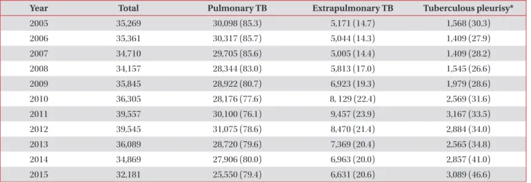
cases were reported in 2015, which accounted for 9.6% of the 32,181 new TB cases and 46.6% of the 6,631 extrapulmonary TB cases (Table 1)
3.
Tuberculous pleurisy is the most common form of extra- pulmonary tuberculosis (TB) and is the main cause of pleural effusion in Korea
1,2. In Korea, 3,089 new tuberculous pleurisy
Progression of Tuberculous Pleurisy: From a Lymphocyte-Predominant Free-Flowing Effusion to a Neutrophil-Predominant
Loculated Effusion
Won-Jung Koh, M.D.
Division of Pulmonary and Critical Care Medicine, Department of Medicine, Samsung Medical Center, Sungkyunkwan University School of Medicine, Seoul, Korea
Address for correspondence: Won-Jung Koh, M.D.
Division of Pulmonary and Critical Care Medicine, Department of Medicine, Samsung Medical Center, Sungkyunkwan University School of Medicine, 81 Irwon-ro, Gangnam-gu, Seoul 06351, Korea
Phone: 82-2-3410-3429, Fax: 82-2-3410-3849, E-mail: [email protected] Received: Dec. 17, 2016, Revised: Dec. 19, 2016, Accepted: Dec. 20, 2016
cc
It is identical to the Creative Commons Attribution Non-Commercial License (http://creativecommons.org/licenses/by-nc/4.0/).
EDITORIAL
https://doi.org/10.4046/trd.2017.80.1.90ISSN: 1738-3536(Print)/2005-6184(Online) • Tuberc Respir Dis 2017;80:90-92
Copyright © 2017
The Korean Academy of Tuberculosis and Respiratory Diseases.
All rights reserved.
Table 1. Tuberculous pleurisy in Korea (2005–2015)
Year Total Pulmonary TB Extrapulmonary TB Tuberculous pleurisy*
2005 35,269 30,098 (85.3) 5,171 (14.7) 1,568 (30.3)
2006 35,361 30,317 (85.7) 5,044 (14.3) 1,409 (27.9)
2007 34,710 29,705 (85.6) 5,005 (14.4) 1,409 (28.2)
2008 34,157 28,344 (83.0) 5,813 (17.0) 1,545 (26.6)
2009 35,845 28,922 (80.7) 6,923 (19.3) 1,979 (28.6)
2010 36,305 28,176 (77.6) 8, 129 (22.4) 2,569 (31.6)
2011 39,557 30,100 (76.1) 9,457 (23.9) 3,167 (33.5)
2012 39,545 31,075 (78.6) 8,470 (21.4) 2,884 (34.0)
2013 36,089 28,720 (79.6) 7,369 (20.4) 2,565 (34.8)
2014 34,869 27,906 (80.0) 6,963 (20.0) 2,857 (41.0)
2015 32,181 25,550 (79.4) 6,631 (20.6) 3,089 (46.6)
Values are presented as number of patients (%).
*Number of patients with tuberculous pleurisy/Number of patients with extrapulmonary TB.
TB: tuberculosis.
Progression of tuberculosis pleurisy
https://doi.org/10.4046/trd.2017.80.1.90 91
www.e-trd.org
Traditionally, tuberculous pleurisy is indicated by predomi- nant lymphocytosis in the pleural fluid and a low yield of effu- sion culture due to the paucibacillary nature of TB
4. However, several recent studies have reported that the lymphocyte counts in pleural fluid were decreased in patients who were diagnosed with tuberculous pleurisy, and 10%–17% of the pa- tients with tuberculous pleurisy had neutrophil-predominant pleural fluid
5-8. In addition, the yield of effusion culture is re- ported to be higher (15%–63%) than previously thought, with the introduction of a liquid culture method, and the lympho- cyte percentage in pleural fluid was negatively associated with the probability of a positive effusion culture
6-9.
The radiographic appearances of tuberculous pleurisy can be subdivided into two types, based on the chest X-ray, chest computed tomography, or chest ultrasonography findings:
free-flowing and loculated effusions
10,11. Residual pleural thick- ening is a common complication of tuberculous pleurisy, and a loculated effusion at the initial presentation was associated with significant residual pleural thickening
12,13. Intrapleural fibrinolytic therapy can reduce this residual pleural thickening in patients with loculated tuberculous pleurisy
10,14. In compari- son, the characteristics of the effusion in loculated tuberculous pleurisy have not been well studied.
In this issue of Tuberculosis and Respiratory Diseases, Ko et al.
15described the pleural fluid characteristics in patients with tuberculous pleurisy to examine the association between loculation and positive mycobacterial cultures of pleural fluid.
Among 219 patients with tuberculous pleurisy, loculation was identified in 86 patients (39%), and 69 patients (32%) had ef- fusion cultures positive for Mycobacterium tuberculosis. The proportion of loculation was much higher in the patients with positive effusion cultures (86%, 59/69) than in the patients with negative effusion cultures (18%, 27/150). In other words, the majority of patients (69%, 59/86) with loculated tuber-
culous pleurisy had positive effusion cultures, whereas posi- tive effusion cultures were found only in 7.5% of the patients (10/133) without loculation. In their study, nine patients had neutrophil-predominant pleural effusions. All of them were culture positive for M. tuberculosis in pleural fluid, and six of them had loculated tuberculous pleurisy. Compared to the patients with negative effusion cultures, those with positive effusion cultures had a lower lymphocyte percentage, pH, and glucose level and a higher neutrophil percentage and higher protein and lactate dehydrogenase (LDH) levels in the pleural effusion, and higher serum C-reactive protein levels. Multiple logistic regression analysis found that loculation of the pleural fluid (adjusted odds ratio [OR], 40.06; 95% confidence interval [CI], 9.36–171.56; p<0.001) was associated, and lymphocyte percentage was inversely associated (adjusted OR, 0.93; 95%
CI, 0.90–0.97; p=0.001) with a positive effusion culture.
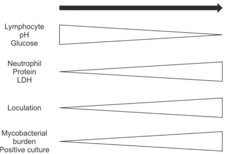
The traditional concept of the pathogenesis of tuberculous pleurisy is rupture of a subpleural caseous focus followed by a delayed hypersensitivity reaction to M. tuberculosis antigens
1. These occurrences result in lymphocyte-predominance and a low yield of effusion culture in tuberculous pleurisy
6. However, tuberculous pleurisy may involve a continuous spectrum of disease processes (Figure 1). In the early phase of tuberculous pleurisy, the pleural effusion could have lymphocyte-predom- inance, a high pH, and high glucose levels. As the tuberculous pleurisy progresses, the pleural effusion could develop neutro- phil-predominance, and high protein and LDH levels, as well as loculation and positive effusion cultures, as found in Ko et al.
15.
From a clinical perspective, Ko et al.
15indicated that sus- picion and the differentiation of tuberculous pleurisy from a parapneumonic effusion are very important in patients with loculated pleural effusions. Loculated pleural effusions, especially neutrophil-predominant effusions, are typically considered to be parapneumonic effusions in clinical prac- tice. The inclusion of tuberculous pleurisy in the differential diagnosis and prompt sputum and pleural fluid examination for possible tuberculous pleurisy are needed, especially in TB- endemic areas.
Conflicts of Interest
No potential conflict of interest relevant to this article was reported.
References
1. Jeon D. Tuberculous pleurisy: an update. Tuberc Respir Dis 2014;76:153-9.
2. Lee JY. Diagnosis and treatment of extrapulmonary tubercu- losis. Tuberc Respir Dis 2015;78:47-55.
Lymphocyte pH Glucose
Neutrophil Protein
LDH
Loculation
Mycobacterial burden Positive culture
Progression of tuberculous pleurisy