ABSTRACT
BACKGROUND: Left atrial (LA) strain is a novel parameter of LA function. However, its reference value has not been established, and the determining factors for LA strain remain elusive. We aimed to present LA strain with reservoir, conduit, and contractile components and associated parameters in a large-sized group of healthy individuals.
METHODS: The present study was from a prospective multicenter registry in South Korea.
Subjects who had no history of cardiovascular disease with adequate images were eligible for inclusion. LA reservoir, conduit, and contractile strains (LAS
RES, LAS
CDand LAS
CT, respectively) were measured. Left ventricular global longitudinal strain (LV GLS) and early
Original Article
Byung Joo Sun , MD, PhD
1, Jae-Hyeong Park , MD, PhD
1, Minyeong Lee , BSc
2, Jin-Oh Choi , MD, PhD
3, Ju-Hee Lee , MD, PhD
4, Mi-Seung Shin , MD, PhD
5, Mi-Jeong Kim , MD, PhD
6, Hae Ok Jung , MD, PhD
7,
Jeong Rang Park , MD, PhD
8, Il Suk Sohn , MD, PhD
9, Hyungseop Kim , MD, PhD
10, Hyung-Kwan Kim , MD, PhD
11, Goo-Yeong Cho , MD, PhD
12, Jin-Sun Park , MD, PhD
13,
Chi Young Shim , MD, PhD
14, Sung Hee Shin , MD, PhD
15, Kye Hun Kim , MD, PhD
16, Woo-Shik Kim , MD, PhD
17, and Seung Woo Park , MD, PhD
31 Division of Cardiology, Department of Internal Medicine, Chungnam National University Hospital, Chungnam National University School of Medicine, Daejeon, Korea
2Cardiovascular Center, Chungnam National University Hospital, Daejeon, Korea
3 Division of Cardiology, Department of Medicine, Samsung Medical Center, Sungkyunkwan University School of Medicine, Seoul, Korea
4 Division of Cardiology, Department of Internal Medicine, Chungbuk National University School of Medicine, Cheongju, Korea
5 Division of Cardiology, Department of Internal Medicine, Gil Hospital, Gachon University of Medicine and Science, Incheon, Korea
6 Division of Cardiology, Department of Internal Medicine, Incheon St. Mary's Hospital, College of Medicine, The Catholic University of Korea, Incheon, Korea
7 Department of Internal Medicine, Seoul St. Mary's Hospital, College of Medicine, The Catholic University of Korea, Seoul, Korea
8 Division of Cardiology, Department of Internal Medicine, Gyeongsang National University Hospital, Gyeongsang National University School of Medicine, Jinju, Korea
9 Department of Cardiology, Kyung Hee University School of Medicine, Kyung Hee University Hospital at Gangdong, Seoul, Korea
10 Division of Cardiology, Keimyung University Dongsan Medical Center, Daegu, Korea
11 Division of Cardiology, Department of Internal Medicine, Cardiovascular Center, Seoul National University College of Medicine, Seoul, Korea
12 Division of Cardiology, Department of Internal Medicine, Seoul National University and Cardiovascular Center, Seoul National University Bundang Hospital, Seongnam, Korea
13Department of Cardiology, Ajou University School of Medicine, Suwon, Korea
14Division of Cardiology, Severance Cardiovascular Hospital, Yonsei University College of Medicine, Seoul, Korea
15Division of Cardiology, Department of Internal Medicine, Inha University College of Medicine, Incheon, Korea
16Department of Cardiology, Chonnam National University Hospital, Gwangju, Korea
17Cardiovascular Center, Department of Internal Medicine, Kyung Hee University Medical Center, Seoul, Korea
Normal Reference Values for Left Atrial Strain and Its Determinants from a
Large Korean Multicenter Registry
Received: Apr 2, 2020 Revised: Apr 21, 2020 Accepted: May 5, 2020 Address for Correspondence:
Jae-Hyeong Park, MD, PhD
Division of Cardiology, Department of Internal Medicine, School of Medicine, Chungnam National University, Chungnam National University Hospital, 282 Munhwa-ro, Jung-gu, Daejeon 35015, Korea.
E-mail: [email protected] Copyright © 2020 Korean Society of Echocardiography
This is an Open Access article distributed under the terms of the Creative Commons Attribution Non-Commercial License (https://
creativecommons.org/licenses/by-nc/4.0/) which permits unrestricted non-commercial use, distribution, and reproduction in any medium, provided the original work is properly cited.
ORCID iDs Byung Joo Sun
https://orcid.org/0000-0001-6019-4343 Jae-Hyeong Park
https://orcid.org/0000-0001-7035-286X Minyeong Lee
https://orcid.org/0000-0002-2228-0440 Jin-Oh Choi
https://orcid.org/0000-0002-2441-2267 Ju-Hee Lee
https://orcid.org/0000-0002-0858-0973 Mi-Seung Shin
https://orcid.org/0000-0002-0273-0109 Mi-Jeong Kim
https://orcid.org/0000-0002-7887-3309 Hae Ok Jung
https://orcid.org/0000-0002-5102-9212 Jeong Rang Park
https://orcid.org/0000-0002-8330-2738
Il Suk Sohn
https://orcid.org/0000-0001-8004-5185 Hyungseop Kim
https://orcid.org/0000-0001-7056-4221 Hyung-Kwan Kim
https://orcid.org/0000-0001-7950-2131 Goo-Yeong Cho
https://orcid.org/0000-0002-7067-5535 Jin-Sun Park
https://orcid.org/0000-0002-7775-4092 Chi Young Shim
https://orcid.org/0000-0002-6136-0136 Sung Hee Shin
https://orcid.org/0000-0002-8306-9622 Kye Hun Kim
https://orcid.org/0000-0002-6885-1501 Woo-Shik Kim
https://orcid.org/0000-0002-3345-1137 Seung Woo Park
https://orcid.org/0000-0002-2941-515X Conflict of Interest
The authors have no financial conflicts of interest.
and late diastolic strain rates (DSR
eand DSR
a, respectively) were also evaluated.
RESULTS: Among a total of 324 subjects (mean age: 49 ± 16 years, 167 females), the mean LAS
RES, LAS
CD, and LAS
CTvalues were 35.9% ± 10.6%, 21.9% ± 9.3%, and 13.9% ± 3.6%, respectively. Mean LV GLS was -20.4% ± 2.2%, and mean DSR
eand DSR
awere 1.6 ± 0.4 s
-1and 0.8 ± 0.3 s
-1, respectively. With aging, LAS
RESand LAS
CDshowed significant decreases.
Factors showing independent associations with LAS
RESwere age (B = -0.425, p < 0.001), DSR
e(B = 4.706, p = 0.001), and LV GLS (B = -1.081, p < 0.001). Age (B = -0.319, p < 0.001), DSR
e(B
= 4.140, p = 0.002), DSR
a(B = -3.409, p = 0.018), and LV GLS (B = -0.783, p < 0.001) showed associations with LAS
CD. With LAS
CT, only DSR
ashowed a correlation (R = 0.277, p < 0.001).
CONCLUSIONS: We presented LA strain in a large-sized group of healthy subjects. Age is a significant determinant of LA function. Associations of LA strain with diastolic strain rates and LV GLS reflect cardiac mechanics.
Keywords: LA function; LA strain; Speckle-tracking echocardiography
INTRODUCTION
The clinical significance of left atrial (LA) remodeling has been shown in a variety of
cardiovascular diseases. However, the dynamic feature of LA has been recently highlighted.
1)2)These studies showed that LA is not simply a conduit chamber but that it has sophisticated actions during a cardiac cycle that performs in close interplay with the left ventricular (LV) mechanics.
1)Currently, LA volume index (LAVI) is a main parameter of LA remodeling.
1)However, as LAVI is based on a static volumetric measurement, it has inherent limitations with respect to reflecting the dynamic aspects of LA.
2)With this background, a novel speckle tracking echocardiography derived index, ‘LA strain,’ was recently introduced. LA strain may be more suitable to represent LA function.
2)LA strain has been tested in several clinical studies and shown to be a more useful diagnostic tool than conventional parameters.
3-5)However, the clinical evidence for using LA strain as a diagnostic tool is insufficient. In addition, applying LA strain in clinical practice is difficult because its reference value has not yet been established for a variety of subject groups. In the present study, we aimed to identify a reference value for LA strain with reservoir, conduit, and contractile components in a large group of healthy Koreans. We also aimed to elucidate determining factors for LA strain.
METHODS
Study outline
Our research group from the Korean Society of Echocardiography recently conducted a prospective nation-wide registry (Normal echOcardiogRaphic Measurements in KoreAn popuLation, NORMAL) including 23 tertiary-referral hospitals in South Korea and constructed a large-sized echocardiographic database of healthy individuals.
6)The primary objective of the study was to present normal reference values of echocardiography in a Korean population. The eligibility criteria were as follows: 1) an adult aged 20–79 years, and 2) no history of cardiovascular diseases such as hypertension, diabetes mellitus, coronary/
peripheral arterial disease, and atrial fibrillation. The NORMAL study required only two-
dimensional and Doppler echocardiographic images. The overall database included 1,003
subjects. In a sub-study focusing on strain analysis, 501 subjects (50%) whose exams were
performed using a single-vendor machine (GE Medical Systems, Horten, Norway) were
selected.
7)In the present study, LA strain analysis was finally available for 324 subjects (32%) for whom LA images included the whole LA roof in both apical four-chamber and two-chamber views. The subject clinical characteristics, conventional echocardiographic data, LV systolic strain, and diastolic strain rate were analyzed, with the main focus on LA strain. This study conformed to the ethical guidelines of the Declaration of Helsinki, and the study protocol was approved by the Ethics Committees of all participating centers. Written informed consent was waived because of the non-invasiveness of this study protocol.
Echocardiographic analysis
All routine echocardiographic studies were performed according to current
recommendations.
8)9)Images for strain analysis were obtained with 60–90 frames/sec from two consecutive cardiac beats.
10)All image data were digitally stored for analysis in the core laboratory of Chungnam National University Hospital. Two dedicated researchers (SBJ and PJH) who were blinded to clinical information analyzed all strain images using EchoPAC
®and BT 201 device (GE Medical Systems, Horten, Norway). LV global longitudinal strain (GLS) and early and late diastolic strain rates (DSR
eand DSR
a) were evaluated according to previously described methods.
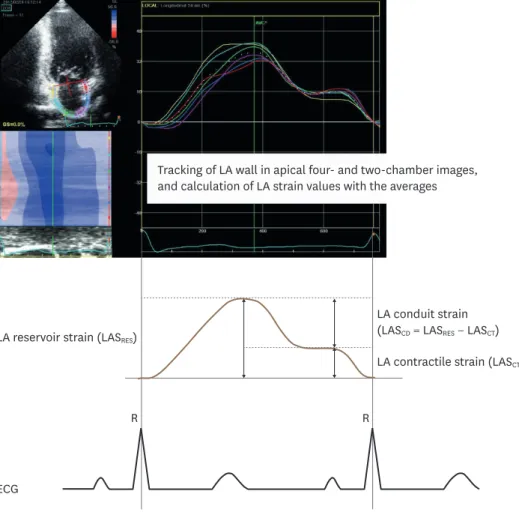
10)11)To measure LA strain, we manually traced the LA endocardium and thus the software automatically tracked the region of interest on the LA wall. A LA strain curve cycle was generated with a configuration by R-R gating that began with the onset of systole.
12)We then defined the following three components of LA function: LA reservoir, conduit, and contractile strains (LAS
RES, LAS
CD, and LAS
CT, respectively). The first peak point was defined as LAS
RES(Figure 1). Another peak point after the P wave on electrocardiography was defined as LAS
CT. We also defined variables LAS
CD(LAS
RES- LAS
CT) and LAS
CD/LAS
CTratio.
We performed this process in both apical 4-chamber and 2-chamber images and calculated average LA strain values.
Statistical analysis
We presented categorical variables as numbers (%) and continuous variables as mean ± standard deviation (SD). We used independent Student's t-tests to compare numeric data between sex groups. To evaluate differences in LA strain values among age groups defined by decade, we used one-way analysis of variance (ANOVA) with Bonferroni correction with non- parametric means as needed. To determine variables associated with LA stain parameters, Pearson's bivariate correlation test and multiple linear regression analysis were performed.
We evaluated intra- and inter-observer measurement variabilities based on intraclass correlation coefficients (ICCs). All reported p-values were two-tailed and a p-value of < 0.05 was accepted as statistically significant. SPSS software version 22 (IBM Corp, Armonk, NY, USA) was used for all statistical analyses.
RESULTS
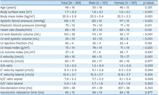
Subject characteristics
This study included a total of 324 subjects; the mean age was 49 ± 16 years, and there were
167 females (52%) (Table 1). The mean systolic and diastolic blood pressures were 120 ± 13
mmHg and 72 ± 10 mmHg, respectively. The mean pulse rate was 68 ± 10 beats/min. For the
echocardiographic parameters, the mean LV ejection fraction (LVEF) was 62 ± 4%, the mean
LV mass index (LVMI) was 75 ± 14 g/m
2, and the mean LAVI was 27 ± 6 mL/m
2. For Doppler
parameters, septal and lateral mitral annular E′ velocity were 9.3 ± 2.9 cm/s and 12.6 ± 3.7 cm/s, respectively. Septal and lateral E/E′ ratios were 7.9 ± 2.3 and 5.9 ± 1.8, respectively.
There was no significant difference in age between the male and female groups. The male group showed a higher body surface area, blood pressure, and LV volumes compared to the female group. However, LVEF and LAVI were not significantly different between male and female groups. The female group presented higher mitral E velocity and E/A ratio without showing significant differences in mitral annular E′ velocity and E/E′ ratios compared to the male group.
LA and LV strain values
In the total subjects, the mean LAS
RES, LAS
CD, and LAS
CTvalues were 35.9% ± 10.6%, 21.9% ± 9.3% and 13.9% ± 3.6%, respectively, and the mean LAS
CD/LAS
CTratio was 1.7 ± 0.8 (Table 2).
The mean LV GLS was -20.4% ± 2.2%. The mean DSR
eand DSR
awere 1.6 ± 0.4 s
-1and 0.8 ± 0.3 s
-1, respectively. The female group showed significantly higher LAS
RES(37.3% ± 11.0% vs. 34.3% ± 10.0%, p = 0.009) and LAS
CD(23.5% ± 9.8% vs. 20.3% ± 8.5%, p = 0.002) than the male group, whereas there was no significant difference in LAS
CTbetween sex groups. The female group showed significantly higher LAS
CD/LASCT ratio (1.8% ± 0.8% vs. 1.5% ± 0.7%, p = 0.001). The female group also showed higher LV GLS (-21.2% ± 2.1% vs. -19.5% ± 2.1%, p < 0.001) and DSRe (1.7 ± 0.4 s-1 vs. 1.4 ± 0.3 s
-1, p < 0.001), whereas DSR
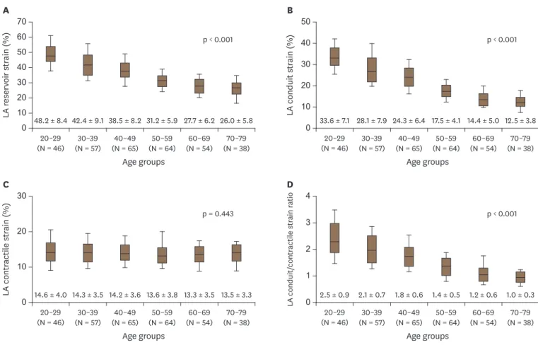
adid not differ between female and male groups. LAS
RES, LAS
CD, and LAS
CD/LAS
CTratio showed significant decreases with increasing age
Tracking of LA wall in apical four- and two-chamber images, and calculation of LA strain values with the averages
LA reservoir strain (LAS
RES)
LA conduit strain (LAS
CD= LAS
RES− LAS
CT) LA contractile strain (LAS
CT)
ECG
R R
Figure 1. Demonstration of LA strain measurement. ECG: electrocardiography, LA: left atrium.
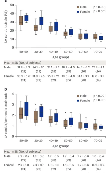
(Figures 2A, 2B, and 2D, respectively; all p < 0.001). However, there was no age-related trend for LAS
CT(Figure 2C). These trends according to age were observed in both male and female groups (Figure 3).
Factors associated with LA strain
In Pearson's bivariate analysis, LAS
RESshowed significant correlations with age (R = -0.705, p < 0.001), LVMI (R = -0.310, p < 0.001), E′ velocity (R = 0.603, p < 0.001), E/E′ ratio (R = -0.304, p < 0.001), DSR
e(R = 0.585, p < 0.001), DSR
a(R = -0.337, p < 0.001), and LV GLS (R = -0.359, p < 0.001) (Table 3). In multiple linear regression analysis, factors showing significant associations with LAS
RESwere age (B = -0.425, p < 0.001), DSR
e(B = 4.706, p = 0.001), and LV GLS (B = -1.081, p < 001). With LAS
CD, age (B = -0.319, p < 0.001), DSR
e(B = 4.140, p = 0.002), DSR
a(B = -3.409, p = 0.018), and LV GLS (B = -0.783, p < 0.001) showed independent associations while septal E′ velocity showed a trend without statistical significance (Table 4).
With LAS
CT, only DSR
ashowed a weak correlation (R = 0.277, p < 0.001). However, there was no other feasible variable for test of independent association (Supplementary Table 1).
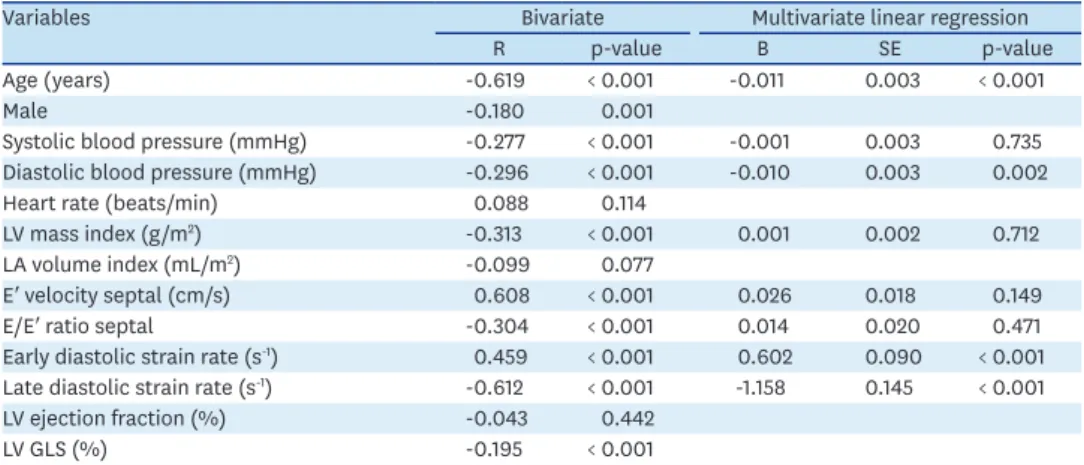
With LAS
CD/LAS
CTratio, age (B = -0.011, p < 0.001), diastolic blood pressure (B = -0.010, p =
Table 1. Clinical and echocardiographic parametersTotal (N = 324) Male (n = 157) Female (n = 167) p-value
Age (years) 49 ± 16 50 ± 16 48 ± 15 0.301
Body surface area (m2) 1.7 ± 0.2 1.8 ± 0.1 1.6 ± 0.1 < 0.001
Body mass index (kg/m2) 22.9 ± 2.9 23.3 ± 2.4 22.5 ± 3.2 0.001
Systolic blood pressure (mmHg) 120 ± 13 123 ± 12 117 ± 13 < 0.001
Diastolic blood pressure (mmHg) 72 ± 10 74 ± 10 71 ± 10 0.011
Heart rate (beats/min) 68 ± 10 67 ± 10 69 ± 10 0.133
LV end diastolic volume (mL) 103 ± 22 113 ± 21 93 ± 17 < 0.001
LV end systolic volume (mL) 39 ± 10 43 ± 10 35 ± 9 < 0.001
LV ejection fraction (%) 62 ± 4 62 ± 4 63 ± 4 0.162
LV mass index (g/m2) 75 ± 14 78 ± 14 71 ± 14 < 0.001
LA volume index (mL/m2) 27 ± 6 27 ± 6 28 ± 7 0.647
E velocity (cm/s) 69 ± 16 66 ± 15 72 ± 17 0.001
A velocity (cm/s) 60 ± 17 60 ± 17 60 ± 16 0.977
E/A ratio 1.2 ± 0.5 1.2 ± 0.4 1.3 ± 0.6 0.030
E′ velocity septal (cm/s) 9.3 ± 2.9 9.1 ± 2.8 9.5 ± 3.0 0.244
E′ velocity lateral (cm/s) 12.6 ± 3.7 12.3 ± 3.7 12.8 ± 3.7 0.254
E/E′ ratio septal 7.9 ± 2.3 7.7 ± 2.2 8.1 ± 2.4 0.090
E/E′ ratio lateral 5.9 ± 1.8 5.7 ± 1.8 6.0 ± 1.8 0.203
Deceleration time (ms) 209 ± 38 211 ± 39 207 ± 38 0.303
Isovolumic relaxation time (ms) 84 ± 15 84 ± 14 84 ± 16 0.677
LA: left atrium, LV: left ventricle.
Table 2. Strain parameter values
Total (N = 324) Male (n = 157) Female (n = 167) p-value
LA reservoir strain (%) 35.9 ± 10.6 34.3 ± 10.0 37.3 ± 11.0 0.009
Four-chamber (%) 35.4 ± 11.3 33.7 ± 10.3 37.0 ± 11.9 0.008
Two-chamber (%) 36.3 ± 10.9 34.9 ± 10.8 37.7 ± 10.8 0.020
LA conduit strain (%) 21.9 ± 9.3 20.3 ± 8.5 23.5 ± 9.8 0.002
Four-chamber (%) 22.3 ± 9.9 20.5 ± 8.9 24.0 ± 10.4 0.001
Two-chamber (%) 21.5 ± 9.5 20.0 ± 9.0 22.9 ± 9.8 0.005
LA contractile strain (%) 13.9 ± 3.6 14.0 ± 3.7 13.8 ± 3.6 0.645
Four-chamber (%) 13.0 ± 4.1 13.2 ± 4.0 12.9 ± 4.1 0.585
Two-chamber (%) 14.8 ± 4.5 14.9 ± 4.6 14.8 ± 4.4 0.801
LA conduit/contractile strain ratio 1.7 ± 0.8 1.5 ± 0.7 1.8 ± 0.8 0.001 Global LV longitudinal strain (%) -20.4 ± 2.2 -19.5 ± 2.1 -21.2 ± 2.1 < 0.001 Early diastolic strain rate (s-1) 1.6 ± 0.4 1.4 ± 0.3 1.7 ± 0.4 < 0.001
Late diastolic strain rate (s-1) 0.8 ± 0.3 0.8 ± 0.3 0.8 ± 0.3 0.966
LA: left atrium, LV: left ventricle.
0.002), DSR
e(B = 0.602, p < 0.001), and DSR
a(B = -1.158, p < 0.001) showed independent associations (Table 5).
Measurement variability
Intra- and inter-observer ICCs for LAS
RESwere 0.974 (95% confidence interval [CI], 0.946–0.988) and 0.948 (95% CI, 0.889–0.976), respectively (Table 6). Intra- and inter- observer ICCs for LAS
CDwere 0.978 (95% CI, 0.953–0.990) and 0.930 (95% CI, 0.852–0.967),
LA reservoir strain (%)
0 20 50 70
40 60
30
10
20–29 (N = 46)
48.2 ± 8.4 42.4 ± 9.1 38.5 ± 8.2 31.2 ± 5.9 27.7 ± 6.2 26.0 ± 5.8 30–39
(N = 57) 40–49
(N = 65) 50–59
(N = 64) 60–69
(N = 54) 70–79 (N = 38)
Age groups
p < 0.001
A
LA conduit strain (%)
0 20 50 40 30
10
20–29 (N = 46)
33.6 ± 7.1 28.1 ± 7.9 24.3 ± 6.4 17.5 ± 4.1 14.4 ± 5.0 12.5 ± 3.8 30–39
(N = 57) 40–49
(N = 65) 50–59
(N = 64) 60–69
(N = 54) 70–79 (N = 38)
Age groups
p < 0.001
B
LA contractile strain (%)
0 20 30
10
20–29 (N = 46)
14.6 ± 4.0 14.3 ± 3.5 14.2 ± 3.6 13.6 ± 3.8 13.3 ± 3.5 13.5 ± 3.3 30–39
(N = 57) 40–49
(N = 65) 50–59
(N = 64) 60–69
(N = 54) 70–79 (N = 38)
Age groups
p = 0.443
C
LA conduit/contractile strain ratio
0 4
3
2
1
20–29 (N = 46)
2.5 ± 0.9 2.1 ± 0.7 1.8 ± 0.6 1.4 ± 0.5 1.2 ± 0.6 1.0 ± 0.3 30–39
(N = 57) 40–49
(N = 65) 50–59
(N = 64) 60–69
(N = 54) 70–79 (N = 38)
Age groups
p < 0.001
D
Figure 2. LA strain values according to age groups. LA: left atrium.
Table 3. Parameters associated with LA reservoir strain
Variables Bivariate Multivariate linear regression
R p-value B SE p-value
Age (years) -0.705 < 0.001 -0.425 0.028 < 0.001
Male -0.144 0.009
Systolic blood pressure (mmHg) -0.187 0.001
Diastolic blood pressure (mmHg) -0.118 0.040
Heart rate (beats/min) 0.169 0.002
LV mass index (g/m2) -0.310 < 0.001 -0.028 0.028 0.325
LA volume index (mL/m2) -0.162 0.004
E′ velocity septal (cm/s) 0.603 < 0.001 0.128 0.215 0.553
E/E′ ratio septal -0.304 < 0.001 0.167 0.246 0.497
Early diastolic strain rate (s-1) 0.585 < 0.001 4.706 1.465 0.001
Late diastolic strain rate (s-1) -0.337 < 0.001 2.711 1.704 0.113
LV ejection fraction (%) 0.036 0.525
LV GLS (%) -0.359 < 0.001 -1.081 0.236 < 0.001
GLS: global longitudinal strain, LA: left atrium, LV: left ventricle.
LA reservoir strain (%) 0 20 50 70
40 60
30
10
20–29
46.7 ± 10.0 (22) 38.6 ± 8.1
(28) 37.6 ± 7.2
(28) 30.2 ± 5.7
(29) 27.7 ± 6.8
(26) 26.2 ± 6.0 (24) 49.6 ± 6.6
(24) 46.1 ± 8.7
(29) 39.2 ± 8.9
(37) 32.0 ± 5.9
(35) 27.7 ± 5.6
(28) 25.8 ± 5.7 (14)
30–39 40–49 50–59 60–69 70–79
Age groups A
Male Female
Mean ± SD (No. of subjects)
p < 0.001 p < 0.001 Male
Female
*
LA conduit strain (%)
0 20 50 40 30
10
20–29
31.8 ± 8.3 (22) 24.1 ± 6.1
(28) 23.1 ± 5.2
(28) 16.2 ± 4.0
(29) 14.6 ± 6.2
(26) 12.8 ± 4.1 (24) 35.3 ± 5.6
(24) 31.9 ± 7.5
(29) 25.3 ± 7.1
(37) 18.6 ± 4.0
(35) 14.1 ± 3.7
(28) 12.0 ± 3.1 (14)
30–39 40–49 50–59 60–69 70–79
Age groups B
Male Female
Mean ± SD (No. of subjects)
p < 0.001 p < 0.001 Male
Female
*
*
LA contractile strain (%)
0 20 30
10
20–29
14.9 ± 3.8
(22) 14.5 ± 4.2
(28) 14.4 ± 3.6
(28) 14.0 ± 3.6
(29) 13.1 ± 3.6
(26) 13.4 ± 3.4 (24) 14.4 ± 4.3
(24) 14.2 ± 2.8
(29) 14.0 ± 3.7
(37) 13.4 ± 4.0
(35) 13.6 ± 3.5
(28) 13.8 ± 3.2 (14)
30–39 40–49 50–59 60–69 70–79
Age groups C
Male Female
Mean ± SD (No. of subjects)
p = 0.507 p = 0.916 Male
Female
LA conduit/contractile strain ratio
0 2 4
1 3
20–29
2.2 ± 0.7
(22) 1.8 ± 0.6
(28) 1.7 ± 0.5
(28) 1.2 ± 0.4
(29) 1.2 ± 0.6
(26) 1.0 ± 0.4 (24) 2.7 ± 1.0
(24) 2.3 ± 0.6
(29) 1.9 ± 0.6
(37) 1.5 ± 0.5
(35) 1.2 ± 0.6
(28) 0.9 ± 0.2 (14)
30–39 40–49 50–59 60–69 70–79
Age groups D
Male Female
Mean ± SD (No. of subjects)
p < 0.001 p < 0.001 Male
Female
*
*
*p < 0.05 between male and female Figure 3. LA strain values according to age and sex groups. LA: left atrium.
Table 4. Parameters associated with LA conduit strain
Variables Bivariate Multivariate linear regression
R p-value B SE p-value
Age (years) -0.760 < 0.001 -0.319 0.031 < 0.001
Male -0.174 0.002
Systolic blood pressure (mmHg) -0.251 < 0.001 -0.025 0.024 0.285
Diastolic blood pressure (mmHg) -0.212 < 0.001
Heart rate (beats/min) 0.163 0.003
LV mass index (g/m2) -0.346 < 0.001 -0.002 0.022 0.939
LA volume index (mL/m2) -0.155 0.005
E′ velocity septal (cm/s) 0.683 < 0.001 0.320 0.171 0.063
E/E′ ratio septal -0.338 < 0.001 0.299 0.193 0.121
Early diastolic strain rate (s-1) 0.610 < 0.001 4.140 1.331 0.002
Late diastolic strain rate (s-1) -0.492 < 0.001 -3.409 1.432 0.018
LV ejection fraction (%) 0.002 0.976
LV GLS (%) -0.335 < 0.001 -0.783 0.192 < 0.001
GLS: global longitudinal strain, LA: left atrium, LV: left ventricle.
respectively. For LAS
CT, intra- and inter-observer ICCs were 0.976 (95% CI, 0.949–0.989) and 0.942 (95% CI, 0.876–0.973), respectively.
DISCUSSION
LA remodeling means cumulative structural and functional alterations due to hemodynamic stress.
1)Given the absence of other structural abnormalities, the major cause of LA remodeling is elevated LV filling pressure. Therefore, the presence of LA remodeling reflects long-term progression of various cardiovascular diseases.
1)Currently, LAVI is the main parameter of LA remodeling and has advantages of simple measurement and clinical evidence from a variety of cardiovascular disease.
1)However, LAVI essentially represents a static volume. Thus, LAVI has limitations to reflect the unique LA mechanics during the cardiac cycle including reservoir, conduit, and contractile functions.
1)2)The diagnostic sensitivity of LAVI has been shown to be limited to detection of subtle changes in LA function.
4)13)LA strain, a new parameter of LA remodeling
With this background, a recently introduced parameter, ‘LA strain,’ has potential advantages to surpass conventional diagnostic tools. Its usefulness has been presented in a few clinical cardiovascular disease entities.
3-5)However, LA strain is still not applied in clinical practice to date. The major limitation of LA strain as a clinical metric is the lack of an established normal reference value.
12)In response to this practical need, here we present reference values for LA strain derived from a large-sized group of healthy subjects and age and sex- specified subgroups.
Table 5. Parameters associated with LA conduit/contractile strain ratio
Variables Bivariate Multivariate linear regression
R p-value B SE p-value
Age (years) -0.619 < 0.001 -0.011 0.003 < 0.001
Male -0.180 0.001
Systolic blood pressure (mmHg) -0.277 < 0.001 -0.001 0.003 0.735
Diastolic blood pressure (mmHg) -0.296 < 0.001 -0.010 0.003 0.002
Heart rate (beats/min) 0.088 0.114
LV mass index (g/m2) -0.313 < 0.001 0.001 0.002 0.712
LA volume index (mL/m2) -0.099 0.077
E′ velocity septal (cm/s) 0.608 < 0.001 0.026 0.018 0.149
E/E′ ratio septal -0.304 < 0.001 0.014 0.020 0.471
Early diastolic strain rate (s-1) 0.459 < 0.001 0.602 0.090 < 0.001 Late diastolic strain rate (s-1) -0.612 < 0.001 -1.158 0.145 < 0.001
LV ejection fraction (%) -0.043 0.442
LV GLS (%) -0.195 < 0.001
GLS: global longitudinal strain, LA: left atrium, LV: left ventricle.
Table 6. Measurement variabilities for strain values
Intraclass correlation coefficient (95% CI)
Intra-observer Inter-observer
LA reservoir strain 0.974 (0.946–0.988) 0.948 (0.889–0.976)
LA conduit strain 0.978 (0.953–0.990) 0.930 (0.852–0.967)
LA contractile strain 0.976 (0.949–0.989) 0.942 (0.876–0.973)
LV GLS (%) 0.972 (0.924–0.989) 0.924 (0.812–0.969)
Early diastolic strain rate 0.965 (0.927–0.983) 0.924 (0.843–0.964)
Late diastolic strain rate 0.957 (0.910–0.979) 0.920 (0.834–0.961)
GLS: global longitudinal strain, LA: left atrium, LV: left ventricle.
The mean LAS
RESin our study was 36.3% ± 8.4%. A few previous studies have reported LAS
RESvalues (also described as peak LA strain) in healthy groups.
12)14-16)Pathan et al.
12)performed a meta-analysis of 2,542 healthy subjects and found that the mean LAS
RESwas 39.4% (95%
CI: 38.0%–40.8%). D'Ascenzi et al.
16)also performed a meta-analysis of 2,087 subjects and reported a mean LAS
RESof 38% ± 3% (95% CI: 32%–43%). Kim et al.
14)reported a mean LAS
RESof 35.7% ± 5.8% in a study of 54 healthy Korean subjects. Although these studies presented LAS
RESvalues similar to our results, one recent international multicenter study by Morris et al.
15)reported a mean LAS
RESof 45.5% ± 11.4% from 329 healthy subjects, which is substantially different from our results. For such a variation in values, different clinical features of study subjects might be the major cause. For example, the mean subject age in our study was 49 ± 16 years in contrast to 36.1 ± 12.7 years in the study of Morris et al.
15)In addition, the definitions of ‘healthy subject’ were not specific. Thus, clinical profiles might differ according to studies. There could be a technical bias because different methods for measuring LA strain have been used.
2)Pathan et al.
12)reported significant heterogeneity in LAS
RESvalues between studies, especially according to the size of study groups (n > 100 vs. n < 100). This suggests that technical fluency might matter. In this regard, our present study examined a large-sized subject group (n = 324). Pathan et al.
12)performed a meta-analysis and reported that mean LAS
CDand LAS
CTwere 23.0% (range of 15.7%–33.4%) and 17.4% (range of 14.0%–25.0%), respectively. However, the numbers of original studies examining LAS
CDand LAS
CTare limited (14 and 18 studies, respectively). In addition, all of these studies included small numbers of subjects (n = 30–64). In addition, measurement values showed a wide variability. Therefore, more data are required to obtain reliable reference values for LAS
CDand LAS
CT. The present study is one of the largest original researches to investigate LAS
CDand LAS
CT.
Determinant factors for LA strain
In the present study, common factors that were associated with LA strain parameters were age, diastolic strain rates, and LV GLS. In particular, the effect of age was evident. Thus, LAS
RESand LAS
CDpresented significant attenuations with aging. Although LAS
CTitself showed no significant difference between age groups, considering the significant decrease of LAS
RESwith age, the contribution of LAS
CTseemed to be augmented by age, which was evident by the parameter LAS
CD/LAS
CT. Liao et al.
17)previously reported that LA contractile strain rate was significantly increased when associated with aging and higher blood pressure.
This result was interpreted as a compensatory mechanism due to decreased LA mechanical function. Our study results are consistent with their findings, suggesting that dominant LAS
CDin early diastole is the core of intact LA function. A gradual reverse of that pattern with age would be interpreted as a physiologic decline in LA function. A decrease in LA strain means an increase in LA stiffness, indicating an increase in LA fibrous content from a pathological view point.
18)Cumulative myocardial fibrosis with aging has been revealed in various cardiovascular diseases.
19)The impact of age on LA function has also been shown in previous studies.
15)20)21)However, we did not observe any sex-difference in any LA strain parameter. This pattern (a significant association of LA strain with age but not with sex) has also been shown in a previous study.
21)Conversely, sex differences regarding LV systolic and diastolic functions have been previously described, with some studies reporting that females present higher LV systolic strain
22)and mitral annular E′ velocity.
23)The different impact of sex on LV and LA functions may have interesting implications and should be reevaluated in future studies.
Our data show an association between LA function and LV diastolic function. This finding
has also been described in several previous studies.
24-26)Singh et al.
27)showed that LA strain
could be a discriminator regarding the class of diastolic dysfunction. However, a new finding in our data is the relationship between novel strain parameters. That is, LAS
RES, LAS
CD,and LAS
CD/LAS
CTeach presented a significant association with diastolic strain rates in multivariate analysis, but not with mitral E′ velocity or E/E′ ratio. Our subject group was distinctive from that of previous studies dealing with diastolic dysfunction
20)27)in that our study subjects had no history of cardiovascular disease. Thus, this study group might have a low probability of clinical diastolic dysfunction. Considering the different subject characteristics, we suggest that the significant association between LA strain parameters and diastolic strain rates in our data might come from the better sensitivity of strain parameters over Doppler indices, although this should be evaluated in future studies.
Another interesting finding was the significant association between LA strain and LV GLS. As mentioned above, LV diastolic dysfunction is the main driver of LA remodeling.
24-26)Certainly, LV GLS is a representative parameter of systolic function. However, in a previous clinical study regarding heart failure with preserved EF, decreased LV GLS was also associated with diastolic dysfunction.
28)From a physiologic point of view, LV systole and diastole are not independent functions, but are phasic motions converting to each other.
1)In addition, intact LV systolic function can induce apical displacement of mitral annulus and full expansion of LA.
1)2)Thus, the association between LA strain and LV GLS seems reasonable. However, LVEF did not show a significant association, and thus our finding again emphasized the usefulness of strain parameters for evaluating cardiac mechanics.
Technical aspects of LA strain
The advantages of LA strain measurement are its non-invasiveness, feasibility in routine examinations, and simple post-processing,
2)which are beneficial for clinical application and data accumulation. Compared with Doppler parameters, LA strain has freedom from angle- dependency, which is the biggest advantage. In addition, LA strain is relatively less affected by loading conditions.
29)However, there are also limitations in LA strain analysis. Currently, several techniques such as speckle-tracking, velocity vector imaging, and edge tracking are available, and technical standardization has not been achieved.
2)As the LA is located at the far field of imaging, the limited acoustic window could be problematic.
1)Tracking for thin-wall LA produces a low signal-to-noise ratio, which is another technical challenge.
1)Nonetheless, as previous studies have reported the high feasibility of LA strain,
14)15)the technical merits of LA strain seem to outweigh its drawbacks.
Limitations
Since the NORMAL registry was originally not intended for LA strain analysis, our present study included only 32% of overall subjects with suitable images, which could be a source of selection bias. In addition, the clinical information for study subjects was limited to basic characteristics. Thus, there was a limitation with respect to evaluating the clinical meaning of LA strain. Although age showed significant associations with LA strain parameters in this cross-sectional analysis, the true chronological change in LA strain should be evaluated prospectively with a serial echocardiographic follow-up. Furthermore, as we used images obtained by a single-vendor machine (GE Medical Systems, Horten, Norway), the present study results would not be directly applicable to those using different systems.
Conclusions
We presented a reference value for LA strain with reservoir, conduit, and contractile
components in a large-sized group of healthy subjects. Age was a physiologic determinant of
LA function. However, the impact of sex was not evident. Significant associations between LA strain and diastolic strain rates and LV GLS reflect cardiac mechanics. They might emphasize the usefulness of strain parameters over conventional metrics such as mitral E′ and LVEF.
ACKNOWLEDGMENTS
This work was supported by the NORMAL research group of the Korean Society of Echocardiography.
SUPPLEMENTARY MATERIAL
Supplementary Table 1
Parameters associated with LA contractile strain Click here to view
REFERENCES
1. Hoit BD. Left atrial size and function: role in prognosis. J Am Coll Cardiol 2014;63:493-505.
PUBMED | CROSSREF
2. Vieira MJ, Teixeira R, Gonçalves L, Gersh BJ. Left atrial mechanics: echocardiographic assessment and clinical implications. J Am Soc Echocardiogr 2014;27:463-78.
PUBMED | CROSSREF
3. Antoni ML, Ten Brinke EA, Marsan NA, et al. Comprehensive assessment of changes in left atrial volumes and function after ST-segment elevation acute myocardial infarction: role of two-dimensional speckle- tracking strain imaging. J Am Soc Echocardiogr 2011;24:1126-33.
PUBMED | CROSSREF
4. Mondillo S, Cameli M, Caputo ML, et al. Early detection of left atrial strain abnormalities by speckle- tracking in hypertensive and diabetic patients with normal left atrial size. J Am Soc Echocardiogr 2011;24:898-908.
PUBMED | CROSSREF
5. Obokata M, Negishi K, Kurosawa K, et al. Incremental diagnostic value of la strain with leg lifts in heart failure with preserved ejection fraction. JACC Cardiovasc Imaging 2013;6:749-58.
PUBMED | CROSSREF
6. Choi JO, Shin MS, Kim MJ, et al. Normal echocardiographic measurements in a Korean population study:
Part I. Cardiac chamber and great artery evaluation. J Cardiovasc Ultrasound 2015;23:158-72.
PUBMED | CROSSREF
7. Park JH, Lee JH, Lee SY, et al. Normal 2-dimensional strain values of the left ventricle: a substudy of the normal echocardiographic measurements in Korean population study. J Cardiovasc Ultrasound 2016;24:285-93.
PUBMED | CROSSREF
8. Nagueh SF, Appleton CP, Gillebert TC, et al. Recommendations for the evaluation of left ventricular diastolic function by echocardiography. J Am Soc Echocardiogr 2009;22:107-33.
PUBMED | CROSSREF
9. Lang RM, Badano LP, Mor-Avi V, et al. Recommendations for cardiac chamber quantification by echocardiography in adults: an update from the American Society of Echocardiography and the European Association of Cardiovascular Imaging. J Am Soc Echocardiogr 2015;28:1-39.e14.
PUBMED | CROSSREF
10. Voigt JU, Pedrizzetti G, Lysyansky P, et al. Definitions for a common standard for 2D speckle tracking echocardiography: consensus document of the EACVI/ASE/Industry Task Force to standardize deformation imaging. Eur Heart J Cardiovasc Imaging 2015;16:1-11.
PUBMED | CROSSREF
11. Sun BJ, Park JH, Kim J, et al. Normal reference values of diastolic strain rate in healthy individuals:
Chronological trends and the comparison according to genders. Echocardiography 2018;35:1533-41.
PUBMED | CROSSREF
12. Pathan F, D'Elia N, Nolan MT, Marwick TH, Negishi K. Normal ranges of left atrial strain by speckle- tracking echocardiography: a systematic review and meta-analysis. J Am Soc Echocardiogr 2017;30:59-70.e8.
PUBMED | CROSSREF
13. Morris DA, Parwani A, Huemer M, et al. Clinical significance of the assessment of the systolic and diastolic myocardial function of the left atrium in patients with paroxysmal atrial fibrillation and low CHADS(2) index treated with catheter ablation therapy. Am J Cardiol 2013;111:1002-11.
PUBMED | CROSSREF
14. Kim DG, Lee KJ, Lee S, et al. Feasibility of two-dimensional global longitudinal strain and strain rate imaging for the assessment of left atrial function: a study in subjects with a low probability of cardiovascular disease and normal exercise capacity. Echocardiography 2009;26:1179-87.
PUBMED | CROSSREF
15. Morris DA, Takeuchi M, Krisper M, et al. Normal values and clinical relevance of left atrial myocardial function analysed by speckle-tracking echocardiography: multicentre study. Eur Heart J Cardiovasc Imaging 2015;16:364-72.
PUBMED | CROSSREF
16. D'Ascenzi F, Piu P, Capone V, et al. Reference values of left atrial size and function according to age:
should we redefine the normal upper limits? Int J Cardiovasc Imaging 2019;35:41-8.
PUBMED | CROSSREF
17. Liao JN, Chao TF, Kuo JY, et al. Age, sex, and blood pressure-related influences on reference values of left atrial deformation and mechanics from a large-scale Asian population. Circ Cardiovasc Imaging 2017;10:e006077.
PUBMED | CROSSREF
18. Kuppahally SS, Akoum N, Burgon NS, et al. Left atrial strain and strain rate in patients with paroxysmal and persistent atrial fibrillation: relationship to left atrial structural remodeling detected by delayed- enhancement MRI. Circ Cardiovasc Imaging 2010;3:231-9.
PUBMED | CROSSREF
19. Horn MA, Trafford AW. Aging and the cardiac collagen matrix: Novel mediators of fibrotic remodelling. J Mol Cell Cardiol 2016;93:175-85.
PUBMED | CROSSREF
20. Otani K, Takeuchi M, Kaku K, et al. Impact of diastolic dysfunction grade on left atrial mechanics assessed by two-dimensional speckle tracking echocardiography. J Am Soc Echocardiogr 2010;23:961-7.
PUBMED | CROSSREF
21. Sun JP, Yang Y, Guo R, et al. Left atrial regional phasic strain, strain rate and velocity by speckle-tracking echocardiography: normal values and effects of aging in a large group of normal subjects. Int J Cardiol 2013;168:3473-9.
PUBMED | CROSSREF
22. Cheng S, Larson MG, McCabe EL, et al. Age- and sex-based reference limits and clinical correlates of myocardial strain and synchrony: the Framingham Heart Study. Circ Cardiovasc Imaging 2013;6:692-9.
PUBMED | CROSSREF
23. Dalen H, Thorstensen A, Aase SA, et al. Segmental and global longitudinal strain and strain rate based on echocardiography of 1266 healthy individuals: the HUNT study in Norway. Eur J Echocardiogr 2010;11:176-83.
PUBMED
24. Tsang TS, Barnes ME, Gersh BJ, Bailey KR, Seward JB. Left atrial volume as a morphophysiologic expression of left ventricular diastolic dysfunction and relation to cardiovascular risk burden. Am J Cardiol 2002;90:1284-9.
PUBMED | CROSSREF
25. Tsang TS, Gersh BJ, Appleton CP, et al. Left ventricular diastolic dysfunction as a predictor of the first diagnosed nonvalvular atrial fibrillation in 840 elderly men and women. J Am Coll Cardiol 2002;40:1636-44.
PUBMED | CROSSREF
26. Yu CM, Lin H, Kum LC, Lam WF, Fung WH, Sanderson JE. Evidence of atrial mechanical dysfunction by acoustic quantification in abnormal relaxation and restrictive filling patterns of diastolic dysfunction in patients with coronary artery disease. Eur J Echocardiogr 2003;4:272-8.
PUBMED | CROSSREF
27. Singh A, Addetia K, Maffessanti F, Mor-Avi V, Lang RM. LA strain for categorization of LV diastolic dysfunction. JACC Cardiovasc Imaging 2017;10:735-43.
PUBMED | CROSSREF
28. Kraigher-Krainer E, Shah AM, Gupta DK, et al. Impaired systolic function by strain imaging in heart failure with preserved ejection fraction. J Am Coll Cardiol 2014;63:447-56.
PUBMED | CROSSREF
29. Genovese D, Singh A, Volpato V, et al. Load dependency of left atrial strain in normal subjects. J Am Soc Echocardiogr 2018;31:1221-8.
PUBMED | CROSSREF