KISEP Special Article Sleep Medicine and Psychophysiology
•••••••••••••••••••••••••••••••••••••••••••••••••••••••••••••••••••••••••
수면·정신생리 8(2): 83-89, 2001
꿈의 신경생물학적, 정신생리적 기초
The Neurobiology and Psychophysiology of Dreaming
정 상 근1
Sang-Keun Chung1
■ABSTRACT
In all ages and countries, dreaming has always been a topic that has interested people. Throughout history, theories about dreaming have been heavily dependent on concurrent theories in related domains. Many researchers have claimed that dreaming occurs during REM and NREM sleep and have rejected the strict association between REM sleep mechanisms and dreams. Although dreams may occur in both REM and NREM periods, they are likely to be produced by different mechanisms during REM and NREM sleep. All physicians managing dreaming-related problems in clinical practice need to understand the multidimensional aspects of dreaming. Therefore, I have reviewed the literature on mechanisms generating and the meaning of dreaming in the neurobiological and psychophysiological perspectives. Sleep Medicine and Psychophysiology 2001;8(2):83-89
Key words: Dreaming·Neurobiology·Psychophysiology.
서 론
꿈은 동서고금을 통해 많은 학자들뿐만 아니라 일반 대 중들로부터도 큰 관심과 흥미를 불러일으키는 주제가 되었 다. 꿈(dreaming)은 수면중 발생하는 인지적 과정으로서, 각성시기에 정신기능(mentation)을 수행하는 것과 동일한 신경 하드웨어(neural hardware)의 일부분에 의해 생성 되고, 동일한 저장된 기억의 일부를 이용하기 때문에, 꿈 을 각성시의 인지, 정동, 그리고 신경 생리적 과정들의 수 정된 형태로 간주하는 것이 합리적이다. 이러한 관점에서, 꿈의 이론은 독립형 이론이 아니라 오히려 신경인지 과정 들의 더 포괄적인 이론의 일부분이어야 한다. 인접영역 특 히 사회, 인지, 그리고 신경과학에 대한 큰 의존성은 바람직
할 뿐만 아니라, 꿈 과정들의 강력한 근거를 구성하는데 필 수적이다. 꿈의 이론들은 관련 영역에서 동시대에 대두되 었던 이론들에 의해 많은 영향을 받았다. 꿈 이론의 역사적 배경을 살펴보면 다음과 같다. 처음에 등장한 영혼 세계 이 론이 아리스토텔레스에 의한 이성론으로 대치되었고, 이것 은 플라톤, 프로이드 및 정신분석가들에 의한 무의식적 욕 동-관련 이론들로 대치되었으며, 이후 1900년대 들어 인 지적 연상이론이 대두되었다. Aserinsky와 Kleitman이 보 고된 꿈과 뇌파상 REM수면사이에 강력한 연관성을 있음 을 발견한 이후, 꿈의 특징들을 정신생리적 과정들의 세트 로 나타냄으로써 꿈을 설명하려는 집중적인 국제적인 연구 노력들이 이루어졌으나 꿈 과정을 이해하는데 그 한계가 있다. 동물연구에서 Hobson과 McCarley는 대뇌 피질하 과정들이 대뇌피질에서 일어나는 꿈을 어떻게 설명할 것인 지에 관한 기본원리를 제안했고, Ellman 등은 Olds가 제안 한 피질하 보상계가 꿈의 동기화 특성들을 어떻게 설명할 것인지를 보여주었다. 최근에는 뇌영상기법들과 컴퓨터를 이용한 신경망 모델의 급속한 발달에 의한 꿈의 신경인지 이론이 소개되었다(1). 이와 같이 꿈에 관한 연구와 이론 들이 지속적으로 발전되어왔다. 여기에서는 꿈의 신경생물 학적, 정신생리적 관점에서 꿈의 생성기전과 그 의미를 고 찰하였다.
••••••••••••••••••••••••••••••••••••••••••••••••••••••••••••••••••••••••••••••••••••••••••••••••••••••••••••••••••••
본 논문은 2001년 10월 12일 서울대학교병원 어린이병원 임상 제 2 강의실에서 열린 대한수면·정신생리학회 2001년도 추계학술대회 에서 발표되었음.
1전북대학교 의과대학 정신과학교실
Department of Psychiatry, Chonbuk National University Medical School, Jeonju, Korea
Corresponding author: Sang-Keun Chung, Department of Psy- chiatry, Chonbuk National University Medical School, San2-20, Geu- mam-dong, Deokjin-gu, Jeonju 561-712, Korea
Tel: 063) 250-1398, 1390, Fax: 063) 275-3157 E-mail: [email protected]
본 론
많은 연구자들은 REM수면 기전과 꿈 사이의 엄격한 연 관성을 거부하고, 꿈이 REM과 NREM 수면 모두에서 발 생한다고 주장하고 있다(2-8). 비록 NREM과 REM 수면 양자의 꿈이 모두 현상학적 용어에서 동일한 꿈으로 간주 되지만, 각 수면기간에 발생한 꿈은 정량적, 정성적 측면에 서 유의한 차이가 있으며, 서로 다른 기전들에 의해 생성될 가능성이 높다(9).
1. 꿈의 생성기전
1) REM수면과 꿈의 신경생물학적 기초
REM수면의 꿈은 REM수면의 생성 및 유지에서 활동적 인 기전과 관련되어 있다.
(1) REM수면의 생성기전(10)
REM수면 발생에서 주요한 뇌의 해부학적 부위는 뇌간 (brainstem) 특히 중뇌(midbrain)의 연수(pons) 및 그 인 접 부위들이다. 중뇌 각성계의 중요한 구성요소는 중뇌 및 인접 dorsal pons의 콜린성 neuron으로부터 기인한다. 이 러한 콜린성 세포 및 인접 다른 세포들이 각성기간과 REM수 면동안 최대로 활동적이고, 그러한 활동의 결과 뇌파의 서 파가 차단된다.
REM수면을 일으키는데 중요한 다른 신경계 장치는 pon- tine 부위의 핵인 nucleus reticularis pontis oralis에 있는 데, 이 부위가 REM수면의 다양한 특징들을 만들어낸다.
이 부위의 neuron들은 인접한 콜린성 세포들로부터 input 을 받아들인다.
Nucleus reticularis pontis oralis에 3종류의 neuron 들이 있다. 콜린성 Ponto-geniculo-occipital(PGO)-on 세포들이 폭발적으로 발화하여(fire) lateral geniculate nu- cleus의 세포에서 PGO spikes를 개시한다. PGO-on 세포 들은 뇌간의 raphe nuclei에 있는 세로토닌성 REM-off 세포에 의해 조절된다. NREM 으로부터 REM 수면으로 변 화될 때, REM-off 세포의 활동이 정지되면 PGO세포가 폭발적으로 방전하기 시작하여 PGO파를 생성하게된다.
Locus ceruleus에 있는 노르아드레날린성 neuron과 후시 상하부에 있는 히스타민성 neuron은 세로토닌성 REM- off세포와 비슷한 활동양상을 갖고 있다. 이들 세 가지 세 포군의 활동이 정지되면, REM수면 동안의 자율신경계 긴 장도, 뇌파, 근육긴장도의 변화가 일어난다. 두번째 종류의
neuron인 REM-waking-on 세포는 각성기간과 REM수면 동안 활동적이며, 이 세포는 척수의 운동성 neuron과 외안 근을 움직이게 하는 운동성 neuron에 돌출해 있다. REM 수면동안 그러한 세포의 발화가 운동 neuron의 억제와 동 시에 급속 안구운동, 근육 경련을 일으킨다. 세번째 종류의 neuron인 REM-on 세포는 각성기간과 NREM 수면 기간 동안 활동이 거의 없으나, REM수면에서 높은 활동수준을 보여준다. 이 세포들은 수는 적지만, REM수면 조절에서 중 요한 역할을 한다. 이 세포의 한 가지 아형은 GABA성인데 REM수면 동안 세로토닌성 및 노르아드레날린성 세포의 활 동을 억제하고 콜린성 세포의 활성화(또는 탈억제)시키며, 다른 아형은 글루탐산성(glutamatergic) 세포인데 콜린성 세 포에 의해 흥분되고 REM수면에서 근육긴장도의 상실을 초 래한다.
REM수면기간의 시작과 종료는 뇌간 neuron들에 의해 조 절되는데, REM수면의 시작 직전에 REM-on 세포들의 발 화 속도가 증가하고, REM수면의 종료직전에 REM-off 세 포들의 발화속도가 증가한다(11).
요약하면, REM수면의 발생에 주요한 뇌의 해부학적 부 위는 뇌간, 특히 중뇌의 연수 및 인접 부위들이다. 이들 부위 는 REM-on 세포로 불리는 REM수면에서 최대의 활동을 나타내는 세포들과, REM-off 세포로 불리는 REM수면에 서 최소의 활동을 나타내는 세포들을 함유하고 있다. REM- on 세포들은 아세틸콜린(Ach), 가바(GABA), 글루탐산(gl- utamate), 글라이신(glycine)을 이용하고, REM-off 세포 들은 노르에피네프린, 에피네프린, 히스타민, 세로토닌을 이 용한다. 아마도 REM-on과 REM-off세포들 사이의 상호작 용이 REM수면의 주요 현상을 조정하는 것으로 보인다(12).
(2) REM수면과 꿈
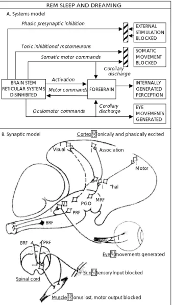
REM수면과 꿈의 관계를 설명하기 위해 활성화와 신경 변조(neuromodu- lation)의 이중 역할을 인지했던 한 가 지 초기 모델이‘활성화-합성 모델(activation-synthesis model)(13)이다. 그림 1은 노르에피네프린과 세로토닌의 결핍으로 인해 탈억제되었던 콜린성 뇌간 neuron으로부터 나온 신호에 의해 시각 피질, 운동 피질, 변연계, 및 기타 전 뇌가 단계적으로 활성화되는 자가 활성화된 뇌의 활성화- 합성 모델을 보여주고 있다(14). 그림 1A은 시스템 모델이 다. 아민성(aminergic) neuron의 발화 중지에 의한 탈억 제 결과로 뇌간 망상계가 자가 활성화된다. 그러한 활성화가 여러 가지 영향(REM 폭발<bursts>동안 단계적 시냅스전 억제와 외부 자극의 차단을 일으키는 afferent terminalis의 탈분극화<depolarization>;운동성 neuron의 긴장성 억제
를 일으켜서 동반하는 운동 명령을 효과적으로 상쇄시키고 그 결과 신체적 움직임이 차단되는 시냅스후의 과다분극화
<hyperpolarization>)을 미친다. 안구운동 명령만이 안구 운 동으로 출력되는데, 그 이유는 이러한 운동 neuron들이 억 제되지 않기 때문이다. 망상계(reticular formation)에 의 해 활성화되고 아민성으로 억제된 전뇌는, 꿈의 정신기능을 정형화시키는 시각적 이미지와 운동감각과 같은 내적으로 생성된 지각들을 합성시킬 수 있는 신체 운동과 안구운동 명령에 대한 원심성 모사나 결과 방출 정보를 받아들인다.
계속해서 전뇌는 망상계에 대한 positive feedback을 통해 상기 과정을 영속화시키도록 도와주는 자체의 운동 명령을 생성한다. 그림 1B는 시냅스 모델이다. 시상으로 돌출하는 중뇌 망상계 neuron은 긴장성 및 단계적 신호들을 하부로
전달한다. PGO 폭발 세포들은 단계적 활성화와 특정한 안구 운동 정보를 geniculate body와 대뇌피질로 전달한다. 연 수의 망상계 neuron은 동안신경(Ⅵ)과 척수로 단계적 활 성화 신호를 전달하여 안구운동, 사지의 경련, 시냅스전 억 제를 일으킨다. 연수의 망상계 neuron은 척수의 운동 neu- ron에 긴장성 과다분극화 신호를 전달한다. 이러한 하행성
EXTERNAL STIMULATION BLOCKED
SOMATIC MOVEMENT BLOCKED
INTERNALLY GENERATED PERCEPTION EYE MOVEMENTS GENERATED BRAIN STEM
RETICULAR SYSTEMS
DISINHIBITED FOREBRAIN
Activation A. Systems model
Tonic inhibitionof motorneurons Somatic motor commands Phasic presynaptic inhibition
Corollary discharge
Motor commands
Oculomotor commands Corollary dischorge
B. Synaptic model Cortex:Tonically and phasically excited Visual + + Association
+ Motor
+ Thal
PGO MRF
PRF BRF
PRF BRF
Spinal cord
Skin:Sensory lnput blocked
Muscle:Tonus lost, motor output blocked Eye:movements generated +
+ +
REM SLEEP AND DREAMING
Fig. 1. The activation-synthesis model. A:Systems model.
B:Synaptic model. MRF:midbrain reticular neurons, PRF:pon- tine reticular formation neurons, BRF:The bulbar reticular for- mation neurons(from Hobson & McCarley, 1977).
Activation
Thalamus basal forebram & amygdala Cholinergically modulated Pons
Aminergic neurons off
↓NE. ↓ SHT Cholinergic neurons on
↑Ach
Cortex Aminergically demoduisted
↓Recent memory
↓Orientation Modulation
Parietal operculum
↑Visuospatial imagery
Prefrontal cortex deactivated
↓Volition
↓Insight & judgemient
↓Working memory
Amygdala & paralimbic cortex
↑Emotion
↑Remote memory Pontine tegmentum
Activates reticutar formation Activates PGO system Activates cholinergic system
+ +
+ +
Sensory input blocked Real world data unavailable PGO system turned on
Fictive visual &
motor data generated
Motor output blocked Real action impossible Input Source
Occipital cortex
Genicuitate
Pons
RN LC
PPTLDT
Fig. 2. Physiological signs and regional brain mechanisms of REM sleep dreaming separated into the activation (A), input source (I), and modulation (M) functional components of the AIM model. These presents a schematic model for the gene- ration of cognitive dream features by combining the findings on state-dependent regional activation with a model of the neuromodulation of conscious states. Dynamic changes during REM sleep dreaming are noted adjacent to each figure (from Hobson 1990, 1992;Hobson, et al. 1998).
영향의 결과, 척수 수준에서 감각성 입력과 운동성 출력이 차단된다. 전뇌 수준에서 시각적 연합 및 운동성 피질 ne- uron은 비특이적 및 특이적 시상의 중계를 위한 시간과 단 계적 활성화 신호를 받아들인다.
그림 2는 REM수면 꿈의 생리적 증후와 국소적 뇌 기전을 활성화(activation=A), 입력(input=I), 변조(modulation=
M)로 나누어 설명하려는 AIM 모델(15-17)이다. 이 모델 은 REM 수면 꿈 기간동안 상태-의존적(state-depend- ent) 국소적 활성화에 관한 소견들을 의식상태의 신경변조 모델과 결합시켜 꿈의 특징들이 생성되는 것을 도식화시킨 모델이다.
2) NREM수면 꿈의 생성기전
(1) 은밀한 REM수면과정<Covert(phantom) REM pro- cess>
NREM 꿈은 NREM수면 동안 은밀한 REM 수면과정의 침입(intrusion)과 연관될 수 있다. REM 수면과정과 기본적 으로 연결되어있는 수면의 정신기능이, REM수면에 대한 수 면 개시 또는 NREM수면의 시간적 근접에 의존하는 어떤 조 건하에 은밀한 방식으로 수면 개시나 NREM수면 동안 발생 할 수도 있다. 따라서, 은밀한 REM 수면과정이 다양한 의식 상태에서 꿈과 같은 활동을 생성할 수 있는 것으로 본다(18).
(2) 단기 각성 침입(Brief arousal intrusions)
NREM수면 기간동안 꿈은 수면과정자체(수면단계 1 또는 2)보다 오히려 수면으로부터의 단기 각성 침습(brief arou- sal intrusions;수면 중 각성 또는 신체 움직임)과 연관되 어 있는 것으로 본다(9). NREM 꿈의 발생은 NREM수면 기간동안 어떤 종류의 각성 과정이 원인일 수도 있다. 뇌파 에서 명백하게 수면 기간동안 각성상태를 보여주는데도 불 구하고, 피검자들이 수면 기간동안 미세 각성 상태를 의식하 지 못한다(19). 게다가 외부자극은 NREM수면 기간동안 정 신기능에 영향을 미치지만, REM수면 기간동안에는 그렇지 않은 것으로 밝혀졌다(20,21). 따라서, 각성과정이 NREM 수면 기간동안의 꿈 생성과 관련될 수 있다. 즉, 단기 각성 동안 사람들은 외부환경으로부터 얻은 어떤 정보를 자신의 기억으로 통합시켜서, 나중에 마치 자기가 수면 기간동안 경 험했던 꿈인 것처럼 이러한 정신기능을 수정하거나 재구성 할지도 모른다.
2. 꿈의 정신생리
1) 수면의 정신생리적 측정 항목
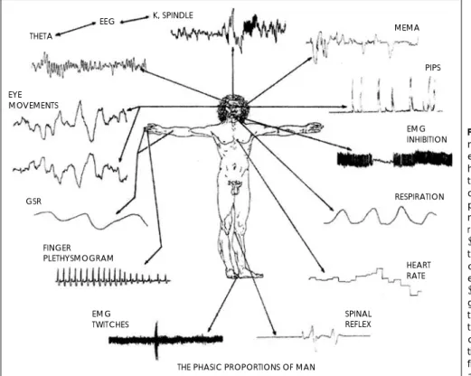
그림 3(22)은 수면의 정신 생리적 연구에서 조사되었던, 단계적으로 발생하는 다양한 전기 생리적 측정 항목들을 보 여주고 있다:뇌파(K complex, spindle, theta 활성도 등);
FINGER PLETHYSMOGRAM
EMG INHIBITION K, SPINDLE
EEG THETA
EYE MOVEMENTS
GSR
EMG TWITCHES
SPINAL REFLEX
HEART RATE MEMA
PIPS
RESPIRATION
THE PHASIC PROPORTIONS OF MAN
Fig. 3. The phasic proportions of man. Various phasically occurring electrophysiological measures that have been investigated in the con- text of psychophysiological studies of sleep. These variables, with ap- proximate designations of anato- mic areas from which they can be recorded, include EEG measures (K complex, spindle, and theta ac- tivity), various expressions of mus- cle activity from auditory (middle ear muscle activity[MEMA], visual (eye movements, periorbital inte- grated potentials [PIPS] and skele- tal musculature (facial EMG inhibi- tion, spinal reflexes, and EMG twit- ches) systems and autonomic ac- tivity (galvanic skin response [GSR], finger plethysmogram, respiration, and heart rate)(from Pivik, 1986).
다양한 근육 활동-귀의 근육(중이 근육 활동, middle ear muscle activity[MEMA]), 눈의 근육(안구운동;안구 주위 근의 통합 전위, periorbital integrated potentials [PIPS]), 골격근(안면근 근전도 억제;척수 반사;근전도 경련, EMG twitches);자유신경계 활동(피부전도반응, galvanic skin response[GSR];수지 혈류량, finger plethys-mogram;
호흡;맥박).
2) 꿈의 단계적(phasic) 사건들과 꿈의 생리적 연관성(23)
(1) REM수면의 꿈
단계적 REM안구운동이 왕성한 기간일수록 꿈의 회상수
준이 더 높고, 시각적 생생함이 더 크며, 비언어적 사진 반응 척도에서 물체의 명쾌함이 더 크고, 더 적극적인 꿈이 나타 나며, 더 기이하고 정서성이 더 증가되어 있다. 톱니 파는 시 각적 생생함 및 정신기능의 불연속성과 관련되어 있다. 중 이근 활성도(MEMA)는 기이한 정신기능 및 청각적 이미지 와 관련되어 있는 것으로 밝혀졌다. 안구 주위 근육의 통합 전위(PIPs)는 기이함과 관련되어있다. 단계적 사지운동은 특정한 꿈 내용과 관련되어 있다.
(2) NREM 수면의 꿈
단계적 척수 반사 억제는 더 많은 꿈의 회상, 청각적 이미 지, 적개심과 관련되어 있고, PIPs는 꿈 회상의 증가와 관
Table 1. The formal features of REM sleep dreaming
Hallucinations Especially visual and meteoric, but occasionally in any and all sensory modalities.
Bizarreness
Incongruity (imagery is strange, unusual or impossible), discontinuity (imagery and plot can change, appear or disappear rapidly), uncertainty (persons, places and events often bizarrely uncertain by waking standards)
Delusion We are consistently duped into believing that we are awke (unless we cultivate lucidity) Self-reflection Self-reflection absent or greatly reduced relative to waking
Lack of orientational stability
Persons, times and places are fused, plastic, incongruous and discontinuous
Narrative story lines Explain and integrate all the dream elements in a confabulatory manner Emotions increased Intensified and dominated by fear-anxiety
Instinctual programs (Especially fight-flight)often incorporated Volitional control Greatly attenuated
Memory deficits Across dream-wake, wake-dream and dream-dream transitions (from Hobson et al., 2000)
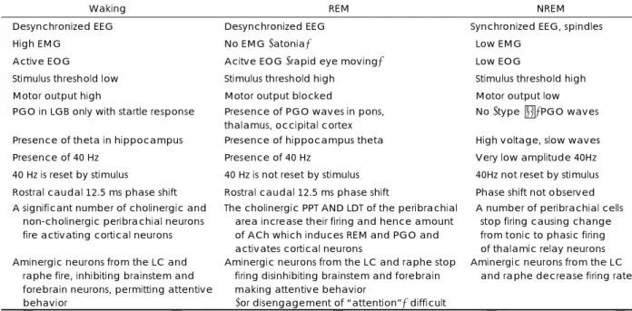
Table 2. Physiological and chemical differences in waking, REM, NREM
Waking REM NREM
Desynchronized EEG Desynchronized EEG Synchronized EEG, spindles
High EMG No EMG (atonia) Low EMG
Active EOG Acitve EOG (rapid eye moving) Low EOG
Stimulus threshold low Stimulus threshold high Stimulus threshold high
Motor output high Motor output blocked Motor output low
PGO in LGB only with startle response Presence of PGO waves in pons, thalamus, occipital cortex
No (type Ⅱ)PGO waves
Presence of theta in hippocampus Presence of hippocampus theta High voltage, slow waves
Presence of 40 Hz Presence of 40 Hz Very low amplitude 40Hz
40 Hz is reset by stimulus 40 Hz is not reset by stimulus 40Hz not reset by stimulus Rostral caudal 12.5 ms phase shift Rostral caudal 12.5 ms phase shift Phase shift not observed A significant number of cholinergic and
non-cholinergic peribrachial neurons fire activating cortical neurons
The cholinergic PPT AND LDT of the peribrachial area increase their firing and hence amount of ACh which induces REM and PGO and activates cortical neurons
A number of peribrachial cells stop firing causing change from tonic to phasic firing of thalamic relay neurons Aminergic neurons from the LC and
raphe fire, inhibiting brainstem and forebrain neurons, permitting attentive behavior
Aminergic neurons from the LC and raphe stop firing disinhibiting brainstem and forebrain making attentive behavior
(or disengagement of “attention”) difficult
Aminergic neurons from the LC and raphe decrease firing rate
REM:rapid eye movement, NREM:non-rapid eye movement, EEG:electroencephalogram, EMG:electromyography, EOG:el- ectrooculography, PGO:ponto-geniculo-occipital, LGB:lateral geniculate body, PPT:pedunculopontine tegmental nucleus, LDT:laterodorsal tegmental nucleus, ACh:acetylcholine, LC:locus ceruleus (from Kahn et al., 1997)
련되어 있으며, 수면 개시 쎄타파다발(theta burst)은 수면 개시 정신기능의 특징과 관련되어 있다.
3) REM수면 꿈의 외형적 특징
표 1(24)은 REM 수면 꿈의 외형적 특징을 정리해놓은 것이다.
3. REM수면, NREM수면의 차이
1) 각성상태(Waking), REM, NREM수면의 차이 표 2는 각성, REM, NREM의 생리적, 화학적 차이를 나 타낸 것이다(23). 그림 4(25)는 각성, NREM, REM 수면 상태의 행동적, 전기 생리적 기록상(polygraphy), 심리적 특 징들을 나타낸 것이다.
2) REM 수면과 NREM수면 꿈의 차이
각 수면기간에 얻어진 꿈은 정량적, 정성적 측면에서 유의 한 차이가 있으며, 서로 다른 기전에 의해 생성되는 것으로 생각한다.
정량적 차이를 보면, REM수면으로부터 깨었을때 꿈 회 상율(76~84%)이 NREM수면으로부터의 꿈 회상율(12~
25%)보다 훨씬 더 높다(18).
정성적 차이를 보면, REM수면의 꿈이 NREM수면의 꿈보 다 훨씬 더 밝고, 유쾌하며, 편안하고, 호감이 더 가며, 분명 하고, 명백하며, 더 많이 기억할 수 있고, 더 생생하며, 소란 스럽고, 역동적이며, 부산한 특징이 있다(9).
생성기전의 차이는 위에서 이미 언급한 바 있다.
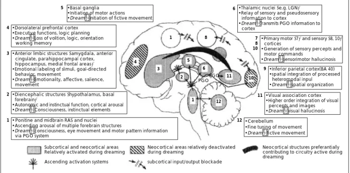
4. REM 수면 꿈의 통합 모델(24)
그림 5는 뇌영상, 신경생리 및 병변 연구들로부터 얻은 소 견들을 통합시키는 정상 꿈의 가설 모델이다. 이 모델에서 꿈은, 선택적으로 촉진되고, 억제되거나 또는 입력/출력이 차
Fig. 4. Behavioral states in humans. States of waking, NREM sleep, and REM sleep have behavioral, polygraphic, and psy- chological manifestations Posture shifts can occur during wak- ing and in concert with phase changes of the sleep cycle.
Two different mechanisms account for sleep immobility:dis- facilitation (NREM sleep), inhibition (REM sleep). In dreams, we imagine that we move but we do not. Sample tracings of EMG, EEG, EOG used to distinguish state are also shown. Three lower channels describe other subjective and objective state varia- bles (from Hobson & Steriade, 1986).
Behavior
Awake Polygraph
EMG EEG EOG Sensation and
perception
Thought
Movement
WAKE NREM SLEEP REM SLEEP
Stages I REM
II III IV
Vivid, Externally generated
Dull or absent
Vivid, Internally generated Logical
progressive
Logical perseverative
Illogical bizarre Continuous
voluntary
Episodic involuntary
Commanded But Inhibited
Fig. 5. Forebrain processes in normal dreaming-an integration of neurophysiological, neuropsychological, and neuroimaging data (from Hobson et al., 2000).
•Thalamic nuclei (e.g. LGN)
• Relay of sensory and pseudosensory information to cortex
• Dream:Transmits PGO information to cortex
•Primary motor (7) and sensory (8, 10) cortices
• Generation of sensory percepts and motor commands
• Dream:sensorimotor hallucinosis
• Inferior parietal cortex(BA 40)
• spatial integration of processed heteromodal inpul
• Dream:spatial organization
• Visual association cortex
• Higher order integration of visual percepts and images
• Dream:visual hallucinosis
• Cerebellum
• Fine tuning of movement
• Dream:fictive movement 7
108
11 9
12
• Basal ganglia 6
• Initiation of motor actions
• Dream:initiation of fictive movement
• Dorsolateral prefrontal cortex
• Executive functions, logic planning
• Dream:Loss of volition, logic, orientation working memory
• Anterior limbic structures (amygdala, anterior cingulate, parahippocampal cortex, hippocampus, medial frontal areas)
• Emotional labeling of slimuli, goal-directed behavior, movement
• Dream:Emotionality, affective, salience, movement
• Diencephalic structures (hypothalamus, basal forebrain)
• Autonomic and instinctual function, cortical arousal
• Dream:Consciousness, instinctual elements
• Ponitine and midbrain RAS and nuclei
• Ascending arousal of multiple forebrain structures
• Dream:consciousness, eye movement and motor pattern information via PGO system
4
3
5
2
1
1 8
6 5 3
4
1 12
11 10
2 PGOPGO
Subcortical and neocortical areas Neocortical areas relatively deactivated Neocortical structures preferantially Relatively activated during dreaming during dreaming contributing to circuitry active during
dreaming
Ascending activation systems subcortical input/output blockade
단된 전뇌 부위에 영향을 미치는 각성 과정들에 기인한다.
정상 꿈의 다양한 구성 요소는 뇌 네트워크에 의해 이루어 진다.
결 론
꿈은 동서고금을 통해 많은 사람들이 관심을 가졌던 주 제 중 하나이다. 역사적으로 꿈의 이론들은 관련 영역에 서 동시대 이론들의 영향을 크게 받았다. 많은 연구자들은 REM수면 기전과 꿈 사이의 엄격한 연관성을 거부하고, 꿈 이 REM과 NREM 수면 모두에서 발생한다고 주장하고 있 다(2-8). 비록 NREM과 REM 수면 양자의 꿈이 모두 현 상학적 용어에서 동일한 꿈으로 간주되지만, 각 수면기간 에 발생한 꿈은 정량적, 정성적 측면에서 유의한 차이가 있 으며, 서로 다른 기전들에 의해 생성될 가능성이 높다(9).
이러한 꿈을 충분히 이해함으로써, 임상현장에서 부딪히는 환자들의 꿈에 관련된 문제들을 보다 쉽게 해결할 수 있을 것이다. 따라서, 저자는 신경생물학적, 정신생리적 관점에 관한 관련 문헌들을 고찰하고, 꿈의 생성기전과 그 의미를 정리하였다.
중심 단어:꿈·신경생물학·정신생리학.
REFERENCES
1. Antrobus J. Theories of dreaming. In: Principles and Practice of Sleep Medicine, 3rd ed, ed by Kryger MH, Roth T, Dement WC, Philadelphia, W.B. Saunders Company;2000. p.472-490
2. Antrobius J. REM and NREM sleep reports: comparison of word frequencies by cognitive classes. Psychophysiology 1983;20:562-568 3. Antrobius J, Kondo T, Reinsel R. Dreaming in the late morning:
summation of REM and diurnal cortical activation. Conscious Cogn 1995;4:275-299
4. Cavallero C, Cicogna P, Natale V, Occhionero M, Zito A. Slow wave sleep dreaming. Sleep 1992;15:562-566
5. Cavellero C, Foulkes D, Hollifield M, Terry R. Memory sources of REM and NREM dreams. Sleep 1990;13:449-455
6. Foulkes D. Dreaming and REM sleep. J Sleep Res 1993;2:199-202 7. Foulkes D, Scmidt M. Temporal sequence and unit composition in
dream reports from different stages of sleep. Sleep 1983;6:265-280 8. Rosenlicht N, Maloney T, Feinberg I. Dream report length is more
dependent on arousal level than prior REM duration. Brain Res Bull 1994;34:99-101
9. Takeuchi T, Miyasita A, Inugami M, Yamamoto Y. Intrinsic dre- ams are not produced without REM sleep mechanisms: evidence through elicitation of sleep onset REM periods. J Sleep Res 2001;
10:43-52
10. Rechtschaffen A, Siegel J. Sleep and dreaming. In: Principles of neural science, 4th ed, ed by Kandel ER, Schwartz JH, Jessell TM, New York, McGraw-Hill Companies;2000. p.936-947
11. McCarley RW, Massaquoi SG. A limit cycle reciprocal interaction model of the REM sleep oscillator system. Am J Physiol 1986;251:
R1011
12. Siegel JM. Brainstem mechanisms generating REM sleep. In: Pr- incipl- es and Practice of Sleep Medicine, 3rd ed, ed by Kryger MH, Roth T, Dement WC, Philadelphia, W.B. Saunders Company;
2000. p.112-131
13. Hobson JA, McCarley RW. The brain as a dream-state generator:
An activation-synthesis hypothesis of the dream process. Am J Psy- chol 1977;134:1335-1368
14. Gritti I, Manville L, Jones BE. Projections of GABAergic and cho- linergic basal forebrain and GABAergic preoptic-anterior hypoth- alamic neurons to the posterior lateral hypothalamus of the rat. J comp Neurol 1994;339:251-268
15. Hobson JA. Activation, input source, and modulation: A neuroco- gnitive model of the state of the brain-mind. In: Sleep and cogni- tion. ed by Bootzin R, Kihlstrom J and Schacter D, Washington DC, American Psychological Association;1990. p.25-40
16. Hobson JA. A new model of brain-mind state: Activation level, in- put source, and mode of processing(AIM). In: The neuropsycho- logy of Sleep and Dreaming. ed by Antrobus J and Bertini M, Ma- hwah, NJ, Lawrence Erlbaum Assoc;1992. p.227-247
17. Hobson JA, Stickgold R, Pace-Schott EF. The neuropsychology of REM sleep dreaming. NeuroReport 1998;9:R1-R14
18. Nielsen TA. A review of mentation in REM and NREM sleep: ‘co- vert’ REM sleep as a possible reconciliation of two opposing mod- els. Behav Brain Sci 2000;23(6):851-66
19. Ogilvie RD, Wilkinson RT, Allison S. The detection of sleep onset:
behavioral, physiological, and subjective convergence. Sleep 1989;
12:458-474
20. Castaldo V, Shevrin H. Different effect of an auditory stimulus as a function of rapid eye movement and non-rapid eye movement sleep.
J Nerv Ment Dis 1970;150:195-200
21. Conduit R, Bruck D, Coleman G. Induction of visual imagery dur- ing NREM sleep. Sleep 1997;20:948-956
22. Pivik RT. Sleep: physiology and psychophysiology. In: Psycho- physio- logy: Systems, Processes and Applications, ed by Coles MGH, Donchin E, Porges SW, New York, NY, Guilford Press;
1986. p.378-406
23. Kahn D, Pace-Schott EF, Hobson JA. Consciousness in waking and dreaming: the roles of neuronal oscillation and neuromodulation in determining similarities and differences. Neuroscience 1997;78:
13-38
24. Hobson JA, Pace-Schott EF, Stickgold R. Consciousness: Its vicissi- tudes in waking and sleep. In: The New Cognitive Neurosciences, 2nd ed, ed by Gazzaniga MS, Champaign, Illinois, Massachusetts Institute of Technology;2000. p.1341-1354
25. Hobson JA, Steriade M. Neuronal basis of behavioral state control.
In: Handbook of Physiology: The Nervous System, Vol 4, ed by Mountcastle V, Bloom FE, Washington: American Physiological So- ciety;1986. p.701- 823