ISSN 2234-3806 • eISSN 2234-3814
http://dx.doi.org/10.3343/alm.2012.32.2.113
Relationship of Oxidative Stress in Hepatitis B Infection Activity with HBV DNA and Fibrosis
Fazilet Duygu, M.D.¹, Hasan Karsen, M.D.², Nurten Aksoy, M.D.³, and Abdullah Taskin, M.D.³
Department of Infectious Disease and Clinic Microbiology1, Tokat State Hospital, Tokat; Department of Infectious Disease and Clinic Microbiology2, Faculty of Medicine, Harran University, Sanliurfa; ³Department of Biochemistry3, Faculty of Medicine, Harran University, Sanliurfa,Turkey
Background: The aim of this study was to evaluate oxidative stress in various clinical forms of hepatitis B infection and to investigate its role in the development of the chronic form of the disease.
Methods: Ninety-three patients with inactive hepatitis B surface antigen (HbsAg) carrier state (IHBCS), 65 patients with chronic hepatitis B infection (CHB), and 42 healthy adults were included in the study. The following values were measured and compared in patient groups: total antioxidant status (TAS), total oxidative stress (TOS), oxidative stress index (OSI), sulfhydryl (SH), lipid peroxidation (LOOH), catalase (CAT), and ceruloplasmin. In patients with chronic hepatitis B, these values were compared with HBV DNA and fibrosis levels.
Results: ALT, TOS, LOOH, and OSI levels were higher in the CHB group compared to the other groups (P <0.001). Catalase levels increased in the CHB and IHBCS groups compared to the control group (P <0.001). Total aminooxidant and ceruloplasmin levels were found to be lowest in the CHB group and highest in the control group (P <0.001). Sulfhyrdyl was higher in the control group compared to the other groups (P <0.001). In the CHB group, there was no correlation between the HBV DNA and OSI (P >0.05).
Conclusions: These finding suggested that oxidative stress is associated with hepatitis B activity.
Key Words: Hepatitis B, HBV DNA, Fibrosis, Oxidative stress, Catalase, Sulfhydryl
Received: July 8, 2011
Revision received: November 15, 2011 Accepted: December 31, 2011 Corresponding author: Fazilet Duygu Gultekin Topcam St. No:76 Tokat 60100, Turkey
Tel: +905324945137 Fax: +903562145400 E-mail: [email protected]
© The Korean Society for Laboratory Medicine.
This is an Open Access article distributed under the terms of the Creative Commons Attribution Non-Commercial License (http://creativecom- mons.org/licenses/by-nc/3.0) which permits un- restricted non-commercial use, distribution, and reproduction in any medium, provided the origi- nal work is properly cited.
INTRODUCTION
The hepatitis B virus (HBV), which is a chronic viral hepatitis factor, is a significant pathogen that causes fibrosis, cirrhosis, hepatocellular cancer as a result of the damage it causes to liver cells. Chronic hepatitis B (CHB) infection is a health issue that affects more than 400 million people globally. Although there is an effective vaccine and advances have been made in diagnos- tic and treatment methods, almost 1,000,000 people die every year due to HBV infection-related complications [1]. Hepatitis B infection exhibits a varied clinical course, which can range from asymptomatic infection to fulminant liver disease [2].
Cells continuously form free radicals and reactive oxygen spe- cies as part of metabolic processes. These free radicals and re- active oxygen species are neutralized by a complex antioxidant system. Oxidative stress is the imbalance that occurs between the reactive oxygen species or free radicals and the antioxidant system, and this imbalance may cause irreversible damage in important cellular compartments.
In chronic viral hepatitis, the role of oxidative stress in cell de- struction and DNA and RNA damage has been established through experimental studies [3, 4]. In the present study, oxida- tive stress was measured in various clinical forms of the chronic hepatitis B and its role was investigated in the development of
ISSN 2234-3806 • eISSN 2234-3814
chronic hepatitis.
METHODS
1. Patient selection and sample preparation
Patients infected with hepatitis B virus, who presented to Tokat State Hospital (in Tokat, Turkey) between June 2010 and May 2011, and healthy adults participated in the study. A total of 200 individuals participated in the study, comprising 65 chronic ac- tive hepatitis B patients in the 15-66 age range (32.5%), 93 pa- tients with inactive hepatitis B infection (46.5%) and 42 healthy adults. All subjects who participated in the study gave informed consent and the study was approved by the Ethics Committee of the Harran University, Faculty of Medicine.
1) Diagnosis criteria
The disease was diagnosed according to the following criteria [5].
Chronic hepatitis B infection (CHB):
(1) HBsAg-positive for more than 6 months,
(2) ALT value greater than 1.5 times the normal value (nor- mally, ALT value is less than 40 IU/mL),
(3) HBV DNA value ≥100,000 copies/mL (20,000 IU/mL) in those who were postitive for the hepatitis Be antigen (HBeAg-positive),
(4) Whereas it was ≥10,000 copies/mL (2,000 IU/mL) in those who were HBeAg-negative, and
(5) Fibrosis ≥2 in the histopathological evaluation of the liver
Inactive HbsAg Carrier State (IHBCS):
(1) HBsAg-positive, (2) Normal ALT values, (3) HBeAg-negative, and
(4) HBV DNA ≤10,000 copies/mL (2,000 IU/mL)
The control group was formed from HBsAg-negative healthy adults whose anti-HBc total value was negative.
Exclusion criteria: Patients with liver cirrhosis, diabetes mellitus, hypertension, coronary artery disease, acute infection, chronic obstructive pulmonary disease, corticosteroid usage, malignancy, morbid obesity, pregnancy, liver and kidney failure, and smok- ers were not included in the study.
Liver biopsy was performed on all patients with CHB prior to treatment. In order to rate the liver damage, histological activity (necroinflammatory lesion) and fibrosis (according to KNODELL fibrosis scoring system) [6] assessment was performed.
In addition to the patient’s age, gender, HBsAg, HBeAg, anti- HBe, ALT, and AST values, HBV DNA levels and liver biopsy, fi- brosis levels were also recorded in chronic hepatitis B patients.
2) Collection and storage of samples
Blood samples were collected in the form of venous blood follow- ing 12 hr of fasting. Physical examination was performed on the patients to exclude other diseases. Ten milliliter blood samples were collected into biochemistry tubes containing heparin. The plasma, which was obtained after centrifuging blood samples at 3,500 rpm for 10 min, was stored at -80ºC until the study date.
2. Measurement of oxidative status
Total oxidative status (TOS), total antioxidant status (TAS), and oxidative stress index (OSI) were measured using the method developed by Erel [7]. In this method, long-lasting, durable, rad- ical monocation of 2,2´-azinobis-(3-ethylbenzothiazoline-6-sul- fonic acid) (ABTS) is formed. The color of this radical, which is characteristically blue-green, is reduced by the antioxidants and disappears. The lightening and/or discoloration of the color of the antioxidants in the samples is considered to be their total antioxidant capacity. Trolox, which is a water-soluble analog of vitamin E, was used as the standard, and the results were ex- pressed in terms of µmol trolox equiv./L [7].
The serum catalase (CAT) activity was determined using Goth’s colorimetric method [8], in which serum is incubated with H2O2, and the enzyme reactions are stopped by the addition of am- monium molybdate. Serum CAT activity was expressed as kU/L.
Free sulfhydryl groups of serum samples were assayed accord- ing to the method of Ellman [9], as modified by Hu et al. [10].
Briefly, 1 mL of buffer containing 0.1 M Tris, 10 mM EDTA, pH 8.2, and 50 µL serum was added to cuvettes, followed by 50 µL 10 mM 5,5′-dithio-bis (2-nitrobenzoic acid) (DTNB) in methanol.
Blanks without DNTB in the methanol were run for each sample as a test. Following incubation for 15 min at room temperature, sample absorbance was measured at 412 nm using a spectro- photometer (CE1011; Cecil Instruments Ltd., Milton Technical Centre, Cambridge, England). The value of reagent blanks were subtracted from those of samples for calibration. The concen- tration of sulfhydryl groups was calculated using reduced gluta- thione as the free sulfhydryl group standard and the result was expressed as mmol/L.
Plasma lipid peroxidation (LOOH) was evaluated by the fluori- metric method based on the reaction between malondialdehyde (MDA) and thiobarbutiric acid (TBA) [11]. Briefly, 50 µL of plasma was added to 1 mL of 10 mmol/L diethythiobarbutiric acid (DETBA)
reagent in phosphate buffer (0.1 mol/L, pH 3). The mixture was mixed for 5 sec and incubated for 60 min at 95ºC. Samples were placed on ice for 5 min and then 5 mL of butanol was added.
The mixture was shaken for 1 min to extract the DETBA-MDA adduct, and then centrifuged at 1,500×g for 10 min at 4ºC. Flu- orescence of the butanol extract was measured at an excitation wavelength of 539 nm and an emission wavelength of 553 nm.
1,1,3,3,tetraethoxypropane (Sigma Chemical Company, St. Louis, MO, USA) was used as a standard solution and the values were presented as mol/L.
Ceruloplasmin levels were assessed by measuring its oxida- tive activity using p-phenylenediamine as the substrate, accord- ing to the method of Sunderman and Nomoto [12].
3. Statistical analysis
Pearson’s chi-square test was used to compare the categorical variables between groups. Categorical variables were presented as counts and percentages. The Shapiro-Wilk test was used to evaluate whether the distribution of variables was normal.
The t-test for two independent samples, or the Kruskal-Wallis
test, was used to compare continuous variables between two groups. Continuous variables were presented as mean (SD) or as median (interquartile range [IQR]). A P value of less than 0.05 was considered to be statistically significant. SPSS software 15.0 for Windows (Chicago, IL, USA) was used for all statistical analyses.
RESULTS
The chronic hepatitis B, inactive hepatitis B, and control groups were similar in terms of mean age of the subjects (P >0.05). There was no significant difference in gender characteristics between the CHB group and the other groups (IHBCS, Control) (P =0.103 and P =0.656, respectively). The gender distribution of the IH- BCS and control groups were also similar (P =0.35).
Of the chronic hepatitis B patients, 32 (49.2%) were HBeAg- positive. The age and gender characteristics and ALT values of the patients are shown in Table 1. The ALT level was higher in the CHB group compared to the other two groups (P <0.001). A positive correlation was found between ALT and HBV DNA (r=
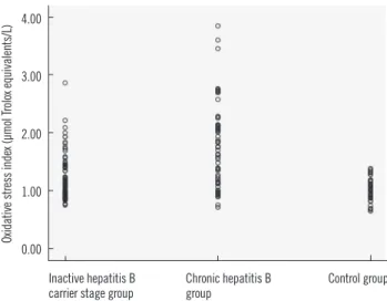
0.308; P =0.012). There was no correlation between ALT and ox- idative stress (P >0.05). The oxidative and antioxidative parame- ters in each group are shown in Table 2. The values of OSI, TAS, and ceruloplasmin of the patient and control groups are shown in Figs. 1-3, respectively. In the CHB group, no correlation was found between the fibrosis levels of the patients and oxidative stress (P >0.05). In patients in the CHB group, HBV DNA was not correlated with OSI (P >0.05). A strong negative correlation was observed between OSI and TAS (r=-0.404; P <0.001) and between OSI and SH (r =-0.333; P =0.015). A strong positive correlation was observed between OSI and LOOH (r =0.731; P <0.001) and TOS (r=0.731; P <0.001).
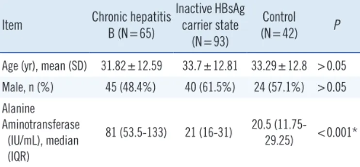
Table 1. The clinical and demographic data of the study groups Item Chronic hepatitis
B (N=65)
Inactive HBsAg carrier state
(N=93)
Control
(N=42) P
Age (yr), mean (SD) 31.82±12.59 33.7±12.81 33.29±12.8 >0.05 Male, n (%) 45 (48.4%) 40 (61.5%) 24 (57.1%) >0.05 Alanine
Aminotransferase (IU/mL), median (IQR)
81 (53.5-133) 21 (16-31) 20.5 (11.75-
29.25) <0.001*
*There was a statistically significant difference between the CHB group and the other groups.
Table 2. Oxidative and antioxidative parameters in each group
Chronic hepatitis B (N=65) Inactive HbsAg carrier state (N=93) Control (N=42) P
TAS (µmol Trolox equivalents/L), mean (SD) 0.74±0.103 0.79±0.107 0.91±0.13 <0.001*
CAT (kU/L), median (IQR) 17.33 (14.77-18.50) 17.95 (16.85-8.82) 8.5 (7.7-9.2) <0.001†
SH, mean (SD) 0.23±0.027 0.24±0.026 0.28±0.027 <0.001‡
Looh (mol/L), median (IQR) 6.82 (5.03-10.31) 5.19 (4.89-5.96) 5.94 (4.72-6.92) <0.001§
TOS (µmol Trolox equivalents/L), median (IQR) 12.87 (8.62-16.93) 8.4 (7.38-10.77) 9.47 (7.61-10.43) <0.001§ Serulop, median (IQR) 510.62 (486.25-544.37) 540 (504.21-588.12) 663.28 (624.60-746.87) <0.001*
OSI (µmol Trolox equivalents/L), median (IQR) 1.75 (1.15-2.13) 1.09 (0.93-1.4) 1.01 (0.88-1.16) <0.001§
*There was a statistically significant difference between all groups; †There was a statistically significant difference between the control group and the other groups (P <0.001) and no statistically significant difference between the chronic hepatitis B group and the inactive HBsAg carrier state group (P <0.005);
‡There was a statistically significant difference between the control group and the other groups but no statistically significant difference between the chronic hepatitis B group and the inactive HBsAg carrier state group; §There was a statistically significant difference between the chronic hepatitis B group and the other groups but no statistically significant difference between the inactive HBsAg carrier state group and the control group.
DISCUSSION
In this study, we showed that in hepatitis B infection, oxidative stress indices such as TOS, LOOH, OSI, and CAT are increased, and TAS, sulfhydryl, and ceruloplasmin levels, which are indica- tive of antioxidant status, are decreased.
The HBV is one of the important factors of acute/chronic hepatitis, cirrhosis and hepatocellular carcinoma. More than 400 million people are known to be chronically infected with HBV [13]. Five percent of the world population are HBV carriers and CHB infection is cited by WHO to be the ninth most com- mon cause of death [1].
Hepatitis B infection exhibits a varied clinical course, which
can range from asymptomatic infection to fulminant liver disease [2]. The disease is diagnosed through clinical, microbiological (ELISA, PCR), biochemical (ALT, AST), and pathological assess- ment [14]. In the present study, oxidative stress parameters in two different forms of the disease (CHB and IHBCS) were com- pared to those in healthy adults.
The liver fibrosis levels of the patients were determined through the histological assessment of the liver biopsy material. The di- agnosis of the patients included in the study was made based on these four methods, i.e. clinical, microbiological, biochemi- cal, and pathological method. HBV DNA analysis and histologi- cal assessments of liver biopsy samples were performed for all CHB patients.
Free radicals are defined as short-lived, unstable, and highly active molecules with one or more unpaired electrons, and with low molecular weight [15, 16]. The substances that can prevent or delay the effects of molecules that may cause oxidation of es- sential substances in an organism are called antioxidants [17].
Oxidative stress is defined simply as the imbalance between the body’s antioxidant defense and the production of the free radi- cals, which can cause peroxidation of the lipid layer of the cells [18, 19]. The main intracellular antioxidants in humans are the superoxide dismutase (SOD), catalase (CAT), and glutathione peroxidase (GPx) enzymes. In contrast to the intracellular envi- ronment, vitamins E and C, transferrin, haptoglobin, ceruloplas- min, albumin, bilirubin, β-carotene, and alpha-l-antitrypsin are responsible for antioxidant defense in the extracellular environ- ment [20].
The free fatty acid (FFA) oxidation products (lipid peroxide, superoxide, and hydrogen peroxide radicals) can generate oxi- Fig. 1. Oxidative stress index values of patient groups and control
group.
Inactive hepatitis B
carrier stage group Chronic hepatitis B
group Control group
Oxidative stress index (μmol Trolox equivalents/L
) 4.00
3.00
2.00
1.00
0.00
Fig. 2. Totally antioxidant status values of patient groups and con- trol group.
Abbreviation: TAX, totally antioxidant status.
Inactive hepatitis B
carrier stage group Chronic hepatitis B
group Control group
TAS (μmol Trolox equivalents/L)
1.40
1.20
1.00
0.80
0.60
0.40
1,000.00
800.00
600.00
400.00
200.00
0.00
Inactive hepatitis B
carrier stage Group Chronic hepatitis B
group Control group
Ceruloplasmin (μmol/L)
Fig. 3. Ceruloplasmin values of patient groups and control group.
dative stress and result in subsequent lipid peroxidation. Lipid hydroperoxide (LOOH), which is a non-radical free oxygen parti- cle, is one of the indicators for oxidative stress [21]. In recent years, the role of the imbalance between oxidative stress and the antioxidant status in cell destruction has become better under- stood [3, 7, 22].
Free oxygen radicals are effective in the pathogenesis of many diseases such as carbon tetrachloride-associated liver damage, glomerulonephritis, arteriosclerosis, diabetes mellitus, vitamin C-E deficiency, malignancy, emphysema, hyperoxia, bronchopul- monary dysplasia, pancreatitis, and rheumatoid arthritis [22-24].
In infectious diseases, inflammatory cells have been shown to become activated and secrete reactive oxygen and nitrogen spe- cies. Studies on oxidative stress have been conducted in many infectious diseases and oxidative stress has been demonstrated to increase in leishmania, measles, Neisseria gonorrhoeae in- fections, sepsis, urinary system infections, and Paracoccidioides brasiliensis infections [25-30].
There have been various studies indicating that oxidative stress is increased in hepatitis B and hepatitis C infections and in liver disease [31-36]. Increased oxidative stress in HBV infection was demonstrated to have an effect on DNA damage and hepato- carcinogenesis [16]. It is known that of the 530,000 hepatocellu- lar carcinoma cases seen each year, 316,000 are associated with HBV [2]. Therefore, we believe oxidative stress, which is known to play a significant role in hepatocarcinogenesis, is important in CHB patients.
Various indicators of oxidative stress have been studied in var- ious clinical forms of hepatitis B and have been demonstrated to increase as the disease becomes chronic [4, 35].
Measurement of the overall antioxidant status may yield more valuable information than the measurement of individual antioxi- dants. In our study, we chose to use the total antioxidant capacity measurement method developed in recent years by Erel, which measures total SH, vitamin C, uric acid, vitamin E, bilirubin, and many other antioxidants precisely, and the total oxidant capacity measurement method, which measures the oxidative stress caused by free radicals in the plasma such as hydroxyl (OH), hydrogen peroxide (H2O2), singlet oxygen (O2↑↓), lipid hydroper- oxide (LOOH), and superoxide (O2.-), which can all be measured with ease due to the fact that these measurements can be per- formed by fully automated colorimetric methods [7, 19]. In the present study, OSI, TOS, and TAS measurements were performed using this method. Additionally, ceruloplasmin, LOOH, and SH, which are also oxidative stress indicators, and SH and CAT en- zymes, which are antioxidant status indicators, were assessed
and compared in IHBCS, CHB and control groups.
The values of OSI, TOS, and LOOH, which are oxidative stress indicators, were higher in the CHB group compared to the other groups (P <0.001). The values of the antioxidant status indicator TAS, the intracellular antioxidant CAT, and the extracellular anti- oxidant ceruloplasmin were different in all three groups, and thus they decreased with increasing severity of the disease (P <
0.001). This suggested that oxidative stress may play a role in the pathogenesis of CHB and in exacerbation of the disease.
This study is the first study to compare oxidative stress with HBV DNA. Our data indicate no correlation between OSI and HBV DNA.
In chronic hepatitis B, the gold standard in assessing fibrosis is the histopathological examination of the liver biopsy material.
The histological activity index (HAI) and fibrosis level in the liver indicates the progression of the disease toward cirrhosis. This study investigated the relationship of oxidative stress with fibro- sis. We did not find a correlation between HAI and fibrosis and the indicators of oxidative stress. We believe this may be in part due to the homogeneity of the patients in whom the biopsies were performed, exclusion of patients with liver cirrhosis from the study, and the presence of moderate chronic active hepatitis in all patients.
In conclusion, oxidative stress increases and antioxidant val- ues decreases as the disease becomes chronic in patients with hepatitis B infection. This study suggests that oxidative stress may be associated with hepatitis B activity.
Authors’ Disclosures of Potential Conflicts of Interest
No potential conflict of interest relevant to this article was re- ported.
REFERENCES
1. Koziel MJ and Siddiqui A. Hepatitis B virus and hepatitis Delta virus. In:
Mandell GL, Douglas RG, et al. eds. Principles and practice of infectious diseases. 6th ed. Philadelphia: Churchill Livingstone, 2005:1864-90. 2. Koff RS. Viral hepatitis. In: Schiff L and Schiff ER, eds. Diseases of the
liver. 7th ed. Philadelphia: JB Lippincott Company, 1993:492-577. 3. Moriya K, Nakagawa K, Santa T, Shintani Y, Fujie H, Miyoshi H, et al.
Oxidative stress in the absence of inflammation in a mouse model for hepatitis C virus-associated hepatocarcinogenesis. Cancer Res 2001;61: 4365-70.
4. Dikici I, Mehmetog(lu I, Dikici N, Bitirgen M, Kurban S. Investigation of oxidative stress and some antioxidants in patients with acute and chron- ic viral hepatitis B and the effect of interferon-α treatment. Clin Biochem 2005;38:1141-4.
5. Anna S. F. Lok L and Brian J. McMahon. AASLD Practıce Guidelines Chronic Hepatitis B: Update 2009.
6. Knodell RG, Ishak KG, Black WC, Chen TS, Craig R, Kaplowitz N, et al.
Formulation and application of numerical scoring system for assessing histological activity in asymptomatic chronic active hepatitis. Hepatology 1981;1:431-5.
7. Erel O. A novel automated method to measure total antioxidant response against potent free radical reactions. Clin Biochem 2004;37:112-229. 8. Góth L. A simple method for determination of serum catalase activity
and revision of reference range. Clin Chim Acta 1991;196:143-51. 9. Ellman GL. Tissue sulfhydryl groups. Arch Biochem Biophys 1959;82:70-7. 10. Hu ML, Louie S, Cross CE, Motchnik P, Halliwell B. Antioxidant protec- tion against hyochlorous acid in human plasma. J Lab Clin Med 1993; 121:257-62.
11. Conti M, Morand PC, Levillain P, Lemonnier A. Improved fluorometric determination of malondialdehyde. Clin Chem 1991;37:1273-5. 12. Sunderman FW Jr and Nomoto S. Measurement of human serum ceru-
loplasmin by its p-phenylenediamine oxidase activity. Clin Chem 1970; 16:903-10.
13. Pawlotsky JM. The concept of hepatitis B virus mutant escape. J Clin Virol 2005;34(S1):125-9.
14. Curry MP and Chopra S. Acute viral hepatitis. In: Mandell GL, Bennett JE, et al. eds. Principles and practice of infectious diseases. 6th ed.
Philadelphia: Churchill Livingstone, 2005:1426-41.
15. Abdollahi M, Bahreini-Moghadam A, Emami B, Fooladian F, Zafariet K.
Increasing intracellular cAMP and cGMP inhibits cadmium-induced ox- idative stress in rat submandibular saliva. Comp Biochem Physol C Tox- icol Pharmacol 2003;135:331-6.
16. Abdollahi M, Ranjbar A, Shadnia S, Nikfar S, Rezaie A. Pesticides and oxidative stress: a review. Med Sci Monit 2004;10:141-7.
17. Yeum KJ, Russell RM, Krinsky NI, Adlini G. Biomarkers of antioxidant capacity in the hydrophilic and lipophilic compartments of human plas- ma. Arch of Biochem Biophys. 2004;430:97-103.
18. Yagi K. Lipid peroxidase and related radicals in clinical medicine. In: Arm- strong D, ed. Free radicals in diagnostic medicine. New York: Plenum Press, 1994:17-27.
19. Erel O. A new automoted colorimetric method for measuring total oxidant status. Clin Biochem 2005;38:1103-11.
20. Halliwell B. Drug antioxidant effects. Drugs 1991;42:569-605.
21. Jain SK, Pemberton PW, Smith A, McMahon RF, Burrows PC, About- werat A, et al. Oxidative stress in chronic hepatitis C: not just a feature of late stage disease. J Hepatol 2002;36:805–11.
22. Aksoy N, Vural H, Sabuncu T, Aksoy S. Effects of melatonin on oxidative- antioxidative status of tissues in streptozotocin-induced diabetic rats. Cell
biochem Funct 2003;21:121-5.
23. Cross CE, Halliwell B, Borish ET, Pryor WA, Ames BN, Saul RL, et al. Ox- ygene radicals and human disease. Ann Intern Med 1987;107:526-45. 24. Vural H, Sabuncu T, Arslan SO, Aksoy N. Melatonin inhibits lipid peroxi-
dation and stimulates the antioxidant status of diabetic rats. J Pineal Res 2001;31:193-8.
25. Swierczynski J, Kochan Z, Mayer D. Dietary α-tocopherol prevents de- hydropiandrosterone-induced lipid peroxidation in rat liver microsomes and mitochondria. Toxicol Lett 1997;91:129-36.
26. Gantt KR, Goldman TL, McCormick ML, Miller MA, Jeronimo SM, Nas- cimento ET, et al. Oxidative responses of human and murine macro- phages during phagocytosis of Leishmania chagasi. J Immunol 2001; 167:893-901.
27. Murray HW and Teitelbaum RF. L-arginine-dependent reactive nitrogen intermediates and the antimicrobial effect of activated human mononu- clear phagocytes. J Infect Dis 1992;165:513-7.
28. Cemek M, Dede S, Bayiroglu F, Caksen H, Cemek F, Mert N. Oxidant and non-enzymatic antioxidant status in measles. J Trop Pediatr 2007; 53:83-6.
29. Draganov D, Teiber J, Watson C. Bisqaier C, Nemzek J, Remick D, et al.
PON1 and oxidative stress in human sepsis and an animal model of sepsis. Adv Exp Med Biol 2010;660:89-97.
30. Bayraktar N, Kilic S, Bayraktar Mr, Aksoy N. Lipid peroxidation and anti- oxidant enzyme activities in cancerous bladder tissue and their relation with bacterial infection: a controlled clinical study. J Clin Lab Anal 2010; 24:25-30.
31. Lin CC, Liu WH, Wang ZH, Yin MC. Vitamins B status and antioxidative defense in patients with chronic hepatitis B or hepatitis C virus infec- tion. Eur J Nutr 2011;50:499-506.
32. Kageyama F, Kobayashi Y, Kawasaki T, Toyokuni S, Uchida K, Nakamu- ra H. Successful interferon therapy reverses enhanced hepatic iron ac- cumulation and lipid peroxidation in chronic hepatitis C. Am J Gastro- enterol 2000;95:1041-50.
33. Mahmood S, Kawanaka M, Kamei A, Izumi A, Nakata K, Niiyama G, et al. Immunohistochemical evaluation of oxidative stress markers in chron- ic hepatitis C. Antioxid Redox Signal 2004;6:19-24.
34. Demirdag K, Yilmaz S, Ozdarendeli A, Ozden M, Kalkan A, Kilic SS. Lev- els of plasma malondialdehyde and erythrocyte antioxidant enzyme ac- tivities in patients with chronic hepatitis B. Hepatogastroenterology 2003; 50:766-70.
35. Bolukbas C, Bolukbas FF, Horoz M, Aslan M, Celik H, Erel O. Increased oxidative stress associated with the severity of the liver disease in vari- ous forms of hepatitis B virus infection. BMC Infect Dis 2005;5:95.