© 2016 Korean Breast Cancer Society. All rights reserved. http://ejbc.kr | pISSN 1738-6756
INTRODUCTION
The use of neoadjuvant chemotherapy for the treatment of patients with locally advanced breast cancer has increased.
Neoadjuvant chemotherapy offers the advantage of reducing primary tumor size, thus allowing for the possibility of breast conservation surgery. Furthermore, evaluation of the tumor response to neoadjuvant chemotherapy also facilitates ther- apeutic regimen modification and the prediction of long-term outcomes [1].
Malignant calcifications may decrease after neoadjuvant chemotherapy, but usually do not completely disappear unless they are few in number [2]. In addition, persistent calcifica- tions on follow-up mammograms after treatment do not nec- essarily indicate residual viable disease [2]. Residual microcal- cifications after chemotherapy could be explained by the cal- cifications of necrotic residual tumor material, hematoma for- mation, or even fat necrosis [3]. To the best of our knowledge, only a few reports have described increased or newly devel- oped malignant microcalcifications after neoadjuvant chemo-
therapy in patients with advanced breast cancer [1,3-5].
We reported a patient with advanced breast cancer who, af- ter undergoing neoadjuvant chemotherapy, presented with paradoxically increased malignant microcalcifications con- comitant with primary tumor regression, which were con- firmed histopathologically as ductal carcinoma in situ (DCIS).
CASE REPORT
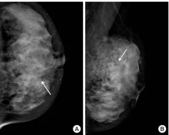
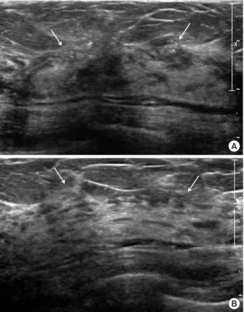
A 36-year-old woman visited our institute for the treatment of newly diagnosed left breast cancer. She had a palpable lump for a month. A mammogram showed fine pleomorphic mi- crocalcifications with segmental distribution in the left upper inner breast (Figure 1). No microcalcifications were observed in the left upper outer quadrant. Masses could not be detected on the mammogram because of the extremely dense fibro- glandular breast tissue. A sonogram revealed multiple, irregu- lar, hypoechoic infiltrative masses involving the upper inner and upper outer quadrants of the left breast (Figure 2). Con- trast-enhanced magnetic resonance imaging (MRI) of the breast showed multicentric, nonmass enhancement lesions with segmental distribution and a heterogeneous internal en- hancement pattern involving nearly all of the left upper inner and outer quadrants of the left breast (Figure 3). The largest lesion was 6 cm in diameter and 1 cm away from the nipple.
The tumors initially showed rapid enhancement and delayed Gi Won Shin, Young Mi Park, Hye Kyoung Yoon, Soo Jin Jung, Tae Hyun Kim, Anbok Lee, Seok Mo Lee
Departments of Radiology, 1Pathology, 2Surgery, and 3Nuclear Medicine, Inje University Busan Paik Hospital, Inje University College of Medicine, Busan, Korea
In patients with advanced breast cancer, most new calcifications detected on a mammogram after neoadjuvant chemotherapy are benign dystrophic calcifications, but this is not always ob- served. We present a patient with advanced breast cancer who had paradoxically increased malignant microcalcifications con- comitant with primary tumor regression after undergoing neoad- juvant chemotherapy. After the neoadjuvant chemotherapy, the follow-up mammogram revealed that local, fine pleomorphic mi- crocalcifications had markedly increased. Pathologically, these
calcifications were ductal carcinoma in situ. We concluded that, in patients with breast cancer undergoing neoadjuvant chemo- therapy, newly developed microcalcifications on follow-up mammo- grams should be carefully evaluated, and any suspicious ma- lignant microcalcifications should be included in surgical exci- sion planning.
Key Words: Breast neoplasms, Calcinosis, Mammography, Neoadjuvant therapy
Correspondence to: Young Mi Park
Department of Radiology, Inje University Busan Paik Hospital, Inje University College of Medicine, 75 Bokji-ro, Busanjin-gu, Busan 47392, Korea
Tel: +82-51-890-6549, Fax: +82-51-896-1085 E-mail: [email protected]
Received: October 30, 2015 Accepted: July 25, 2016
Figure 3. Initial magnetic resonance imaging (MRI) of the left breast.
The early phase of the dynamic sagittal MRI with subtraction shows a nonmass enhancement lesion with segmental distribution and hetero- geneous internal enhancement pattern in the left upper outer (arrow) and upper inner (not shown) quadrants.
Figure 1. Initial mammograms. The cranio-caudal (A) and medio-later- aloblique (B) mammogram show fine pleomorphic microcalcifications in the left upper inner breast (arrows). However, masses are not detected on the mammogram because of the extremely dense fibroglandular breast tissue.
A B
Figure 2. Initial ultrasonograms. The transverse (A) and longitudinal (B) ultrasonograms show an irregular hypoechoic infiltrative masses in the left upper inner and outer breast. Microcalcifications, however, are not detected on ultrasonography.
A
B
Figure 4. Follow-up left mammograms after neoadjuvant chemother- apy. The cranio-caudal (A) and medio-lateraloblique (B) mammograms show markedly increased number of fine pleomorphic microcalcifica- tions (arrows), involving both upper inner and upper outer quadrants of the left breast.
A B
Figure 5. Follow-up ultrasonograms after neoadjuvant chemotherapy.
The transverse (A) and longitudinal (B) ultrasonograms show a reduc- tion in tumor size (arrows).
A
B Figure 6. Follow-up breast magnetic resonance imaging (MRI) after chemotherapy. The early phase of the dynamic sagittal MRI with sub- traction shows the decrease in the tumor extent in left upper outer quadrant (arrow).
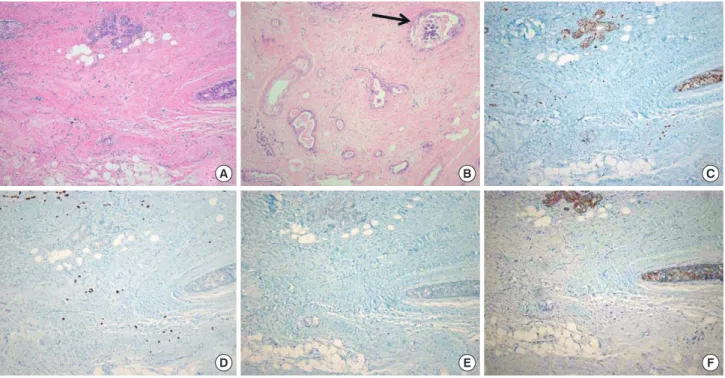
Figure 7. Histopathologic features of the core biopsy specimen of the left breast mass. (A) Invasive ductal carcinoma showing high histologic grade and small portion of ductal carcinoma in situ with necrosis and calcification (arrow) are observed (H&E stain). Invasive cancer tissue is luminal type B, showing positive reaction for estrogen receptor (B), low progesterone receptor expression (<20%) (C), high Ki-67 labelling index (>20%) (D), and 1+
reaction for human epidermal growth factor receptor 2 (E) (A–E, magnification, ×40).
A
D
B
E
C
washout kinetics on dynamic MRI scans, which is typical of malignancy. No abnormal lymph node was detected on mammo- grams and sonograms. However, an abnormal lymph node with a maximum standard uptake value of 2.6 was noted on initial positron emission tomography-computed tomography (PET-CT). The stage was clinically assessed as T3N1M0.
The patient underwent four cycles of neoadjuvant chemo- therapy with doxorubicin (60 mg/m2 intravenous injection) on day 1 plus cyclophosphamide (600 mg/m2 intravenous injec- tion) on day 1, every 21 days, for 3 months. Follow-up imaging studies were performed and her mammograms revealed a marked increase in fine pleomorphic microcalcifications with regional distribution in both upper inner and upper outer quad- rants of the left breast (Figure 4). However, a follow-up breast sonogram and MRI scan (Figures 5 and 6) showed partial regression of the size and extent of multifocal and multicentric tumors. An abnormal lymph node observed on initial PET-CT was normalized on follow-up PET-CT after completion of one cycle of neoadjuvant chemotherapy. The patient underwent a skin-sparing mastectomy with implant insertion. A histopath- ological examination revealed that most of the residual lesions were DCIS with microcalcifications, and invasive carcinoma cell nests were scattered among the DCIS lesions. On dissec- tion of the axillary lymph node, we found that three of 10
Figure 8. Histopathologic features of the surgical specimen of the left mastectomy after neoadjuvant chemotherapy. (A) Small amount of residual in- vasive carcinoma tissues are scattered, and relatively conspicuous ductal carcinoma in situ lesions (H&E stain, ×40) (B) with necrosis and calcification (arrow) are seen (H&E stain, ×100). Invasive cancer tissue is changed to luminal type A after neoadjuvant therapy, showing positive reaction for estro- gen receptor (C) and progesterone receptor (D), low Ki-67 labelling index (E), and 1+ reaction for human epidermal growth factor receptor 2 (F) (C-F, magnification, ×40).
D E F
lymph nodes from the left axilla had metastatic foci less than 1 mm in a diameter. The postoperative stage was ypT1N1miM0.
Immunohistochemical analyses of the core biopsy specimen prior to neoadjuvant chemotherapy showed that both invasive ductal carcinoma and DCIS were estrogen receptor (ER) posi- tive, progesterone receptor (PR) positive, and human epider- mal growth factor receptor 2 (HER2) negative, and had a high Ki-67 labeling index (75%) (Figure 7). After chemotherapy, the surgical specimen of invasive ductal carcinoma was ER posi- tive, PR positive, and HER2 negative and had a low Ki-67 in- dex (5%–10%), while the DCIS was ER positive, PR negative, and HER2 positive and had a low Ki-67 index (5%–10%) (Figure 8).
DISCUSSION
In the present case, a patient with advanced breast cancer underwent neoadjuvant chemotherapy and showed a marked decrease in the size of multiple malignant masses, but also ex- hibited the development of suspicious local, fine pleomorphic malignant microcalcifications. These new calcifications were pathologically confirmed as extensive DCIS.
There are two types of calcification processes in breast pa- thology: a secretory process that occurs in benign lesions and
cytes phagocytose calcium deposits to decrease calcifications, whereas cancer cell necrosis can increase calcifications [6].
Mammography plays an important role in monitoring tu- mor response during neoadjuvant chemotherapy, especially if the tumor is associated with microcalcifications. Only mam- mography can enable an accurate assessment of the extent of microcalcifications, which is essential to determine the extent of excision. Several studies have reported that decreases in the size and density of the tumor mass on a mammogram were the most reliable indicators of treatment response [1,3,7]. By contrast, the appearance of and changes in microcalcifications associated with malignancy have been deemed inaccurate for evaluating tumor response [1,3,7].
Fadul et al. [4] showed the development of malignant-ap- pearing microcalcifications after neoadjuvant chemotherapy in locally advanced breast cancer. These microcalcifications were histopathologically confirmed as microcalcifications of dystrophic and psammomatous types. Li et al. [5] reported an increased density of local microcalcifications after neoadju- vant chemotherapy in the absence of progressive disease.
Vinnicombe et al. [8] reported the microcalcification status in 44 of 95 patients with breast cancer who underwent neoadju- vant chemotherapy. These microcalcifications decreased in four patients, were stable in 21, were more conspicuous in 15, and were newly developed in four. Adrada et al. [1] found that the extent of calcification after neoadjuvant chemotherapy had decreased in 35 of 106 patients with advanced breast cancer, but it had remained stable in 41 patients and had increased or was newly developed in 30 patients. They did not find any correlation between calcification changes and pathologic complete remission. However, none of these studies reported whether the newly developed calcifications were pathological- ly benign or malignant. In this case, new calcifications were pathologically confirmed as extensive DCIS. Our findings suggested that newly developed microcalcifications after che- motherapy are not always benign and dystrophic.
Adrada et al. [1] showed that ER-positive cancers had a sig- nificantly higher proportion of residual malignant calcifica- tions after neoadjuvant chemotherapy compared to ER-nega- tive cancers. Therefore, they suggested that residual calcifica- tions related to incomplete excision might be less of a concern
pearing microcalcifications in a patient receiving neoadjuvant chemotherapy plus trastuzumab. Our case showed an increase in malignant-appearing microcalcifications and the presence of an extensive intraductal component in the postoperative pathological result. We can report that the DCIS component in this case showed no response to the chemotherapy regi- men, but the invasive cancer component revealed partial re- gression.
In conclusion, we present a patient with advanced breast cancer having paradoxically increased malignant microcalci- fications accompanying primary tumor regression after neo- adjuvant chemotherapy. The majority of new calcifications during chemotherapy are benign and dystrophic; however, as we demonstrated here, this is not always the case. Careful evaluations of microcalcifications through follow-up mam- mography are needed for patients who undergo neoadjuvant chemotherapy for locally advanced breast cancer. Any newly developed suspicious malignant microcalcifications in these patients should be excised during surgery.
CONFLICT OF INTEREST
The authors declare that they have no competing interests.
REFERENCES
1. Adrada BE, Huo L, Lane DL, Arribas EM, Resetkova E, Yang W. Histo- pathologic correlation of residual mammographic microcalcifications after neoadjuvant chemotherapy for locally advanced breast cancer. Ann Surg Oncol 2015;22:1111-7.
2. Segel MC, Paulus DD, Hortobagyi GN. Advanced primary breast can- cer: assessment at mammography of response to induction chemothera- py. Radiology 1988;169:49-54.
3. Moskovic EC, Mansi JL, King DM, Murch CR, Smith IE. Mammogra- phy in the assessment of response to medical treatment of large primary breast cancer. Clin Radiol 1993;47:339-44.
4. Fadul D, Rapelyea J, Schwartz AM, Brem RF. Development of malig- nant breast microcalcifications after neoadjuvant chemotherapy in ad- vanced breast cancer. Breast J 2004;10:141-5.
5. Li JJ, Chen C, Gu Y, Di G, Wu J, Liu G, et al. The role of mammographic calcification in the neoadjuvant therapy of breast cancer imaging evalu- ation. PLoS One 2014;9:e88853.
6. Esserman LE, d’Almeida M, Da Costa D, Gerson DM, Poppiti RJ Jr.