http://e-jbm.org/
183
Copyright © 2017 The Korean Society for Bone and Mineral Research
This is an Open Access article distributed under the terms of the Creative Commons Attribution Non-Commercial Li- cense (http://creativecommons.org/licenses/by-nc/4.0/) which permits unrestricted non-commercial use, distribu- tion, and reproduction in any medium, provided the original work is properly cited.
Change of Bone Mineral Density Measurement among Patients with Osteoporotic Fractures in Korean Population Using National Claim Database
Chan Ho Park1, Young-Kyun Lee2, Yong-Chan Ha3
1Department of Orthopedic Surgery, Yeungnam University Medical Center, Daegu;
2Department of Orthopedic Surgery, Seoul National University Bundang Hospital, Seongnam;
3Department of Orthopaedic Surgery, Chung-Ang University College of Medicine, Seoul, Korea
Background: Prior osteoporotic fractures are strongly associated with subsequent frac- tures. To prevent this, the diagnosis of osteoporosis following an osteoporotic fracture is important. The measurement of bone mineral density (BMD) is the first step in the diag- nosis and management of osteoporosis. Therefore, this study aimed 1) to evaluate the rate of BMD measurement after osteoporotic fracture in the Korean population, and 2) to determine whether the rate of BMD measurement after osteoporotic fracture changed between 2005 and 2010. Methods: Using the database of the Health Insurance Review Assessment Service (HIRA), we identified patients with osteoporotic fractures (hip, spine, humerus, and wrist fractures) in 2005 and 2010. BMD examinations were evaluated by using procedure codes and medicines, exclusively approved for osteoporosis treatment.
Results: During the study period, about half of all patients with osteoporotic fractures had BMD measurement. Between 2005 and 2010, the rate of BMD measurement signifi- cantly increased from 42.0% (65,556/156,190) to 53.9% (103,785/192,556) (P<0.001).
Conclusions: Our results showed that about half of all patients with osteoporotic frac- tures had BMD measurement, and that screening for osteoporosis in patients with os- teoporotic fractures increased between 2005 and 2010.
Key Words: Osteoporotic fractures, Bone density, Absorptiometry, Photon
INTRODUCTION
Osteoporosis is a common health care concern in elderly populations that is characterized by compromised bone strength.[1,2] Osteoporosis results in osteo- porotic fracture in hip, spine, humerus, and wrist.[3,4] The lifetime risk of these os- teoporotic fractures is about 60% in Korean women.[5]
It is well-known that patients with an osteoporotic fracture have higher risk of a subsequent fracture than those with no previous fracture.[6,7] That is osteoporot- ic fracture offers physicians an important opportunity to initiate secondary preven- tion.[8,9] Thus, secondary prevention has been recommended by several guide- lines for osteoporosis.
Bone mineral density (BMD) measurement is the first important step to investi- gate and manage patients with osteoporosis. There were few studies on the rate Corresponding author
Young-Kyun Lee
Department of Orthopedic Surgery, Seoul National University Bundang Hospital, 82 Gumi-ro 173 beon-gil, Bundang-gu, Seongnam 13620, Korea
Tel: +82-31-787-7204 Fax: +82-31-787-4056 E-mail: [email protected] Received: June 23, 2017 Revised: July 20, 2017 Accepted: July 23, 2017
No potential conflict of interest relevant to this article was reported.
Original Article
J Bone Metab 2017;24:183-186 https://doi.org/10.11005/jbm.2017.24.3.183 pISSN 2287-6375 eISSN 2287-7029
Chan Ho Park, et al.
184
http://e-jbm.org/ https://doi.org/10.11005/jbm.2017.24.3.183of BMD measurement after osteoporotic fracture in Korea, and whether there was a change of rate of BMD measure- ment after fractures.
Therefore, our purpose was 1) to evaluate the rate of BMD measurement after osteoporotic fracture in Korean popu- lation, and 2) determine whether the rate of BMD measure- ment after osteoporotic fracture changed between 2005 and 2010 in Korea.
METHODS
1. SubjectsWe used data from nationwide claims database of Health Insurance Review Assessment Service (HIRA). About 97%
of the Korean populations are included in this national in- surance system. Patients pay about 30% of total medical cost, and Korean governments reimburse the remaining 70% of medical cost to medical institute after the HIRA re- views all the medical claims data. The medical claims data include demographic information (age and gender), diag- noses using the International Classification of Disease, Tenth Revision (ICD-10) codes and procedures for diagnosis and treatment using codes in both of inpatients and outpatients care.
Thus, virtually all information about health care utiliza- tion is available from the HIRA database. Several epidemio- logic studies have used this national claim database.[10-12]
We analyzed patients aged over 50 years who were di- agnosed with osteoporotic fracture by physician at 2005 and 2010.
2. Identification of patients with osteoporotic fractures
We identified patients with hip, spine, humerus and wrist fractures on 2005 and 2010. To identify patients with these fractures, we used the diagnostic codes using the ICD-10 (hip [S720 and S721], spine [M484, M485, S220, S221, and S320], humerus [S422 and S423] and wrist fractures [S525 and S526]) and the procedure codes according to each an- atomic site.[13,14]
If an individual with fracture had more than one outpa- tient visits or admissions within the time period of six mon- ths, the cases were not counted as separate.[15,16]
Double recording was avoided by including only one re- cord when a person had more than one record in the HIRA
database. If a patient had both spine and wrist fractures, only the first episode was counted.[17]
3. BMD examination rates
Data were obtained from the HIRA on patients who had experienced a hip, spine, humerus or wrist fracture and had undergone BMD examinations within 6 months be- fore and after osteoporotic fractures.
The procedure codes (HC 341 to HC 344) for these exam- inations included dual energy X-ray absorptiometry (DXA) scans (single site, HC 341; multiple sites, HC 342), quantita- tive computed tomography scans (HC 343), and other meth- ods, including ultrasound (HC 344).
In addition, patients who administrated with at least one of the exclusive medicines approved for osteoporosis treat- ment were also considered as patients who were measured with BMD, because the medicines for osteoporosis was available only after BMD measurement during study peri- od in Korea. These medications included bisphosphonates (alendronate, etidronate, pamidronate, risedronate, zole- dronate), selective estrogen receptor modulator (raloxi- fene), and calcitonin. Estrogen replacement therapy, calci- um and vitamin D supplements were not included because they had another indication such as osteopenia.
The rates of BMD examinations were estimated within 6 months before and after osteoporotic fractures. Significance of differences was determined with use of a χ2 test. Statisti- cal analyses were performed using SAS for windows, ver- sion 9.4 (SAS Inc., Cary, NC, USA).
RESULTS
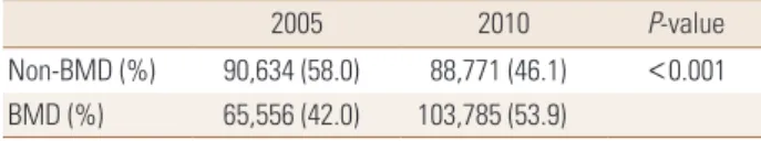
The 156,190 patients with osteoporotic fractures were identified in 2005, and 192,556 in 2010, respectively. Be- tween 2005 and 2010, the rate of BMD measurement sig- nificantly increased from 42.0% to 53.9% (P<0.001) (Table 1).
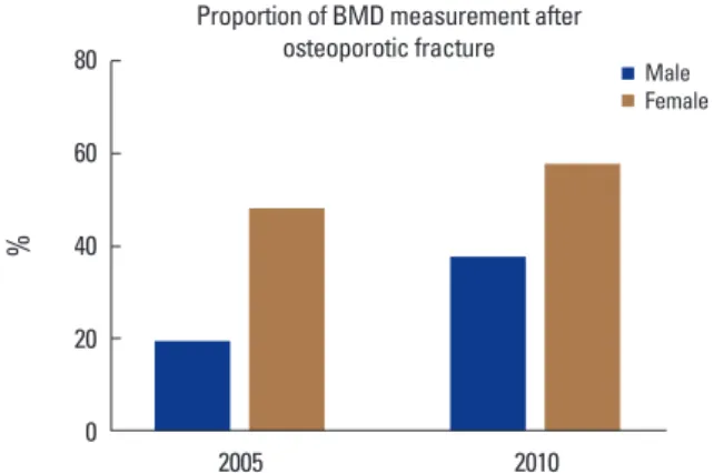
The rate of BMD measurement dramatically increased from 19.7% to 37.9% in men, while the rate of BMD mea- surement changed from 48.3 to 57.9 % in women (Fig. 1).
Table 1. Comparison of the rate of BMD measurement by χ2 test
2005 2010 P-value
Non-BMD (%) 90,634 (58.0) 88,771 (46.1) <0.001 BMD (%) 65,556 (42.0) 103,785 (53.9)
BMD, bone mineral density.
Change of BMD Measurement in South Korea
https://doi.org/10.11005/jbm.2017.24.3.183 http://e-jbm.org/
185
The percentage of women who received a bone density examination was significantly higher for those of men in both 2005 and 2010.
DISCUSSION
The present study demonstrates that the rate of BMD measurement after osteoporotic fractures was around 50%, and significantly increased between 2005 and 2010, espe- cially in men.
Many studies have indicated that BMD measurement following an osteoporotic fracture was performed in less than 15% of patients with osteoporotic fracture.[18,19]
The rate of BMD measurement after fracture seems to be unsatisfactory in Korea. It might be the reason the limita- tion of Korean health-care reimbursement system during the study period. The reimbursement system has allowed the screening of osteoporosis in patients just after fracture since 2015. It might suppress the clinical inertia for initia- tion of a required action such as screening of osteoporosis in patients with fracture before 2015.
However, the rate of BMD measurement increased be- tween 2005 and 2010, which is sure to be positive direc- tion for health care. There are some possible explanations on our results.
First, physician’s awareness on osteoporosis might incre- ase, although we could not evaluate the level of physician’s awareness on osteoporosis. Some studies showed that the increase of awareness in physicians lead to increase of screen- ing of osteoporosis in patients with fracture.[20,21] Second, patients’ awareness on osteoporosis might increase as well.
The strength of this study is that we evaluated the rate
of osteoporosis screening in patients with osteoporotic frac- tures in a large, population-based national cohort.
There were limitations in this study. First, we included patients who had undergone BMD examinations within 6 months ‘before’ as well as ‘after’ osteoporotic fractures, be- cause the obtained dataset from HIRA could not be distin- guished between before and after osteoporotic fractures.
Although it might result in bias, BMD measurement is most- ly taken after osteoporotic fractures.
Second, we could not perform comprehensive analyses to define the factors associated with low rate of BMD mea- surement after fracture, because we could not access to in- dividual records of subjects. Further studies are necessary to identify the risk factors of low investigation of osteopo- rosis in these patients.
Despite these limitations, this study would be helpful in terms of understanding current practice patterns after frac- ture in Korea. Our results showed that about a half of pa- tients with osteoporotic fractures had BMD measurement, and the rate of screening for osteoporosis increased be- tween 2005 and 2010.
ACKNOWLEDGEMENT
This research was supported by grants (HI15C1189) of the Korea Health Technology R&D Project through the Ko- rea Health Industry Development Institute (KHIDI) funded by the Ministry of Health & Welfare, Republic of Korea.
REFERENCES
1. NIH Consensus Development Panel on Osteoporosis Pre- vention, Diagnosis, and Therapy. Osteoporosis prevention, diagnosis, and therapy. NIH Consens Statement 2000;17:
1-45.
2. Peck WA. Consensus development conference: diagnosis, prophylaxis, and treatment of osteoporosis. Am J Med 1993;
94:646-50.
3. Johnell O, Kanis J. Epidemiology of osteoporotic fractures.
Osteoporos Int 2005;16 Suppl 2:S3-7.
4. Kanis JA, Oden A, Johnell O, et al. The burden of osteopo- rotic fractures: a method for setting intervention thresh- olds. Osteoporos Int 2001;12:417-27.
5. Kim JW, Jeon YJ, Baek DH, et al. Percentage of the popula- tion at high risk of osteoporotic fracture in South Korea:
Fig. 1. Change of proportion of bone mineral density (BMD) measure- ment according to gender.
80 60 40 20 0
%
2005 2010
MaleFemale Proportion of BMD measurement after
osteoporotic fracture
Chan Ho Park, et al.
186
http://e-jbm.org/ https://doi.org/10.11005/jbm.2017.24.3.183analysis of the 2010 Fifth Korean National Health and Nu- trition Examination survey data. Osteoporos Int 2014;25:
1313-9.
6. Center JR, Bliuc D, Nguyen TV, et al. Risk of subsequent frac- ture after low-trauma fracture in men and women. JAMA 2007;297:387-94.
7. Langsetmo L, Goltzman D, Kovacs CS, et al. Repeat low- trauma fractures occur frequently among men and wom- en who have osteopenic BMD. J Bone Miner Res 2009;24:
1515-22.
8. Ganda K, Schaffer A, Pearson S, et al. Compliance and per- sistence to oral bisphosphonate therapy following initia- tion within a secondary fracture prevention program: a randomised controlled trial of specialist vs. non-specialist management. Osteoporos Int 2014;25:1345-55.
9. Nakayama A, Major G, Holliday E, et al. Evidence of effec- tiveness of a fracture liaison service to reduce the re-frac- ture rate. Osteoporos Int 2016;27:873-9.
10. Yoon HK, Lee YK, Ha YC. Characteristics of patients diag- nosed with osteoporosis in South Korea: results from the national claim registry. J Bone Metab 2017;24:59-63.
11. Park C, Jang S, Lee A, et al. Incidence and mortality after proximal humerus fractures over 50 years of age in South Korea: national claim data from 2008 to 2012. J Bone Metab 2015;22:17-21.
12. Yoon HK, Park C, Jang S, et al. Incidence and mortality fol- lowing hip fracture in Korea. J Korean Med Sci 2011;26:
1087-92.
13. Park C, Ha YC, Jang S, et al. The incidence and residual life-
time risk of osteoporosis-related fractures in Korea. J Bone Miner Metab 2011;29:744-51.
14. Yoo JH, Moon SH, Ha YC, et al. Osteoporotic fracture: 2015 position statement of the Korean society for bone and min- eral research. J Bone Metab 2015;22:175-81.
15. Lau E, Ong K, Kurtz S, et al. Mortality following the diag- nosis of a vertebral compression fracture in the Medicare population. J Bone Joint Surg Am 2008;90:1479-86.
16. Kang HY, Yang KH, Kim YN, et al. Incidence and mortality of hip fracture among the elderly population in South Ko- rea: a population-based study using the national health insurance claims data. BMC Public Health 2010;10:230.
17. Gong HS, Oh WS, Chung MS, et al. Patients with wrist frac- tures are less likely to be evaluated and managed for os- teoporosis. J Bone Joint Surg Am 2009;91:2376-80.
18. Giangregorio LM, Leslie WD. Time since prior fracture is a risk modifier for 10-year osteoporotic fractures. J Bone Min- er Res 2010;25:1400-5.
19. Metge CJ, Leslie WD, Manness LJ, et al. Postfracture care for older women: gaps between optimal care and actual care. Can Fam Physician 2008;54:1270-6.
20. Giangregorio L, Dolovich L, Cranney A, et al. Osteoporosis risk perceptions among patients who have sustained a fra- gility fracture. Patient Educ Couns 2009;74:213-20.
21. Kim SR, Ha YC, Park YG, et al. Orthopedic surgeon’s aware- ness can improve osteoporosis treatment following hip fracture: a prospective cohort study. J Korean Med Sci 2011;
26:1501-7.