© 2018 Korean Breast Cancer Society. All rights reserved. http://ejbc.kr | pISSN 1738-6756
INTRODUCTION
Intraductal papilloma (IDP) originates from both the large ducts of the subareolar region and the terminal duct lobular unit in the periphery, and it is histologically characterized by a fibrovascular core covered with epithelial and myoepithelial cells. It is a relatively common lesion found in breast biopsies.
However, IDP often accompanies a variety of changes, includ- ing sclerosis, epithelial or myoepithelial hyperplasia, squa- mous or apocrine metaplasia, and even atypical proliferation [1].
A papilloma with atypia, which encompasses atypical duc- tal hyperplasia (ADH) or small foci of low-grade ductal carci- noma in situ (DCIS) within the papilloma, has an increased risk of developing malignancy and has been reported to show a high rate of upgrading on subsequent excision [2-5]. Thus, surgical excision is usually recommended for IDP with atypia diagnosed by core needle biopsy (CNB) as standard manage- ment. On the contrary, no consensus has been met regarding the management of benign IDP without atypia diagnosed by CNB harboring no clinical symptoms. Some studies have shown that IDPs, even those without atypia, are significantly associated with higher-grade lesions [4,6], and surgical exci- sion is recommended in all cases for accurate diagnosis. In contrast, other studies have shown low rates of upgrading for IDP without atypia, suggesting careful observation rather than surgical excision [7-11]. However, most of the previous studies were performed using small samples, and some stud- ies included IDP with atypia or even malignancy in their
Benign Intraductal Papilloma without Atypia on Core Needle Biopsy Has a Low Rate of Upgrading to Malignancy after Excision
Song-Hee Han1,2, Milim Kim1, Yul Ri Chung1, Bo La Yun3, Mijung Jang3, Sun Mi Kim3, Eunyoung Kang4, Eun-Kyu Kim4, So Yeon Park1
1Department of Pathology, Seoul National University Bundang Hospital, Seoul National University College of Medicine, Seongnam; 2Department of Pathology, Konyang University Hospital, Daejeon; Departments of 3Radiology and 4Surgery, Seoul National University Bundang Hospital, Seoul National University College of Medicine, Seongnam, Korea
ORIGINAL ARTICLE
Purpose: The management of benign intraductal papilloma (IDP) without atypia diagnosed on core needle biopsy (CNB) remains controversial. This study was performed to evaluate the rate of upgrading to malignancy or high-risk lesions after excision and to identify factors associated with upgrading using a large series of benign IDP cases without atypia. Methods: We included pa- tients who were diagnosed as having benign IDP without atypia on CNB and underwent surgical or vacuum-assisted excision between 2010 and 2015. We analyzed the clinical, radiologic, and histopathologic features of IDPs that were upgraded to ma- lignancy or high-risk lesions after excision. Results: A total of 511 benign IDPs without atypia diagnosed via CNB were identified, of which 398 cases were treated with excision. After reviewing these cases, four cases of high-risk lesions in adjacent tissue on CNB, two cases which were revealed as papilloma with atypia,
and nine cases of malignancy in the same breast were excluded.
In the remaining 383 cases, the rate of upgrading to malignancy and high-risk lesions after excision was 0.8% and 4.4%, respec- tively. The presence of concurrent contralateral breast cancer, the presence of symptoms, and multifocality were factors signifi- cantly associated with upgrading to malignancy on subsequent excision. Surgical excision rather than vacuum-assisted excision was significantly associated with upgrading to high-risk lesions or malignancy. Conclusion: The rate of upgrading to malignancy for benign IDP without atypia was very low, suggesting that close clinical and radiologic observation may be sufficient for patients with benign IDP without atypia on CNB under proper settings.
Key Words: Breast neoplasms, Large-core needle biopsy, Observation, Papilloma
Correspondence to: So Yeon Park
Department of Pathology, Seoul National University Bundang Hospital, Seoul National University College of Medicine, 82 Gumi-ro 173beon-gil, Bundang- gu, Seongnam 13620, Korea
Tel: +82-31-787-7712, Fax: +82-31-787-4012 E-mail: sypmd@snu.ac.kr
Received: November 8, 2017 Accepted: March 5, 2018
Cancer
analysis.
In the present study, we restricted our analysis to a large co- hort of benign IDPs without atypia, and we evaluated the up- grade rate to malignancy, including DCIS and invasive carci- noma. In addition, evaluation of the presence of proliferative lesions with atypia, such as ADH, atypical lobular hyperplasia, and lobular carcinoma in situ, which are associated with a high risk for developing breast cancer, is also important in as- sessing breast cancer risk and determining patient manage- ment. Thus, we evaluated the upgrade rate to these high-risk lesions as well. Furthermore, we analyzed the clinicopathol- ogic features associated with upgrading on excision.
METHODS
Case selection
We performed a retrospective search of the pathology data- base to identify IDPs without atypia diagnosed via CNB be- tween January 2010 and December 2015 at the Department of Pathology, Seoul National University Bundang Hospital, Korea. All CNBs were performed using a 14-gauge automated biopsy gun (STERICUT®; TSK Laboratory, Tochigi, Japan) under ultrasound guidance by radiologists specialized in breast imaging. We selected cases that were excised with lumpectomy, excisional biopsy, or vacuum-assisted excision (VAE) at our institution. When patients presented with symp- toms such as bloody nipple discharge or a palpable mass or when IDP was located near the skin or nipple, surgical man- agement was recommended. When the lesion was single with a size less than 3 cm and located far from the skin or nipple, patients had an option of surgery or VAE. VAE was per- formed using an 8- or 11-gauge vacuum-assisted biopsy nee- dle (Mammotome®; Devicor Medical Products, Cincinnati, USA).
Benign IDPs with benign proliferative lesions, such as usual ductal hyperplasia or adenosis, and those associated with scle- rosis, radial scars, fibrocystic changes, and columnar cell changes, were included. IDPs coexisting with a malignant le- sion, including invasive carcinoma and DCIS, or other high- risk lesions, such as ADH and lobular neoplasia (atypical lob- ular hyperplasia and lobular carcinoma in situ), in the same side of the breast were excluded. This study was approved by the Institutional Review Board of Seoul National University Bundang Hospital (protocol number: B-1708-414-107) and informed consent was waived.
Evaluation of clinical and radiologic features
Clinical variables, such as the age at the time of diagnosis;
gender; cause of detection; clinical symptoms, including nip-
ple discharge, palpable mass, and local pain; and history of breast cancer, were recorded. Radiologic findings, including the location of the lesion, size of the lesion defined as the larg- est dimension recorded on imaging, Breast Imaging Report- ing and Data System (BI-RADS) classification of the lesion, multifocality, and the number of tissue cores in CNB, were re- trieved from the medical records. Multifocality was defined as two or more lesions separated by normal breast tissue in im- aging that were eventually proven to be benign IDPs on pathol- ogic examination.
To analyze the radiologic-pathologic concordance, we re- viewed all radiologic findings in each case and matched them with the pathologic diagnosis by the location of the CNB and the size of the lesion. Radiologic-pathologic discordance was considered to be present when the lesion was more than mod- erately suspicious for malignancy in radiologic findings, i.e., BI-RADS category 4b, 4c, or 5, but for which the histologic findings of IDP without atypia did not account for the imag- ing pattern.
Evaluation of excision specimens
We assessed accompanying lesions within the papilloma or in the adjacent breast tissue using excision specimens, and we classified them according to the World Health Organization classification of Tumours of the Breast, 4th edition. Upgrade to malignancy was defined as the presence of invasive carci- noma or DCIS in the excision specimen. The presence of atypical proliferative lesions such as ADH and lobular neopla- sia, which are associated with a high risk of malignancy, was also assessed. Upgrade to high-risk lesions was defined as the presence of ADH or lobular neoplasia.
Statistical analysis
Statistical analysis was performed using SPSS version 21.0 software (IBM Corp., Armonk, USA). For patients with more than one IDP, analyses of the radiologic and pathologic fea- tures were based on each IDP. Chi-square tests or Fisher exact tests were used to compare the clinicopathologic variables be- tween upgraded lesions and nonupgraded lesions. Statistical significance was defined as a p-value <0.05. All reported p- values were two-sided.
RESULTS
Clinical, radiologic, and pathologic characteristics of IDP on CNB
A total of 511 benign IDPs without atypia diagnosed by CNB were identified, of which 398 cases were treated with surgical excision or VAE at our institution. After reviewing
the medical records, pathologic reports, and hematoxylin and eosin stained slides of the CNBs, four cases that had high-risk lesions in the adjacent tissue, two cases that were re-diagnosed as papilloma with atypia, and nine cases of concurrent breast cancer in the same breast were excluded. Finally, 383 cases of benign IDP without atypia were included in the analyses.
The median age of the patients at the time of biopsy was 48 years (range, 23–82 years). In this cohort, the two major causes of initial detection were routine mammographic screening on medical checkup (n=224, 58.5%) and the pres- ence of a symptom (n=108, 28.2%). All breast CNBs were performed under ultrasound guidance. The median number of core biopsy samples was 5 (range, 1–15). The other baseline characteristics of the patients are described in Table 1.
Characteristics of IDPs with a likelihood of upgrading to malignancy or high-risk lesions on excision
Among the 383 IDPs without atypia on CNB, 20 cases (5.2%) were upgraded to malignancy or high-risk lesions, in- cluding ADH and lobular neoplasia, on excision; 10 cases were upgraded to ADH, five to atypical lobular hyperplasia, two to lobular carcinoma in situ, and three to DCIS. The rate of upgrading to malignancy and high-risk lesions after exci- sion was 0.8% (n=3) and 4.4% (n=17), respectively. We also
Table 1. Baseline characteristics (n=383)
Characteristic No. (%)
Age (yr)* 48 (23–82)
Cause of detection
Medical checkup 224 (58.5)
Presence of symptom 108 (28.2)
Nipple discharge 67
Palpable mass 34
Local pain 7
R adiologic abnormality detected during
examination of other lesions 51 (13.3)
Concurrent contralateral breast cancer
Yes 32 (8.4)
No 351 (91.6)
Side
Right 178 (46.5)
Left 205 (53.5)
Subareolar location
Yes 50 (13.1)
No 333 (86.9)
Radiologic size (cm)* 0.8 (0.3–4.2)
BI-RADS classification
C3 2 (0.5)
C4a 348 (90.9)
C4b 29 (7.6)
C4c 4 (1.0)
Radiologic identification of intraductal lesion
Yes 72 (18.8)
No 311 (81.2)
Multifocality
No 292 (76.2)
Yes 91 (23.8)
No. of cores in CNB* 5 (1–15)
Method of procedure after CNB
VAE 226 (59.0)
Surgical excision 157 (41.0)
BI-RADS=Breast Imaging Reporting and Data System; CNB=core needle biopsy; VAE=vacuum-assisted excision.
*Median (range).
Table 2. Characteristics of intraductal papillomas without atypia upgraded to malignancy on subsequent excision
Characteristic Not upgraded
to malignancy (n=380)
Upgraded to malignancy
(n=3) p-value
Age (yr) 0.260
<50 213 (56.1) 3 (100)
≥50 167 (43.9) 0
Side 0.099
Right 175 (46.1) 3 (100)
Left 205 (53.9) 0
Subareolar area 0.344
Yes 49 (12.9) 1 (33.3)
No 331 (87.1) 2 (66.7)
Presence of symptom 0.022
Yes 105 (27.6) 3 (100)
No 275 (72.4) 0
Concurrent contralateral breast cancer
0.019
Yes 30 (7.9) 2 (66.7)
No 350 (92.1) 1 (33.3)
Radiologic size (cm) 0.271
<0.8 158 (41.6) 0
≥0.8 222 (58.4) 3 (100)
BI-RADS classification 0.409
C3 2 (0.5) 0
C4a 346 (91.1) 2 (66.7)
C4b 28 (7.4) 1 (33.3)
C4c 4 (1.0) 0
Radiologic identification of intraductal lesion
1.000
Yes 72 (18.9) 0
No 308 (81.1) 3 (100)
Multifocality 0.013
No 292 (76.8) 0
Yes 88 (23.2) 3 (100)
No. of cores in CNB* 0.569
<5 151 (40.5) 2 (66.7)
≥5 222 (59.5) 1 (33.3)
Method of procedure after CNB 0.068
Surgical excision 154 (40.5) 3 (100)
VAE 226 (59.5) 0
p-values were calculated by Fisher exact test.
BI-RADS=Breast Imaging Reporting and Data System; CNB=core needle biopsy; VAE=vacuum-assisted excision.
*Information was available for 376 lesions.
evaluated the characteristics of the lesions upgraded to malig- nancy on excision. Interestingly, we found that the presence of a symptom (p=0.022), the presence of concurrent contralat- eral breast cancer (p=0.019), and multifocality of the lesions (p=0.013) were predictive factors for upgrading to malignan- cy (Table 2). In addition, cases upgraded to malignancy or high-risk lesions were associated with surgical excision after
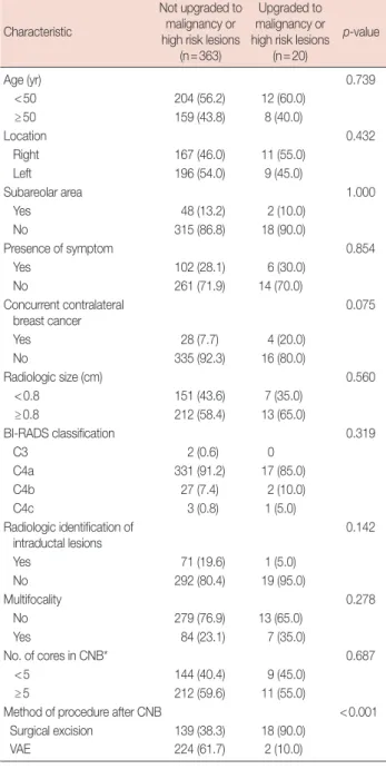
CNB (p<0.001). The presence of concurrent contralateral breast cancer tended to have an association with upgrading to malignancy or high-risk lesions (p=0.075) (Table 3).
Characteristics of the cases upgraded to malignancy on excision
The clinical and pathologic characteristics of the cases that were upgraded to malignant lesions are summarized in Table 4. All of these cases were upgraded to DCIS, which was found around the papilloma (Figure 1). The extent of the DCIS was 0.5 cm, 2.5 cm, and 2.5 cm in each case. Of these three cases, two were from the same patient. All of the cases had clinical symptoms: one manifested as nipple discharge and the others as a palpable mass. Radiologic size of the lesions was 0.8 cm or greater and showed multifocality. Two cases had concur- rent contralateral breast cancer.
DISCUSSION
Advances in screening and imaging techniques for breast cancer have resulted in an increased number of suspicious le- sions, which in turn, have led to an increased number of breast biopsies. The incidence of papillary lesions, including IDPs, has increased steadily over the past decade [12]. How- ever, management of benign IDPs without atypia diagnosed Table 3. Characteristics of intraductal papillomas without atypia up-
graded to malignancy or other high-risk lesions on subsequent excision Characteristic
Not upgraded to malignancy or high risk lesions
(n=363)
Upgraded to malignancy or high risk lesions
(n=20)
p-value
Age (yr) 0.739
<50 204 (56.2) 12 (60.0)
≥50 159 (43.8) 8 (40.0)
Location 0.432
Right 167 (46.0) 11 (55.0)
Left 196 (54.0) 9 (45.0)
Subareolar area 1.000
Yes 48 (13.2) 2 (10.0)
No 315 (86.8) 18 (90.0)
Presence of symptom 0.854
Yes 102 (28.1) 6 (30.0)
No 261 (71.9) 14 (70.0)
Concurrent contralateral breast cancer
0.075
Yes 28 (7.7) 4 (20.0)
No 335 (92.3) 16 (80.0)
Radiologic size (cm) 0.560
<0.8 151 (43.6) 7 (35.0)
≥0.8 212 (58.4) 13 (65.0)
BI-RADS classification 0.319
C3 2 (0.6) 0
C4a 331 (91.2) 17 (85.0)
C4b 27 (7.4) 2 (10.0)
C4c 3 (0.8) 1 (5.0)
Radiologic identification of intraductal lesions
0.142
Yes 71 (19.6) 1 (5.0)
No 292 (80.4) 19 (95.0)
Multifocality 0.278
No 279 (76.9) 13 (65.0)
Yes 84 (23.1) 7 (35.0)
No. of cores in CNB* 0.687
<5 144 (40.4) 9 (45.0)
≥5 212 (59.6) 11 (55.0)
Method of procedure after CNB <0.001
Surgical excision 139 (38.3) 18 (90.0)
VAE 224 (61.7) 2 (10.0)
p-values were calculated by the chi-square or Fisher exact test.
BI-RADS=Breast Imaging Reporting and Data System; CNB=core needle biopsy; VAE=vacuum-assisted excision.
*Information was available for 376 lesions.
Table 4. Summary of the characteristics of the lesions upgraded to ma- lignancy on excision
Characteristic Case 1 Case 2 Case 3
Age (yr) 34 40 40
Symptoms Nipple
discharge Palpable
mass Palpable mass
Side Right Right Right
Radiologic features
Size (cm) 1.9 2.9 0.8
BI-RADS category C4a C4b C4a
Multifocality Yes Yes Yes
No. of cores 4 4 6
Excisional specimen
Method of excision Surgical
excision Surgical
excision Surgical excision
Diagnosis DCIS DCIS DCIS
Extent of malignancy (cm) 0.5 2.5 2.5
Location of malignancy Around papilloma
Around papilloma
Around papilloma Concurrent contralateral breast
cancer
No Yes Yes
Pathologic diagnosis NA MC MC
Incidental upgrade No No No
Case 2 and 3 were of the same patient.
BI-RADS=Breast Imaging Reporting and Data System; DCIS=ductal carci- noma in situ; NA=not applicable; MC=mucinous carcinoma.
by CNB remains controversial with some suggesting follow- up imaging and others excision. Moreover, in previous stud- ies, the reported upgrading rates of benign IDP following ex- cision vary widely [4,7-11,13-15]. This difference may have resulted from the study design, definition of upgrade lesions, and methodological details. Thus, we carefully selected our cohort to include only benign IDP cases diagnosed by CNB that had no malignant or high-risk lesions in the same breast.
In this study, we showed that the rate of upgrading to malig- nancy was 0.8% at our institution. In addition, we found that the presence of a symptom, concurrent contralateral breast cancer, and multifocality were significant factors predictive of upgrading to malignancy. We also showed that upgrading to malignancy or high-risk lesions was associated with surgical excision rather than VAE. However, this may be caused by se- lection bias since patients with symptoms or multiple lesions were generally treated with surgical excision.
Several studies have attempted to identify factors that are associated with upgrading of benign IDP on subsequent exci- sion [7,9,16-22]. It was reported that old age at diagnosis was associated with upgrading to malignancy in benign IDPs [16,22]. Regarding the size of the lesion, IDPs upgraded to malignancy have been reported to be larger [7,17,22]. Previ- ously, we also showed that large-sized IDP was a significant predictor of an upgrade by analyzing solitary IDPs diagnosed at different time points at our institution, excluding patients with breast cancer [9]. Although the size of the IDP was not a significant factor associated with upgrading to malignancy in the present study, all upgraded cases were above the median in size. Besides the size of the lesion, Kil et al. [18] have sug- gested that peripherally located IDPs require additional surgi- cal excision. Holley et al. [19] reported that a reduced amount of tissue collected at biopsy (three cores vs. five cores; 14- gauge vs. 9-gauge needle) was associated with an upgraded
lesion. Additionally, previous studies have reported varying results regarding the impact of the BI-RADS category on up- grade rates in benign IDP [20,21].
Contrary to the previous studies, patient age, size, and loca- tion of the lesion, number of tissue cores, and BI-RADS score were not proven to be significant factors in our study. The most noticeable predictor of upgrading to malignancy was the presence of concurrent contralateral breast cancer, a finding that has never been reported in previous studies. While only 7.9% (30/380) of the nonupgraded cases had concurrent con- tralateral breast cancer, 66.7% (2/3) of upgraded cases had concurrent contralateral breast cancer. This association may be explained by the fact that the risk of developing new breast cancer is increased in patients with a history of breast cancer [23]. We also found that multifocality of the lesion was a sig- nificant predictor for upgrading to malignancy, similar to pre- vious studies that showed that multiple papillomas were more likely to be associated with breast cancer than solitary papillo- mas [24,25].
The mismatch between radiologic findings and pathologic diagnosis is an important issue that requires further evalua- tion. Some studies have documented that the discordance be- tween radiographic and histologic findings was an important factor that should be evaluated by surgical excision. Thus, in- clusion or exclusion of pathologic-radiologic discordant cases may be an important factor leading to variability in upgrade rates. After analyzing 234 benign IDPs, Rizzo et al. [26] re- ported the rate of upgrading to DCIS and invasive carcinoma as 8.9% and to ADH as 17.9%. Shin et al. [27] showed that an upgrade rate to DCIS and invasive carcinoma was 14% of 86 benign IDPs diagnosed by CNB. Mercado et al. [28] reported that 17% of 36 IDPs were upgraded to ADH. Other studies have reported similar high upgrade rates though these results may raise doubts about the definition of an upgrade [1,29,30].
Figure 1. A representative case upgraded to ductal carcinoma in situ on excision. (A) Core needle biopsy shows typical benign intraductal papilloma without atypia (H&E stain, ×100). (B) Excision specimen reveals small foci of ductal carcinoma in situ with intermediate nuclear grade and a cribriform architectural pattern (H&E stain, ×200). (C) Residual papilloma in the excision specimen does not show any atypical proliferative change. Postbiopsy changes are seen in the lower portion of the papilloma (H&E stain, ×100).
A B C
Collectively, these studies included pathologic-radiologic dis- cordant cases in their analyses or failed to document pathol- ogic-radiologic concordance [1,26-30], leading to falsely high upgrade rates. Shin et al. [27] reported that the upgrade rate to malignancy was higher in imaging-pathologic discordant lesions than in concordant lesions. However, recent studies re- ported that the upgrade rate for benign IDP is low when the pathologic-radiologic discordant cases are excluded [10,11].
Although physical findings such as a palpable mass and nipple discharge may not predict the risk of upgrading, lesions with such manifestations require careful evaluation and close observation and eventually need excision even if the pathol- ogic diagnosis on CNB was benign. Nakhlis et al. [10] showed that the actual rate of upgrading in their study was 0% when they excluded both of the two patients from their cohort who presented with a clinically suspicious palpable mass. Other studies shared the same perspective, excluding symptomatic patients from their analyses of upgrading of benign IDP [8,19]. We agree that any patient with symptoms such as a palpable mass or nipple discharge should be treated by exci- sion. Thus, of the 383 benign IDP diagnosed by CNB in our study, none of the cases were truly upgraded lesions when we excluded all three cases with clinical symptoms for which sur- gical treatment is indicated. Of the 17 cases upgraded to high- risk lesions, two cases had pathologic-radiologic discordance, and three cases had clinical symptoms. Therefore, the true up- grade rate to high-risk lesions was 3.1%. Given our results, close clinical and radiologic observation appear to be ade- quate for patients with benign IDP on CNB under proper clinical settings.
Although IDP has a relatively simple histologic definition, it encompasses a wide variety of lesions. Papillary lesions are of- ten diagnostically challenging for pathologists. IDP may have a complex glandular architecture and epithelial hyperplasia;
histologic distinctions between luminal epithelial and myo- epithelial cells or benign hyperplasia and atypical hyperplasia can be subtle, leading to misinterpretations on CNB. Thus, ac- curate interpretation by an experienced pathologist and an adequate amount of an accurately-targeted specimen are es- sential for correct diagnosis. In our institution, all breast CNBs were diagnosed by an experienced breast pathologist (S.Y.P.), which can affect diagnostic accuracy. It was reported that one of the reasons for such diagnostic difficulties might be the limited amounts and fragmentation of the samples [16].
Renshaw et al. [3] pointed out that the incidence of upgrading after excision was associated with the adequacy of sampling in biopsy specimens. Several studies have shown different up- grading rates of benign IDP to malignancy depending on the needle gauge used during biopsy [18,19]. In the current study,
the number of tissue cores was sufficient with a median value of 5.0, which appears to have contributed to the low rate of upgrades.
In conclusion, the most significant finding of our study is that the rate of upgrading to malignancy of benign IDP with- out atypia diagnosed by CNB is very low. Moreover, when we excluded cases with suspicious clinical symptoms, the true rate of under-diagnosis in our cohort was 0%. For this reason, caution should be exercised in recommending surgical man- agement for all benign IDPs. Our study suggests that patients with benign IDPs diagnosed by CNB who have solitary le- sions without clinically suspicious symptoms and, in particu- lar, no concurrent contralateral breast cancer may be candi- dates for close observation rather than prompt excision. How- ever, due to the small number of cases upgraded to malignan- cy in this study, further large-scale studies are warranted to support this suggestion.
CONFLICT OF INTEREST
The authors declare that they have no competing interests.
REFERENCES
1. Cyr AE, Novack D, Trinkaus K, Margenthaler JA, Gillanders WE, Eberlein TJ, et al. Are we overtreating papillomas diagnosed on core needle biopsy? Ann Surg Oncol 2011;18:946-51.
2. Gendler LS, Feldman SM, Balassanian R, Riker MA, Frencher SK, Whelan DB, et al. Association of breast cancer with papillary lesions identified at percutaneous image-guided breast biopsy. Am J Surg 2004;188:365-70.
3. Renshaw AA, Derhagopian RP, Tizol-Blanco DM, Gould EW. Papillomas and atypical papillomas in breast core needle biopsy specimens: risk of carcinoma in subsequent excision. Am J Clin Pathol 2004;122: 217-21.
4. Rizzo M, Lund MJ, Oprea G, Schniederjan M, Wood WC, Mosunjac M.
Surgical follow-up and clinical presentation of 142 breast papillary lesions diagnosed by ultrasound-guided core-needle biopsy. Ann Surg Oncol 2008;15:1040-7.
5. McGhan LJ, Pockaj BA, Wasif N, Giurescu ME, McCullough AE, Gray RJ. Atypical ductal hyperplasia on core biopsy: an automatic trigger for excisional biopsy? Ann Surg Oncol 2012;19:3264-9.
6. Shiino S, Tsuda H, Yoshida M, Jimbo K, Asaga S, Hojo T, et al. Intra- ductal papillomas on core biopsy can be upgraded to malignancy on subsequent excisional biopsy regardless of the presence of atypical fea- tures. Pathol Int 2015;65:293-300.
7. Ahmadiyeh N, Stoleru MA, Raza S, Lester SC, Golshan M. Manage- ment of intraductal papillomas of the breast: an analysis of 129 cases and their outcome. Ann Surg Oncol 2009;16:2264-9.
8. Bennett LE, Ghate SV, Bentley R, Baker JA. Is surgical excision of core biopsy proven benign papillomas of the breast necessary? Acad Radiol 2010;17:553-7.
9. Ko D, Kang E, Park SY, Kim SM, Jang M, Yun B, et al. The management
strategy of benign solitary intraductal papilloma on breast core biopsy.
Clin Breast Cancer 2017;17:367-72.
10. Nakhlis F, Ahmadiyeh N, Lester S, Raza S, Lotfi P, Golshan M. Papilloma on core biopsy: excision vs. observation. Ann Surg Oncol 2015;22:
1479-82.
11. Pareja F, Corben AD, Brennan SB, Murray MP, Bowser ZL, Jakate K, et al. Breast intraductal papillomas without atypia in radiologic-pathologic concordant core-needle biopsies: rate of upgrade to carcinoma at exci- sion. Cancer 2016;122:2819-27.
12. Carder PJ, Garvican J, Haigh I, Liston JC. Needle core biopsy can reli- ably distinguish between benign and malignant papillary lesions of the breast. Histopathology 2005;46:320-7.
13. Liberman L, Bracero N, Vuolo MA, Dershaw DD, Morris EA, Abramson AF, et al. Percutaneous large-core biopsy of papillary breast lesions. AJR Am J Roentgenol 1999;172:331-7.
14. Liberman L, Drotman M, Morris EA, LaTrenta LR, Abramson AF, Zakowski MF, et al. Imaging-histologic discordance at percutaneous breast biopsy. Cancer 2000;89:2538-46.
15. Mercado CL, Hamele-Bena D, Singer C, Koenigsberg T, Pile-Spellman E, Higgins H, et al. Papillary lesions of the breast: evaluation with stereo- tactic directional vacuum-assisted biopsy. Radiology 2001;221:650-5.
16. Jakate K, De Brot M, Goldberg F, Muradali D, O’Malley FP, Mulligan AM. Papillary lesions of the breast: impact of breast pathology subspe- cialization on core biopsy and excision diagnoses. Am J Surg Pathol 2012;36:544-51.
17. Chang JM, Moon WK, Cho N, Han W, Noh DY, Park IA, et al. Risk of carcinoma after subsequent excision of benign papilloma initially diag- nosed with an ultrasound (US)-guided 14-gauge core needle biopsy:
a prospective observational study. Eur Radiol 2010;20:1093-100.
18. Kil WH, Cho EY, Kim JH, Nam SJ, Yang JH. Is surgical excision neces- sary in benign papillary lesions initially diagnosed at core biopsy? Breast 2008;17:258-62.
19. Holley SO, Appleton CM, Farria DM, Reichert VC, Warrick J, Allred DC, et al. Pathologic outcomes of nonmalignant papillary breast lesions diagnosed at imaging-guided core needle biopsy. Radiology 2012;265:
379-84.
20. Youk JH, Kim EK, Kwak JY, Son EJ, Park BW, Kim SI. Benign papilloma
without atypia diagnosed at US-guided 14-gauge core-needle biopsy:
clinical and US features predictive of upgrade to malignancy. Radiology 2011;258:81-8.
21. Puglisi F, Zuiani C, Bazzocchi M, Valent F, Aprile G, Pertoldi B, et al.
Role of mammography, ultrasound and large core biopsy in the diag- nostic evaluation of papillary breast lesions. Oncology 2003;65:311-5.
22. Hong YR, Song BJ, Jung SS, Kang BJ, Kim SH, Chae BJ. Predictive factors for upgrading patients with benign breast papillary lesions using a core needle biopsy. J Breast Cancer 2016;19:410-6.
23. Schacht DV, Yamaguchi K, Lai J, Kulkarni K, Sennett CA, Abe H. Im- portance of a personal history of breast cancer as a risk factor for the de- velopment of subsequent breast cancer: results from screening breast MRI. AJR Am J Roentgenol 2014;202:289-92.
24. Harjit K, Willsher PC, Bennett M, Jackson LR, Metcalf C, Saunders CM. Multiple papillomas of the breast: is current management ade- quate? Breast 2006;15:777-81.
25. Lewis JT, Hartmann LC, Vierkant RA, Maloney SD, Shane Pankratz V, Allers TM, et al. An analysis of breast cancer risk in women with single, multiple, and atypical papilloma. Am J Surg Pathol 2006;30:665-72.
26. Rizzo M, Linebarger J, Lowe MC, Pan L, Gabram SG, Vasquez L, et al.
Management of papillary breast lesions diagnosed on core-needle biopsy: clinical pathologic and radiologic analysis of 276 cases with sur- gical follow-up. J Am Coll Surg 2012;214:280-7.
27. Shin HJ, Kim HH, Kim SM, Yang HR, Sohn JH, Kwon GY, et al. Papillary lesions of the breast diagnosed at percutaneous sonographically guided biopsy: comparison of sonographic features and biopsy methods. AJR Am J Roentgenol 2008;190:630-6.
28. Mercado CL, Hamele-Bena D, Oken SM, Singer CI, Cangiarella J.
Papillary lesions of the breast at percutaneous core-needle biopsy.
Radiology 2006;238:801-8.
29. Skandarajah AR, Field L, Yuen Larn Mou A, Buchanan M, Evans J, Hart S, et al. Benign papilloma on core biopsy requires surgical excision. Ann Surg Oncol 2008;15:2272-7.
30. Tokiniwa H, Horiguchi J, Takata D, Kikuchi M, Rokutanda N, Nagaoka R, et al. Papillary lesions of the breast diagnosed using core needle biop- sies. Exp Ther Med 2011;2:1069-72.