79
서 론
1962년 처음 보고된 heat shock proteins (hsps)은 열, 저산 소증, 방사능 등과 같은 스트레스 환경에서 molecular chap- erone으로 작용하면서 세포를 보호하는 역할을 하는 세포 구성 단백질로 알려져 왔다.(1) 최근 들어 heat shock proteins 이 세포 성장과 세포자멸사 등에 관여하고(2) 여러 종양 조 직에서 과발현되어 있다는 사실이 밝혀지면서, heat shock proteins이 종양발생 과정에서도 일련의 역할을 하는 것으 로 추측되었다. 이는 molecular chaperone의로서의 역할이 세포 성장과 세포자멸사의 조절 단백질 활성도를 조절하는 기능과 연관되어 있기 때문이라고 생각된다.
Heat shock proteins은 분자량의 크기에 따라 small hsps family, hsp60 family, hsp70 family, hsp90 family 등으로 구분 된다. Hsp70은 ATP-dependent molecular chaperone으로 정상 환경에서는 새로 합성된 단백질의 folding에 관여하고 여러 단백질들을 모아 단백질 복합체로 조합하는 등의 기능을 가지는 것으로 밝혀져 있으며, 스트레스 상황에서는 스트 레스로 인한 세포 단백질의 unfolding, 변성을 방지하여 세 포의 정상기능을 유지시키는 역할을 하는 것으로 알려져 있다.(3) 종양과 관련하여 hsp70은 유방암, 자궁내막암, 신 장암 등에서 발현이 증가되어 있는 것으로 보고되고 있으 며,(4-6) 이는 hsp70의 antiapoptotic function과 관련이 있을
갑상선 종양에서 Heat Shock Protein70과 Heat Shock Protein90의 발현 양상
한림대학교 의과대학 외과학교실, 1내과학교실, 2병리학교실, 3진단방사선과학교실
최진욱․김진영․박철영1․오기원1․임성희1․박성우1․조현득2․이명준3․김이수
Expression of Heat Shock Proteins hsp70 and hsp90 in Thyroid Neoplasm
Jin Wook Choi, M.D., Jin Yong Kim, Cheol Young Park,
M.D.1, Ki Won Oh, M.D.1, Sung Hee Ihm, M.D.1, Sung WooPark, M.D.
1, Hyun Deuk Cho, M.D.2, Myung Hoon Lee,
M.D.3 and Lee Su Kim, M.D.Purpose: Heat shock proteins (hsps) are synthesized by
cells in response to various stress conditions, including carcino- genesis. The expression of hsps in neoplasia has been implicated in the regulation of cell signaling pathway such as cell survival and apoptosis. This study aimed to determine whether hsps expression in various thyroid neoplasia are significant and to identify the possibility as a therapeutic molecular target.Methods: We examined the expression of the hsp70 and
hsp90 on tissue section from 53 thyroid tissues (16 normal tissues; 11 nodular hyperplasia; 12 follicular adenomas; 14 papillary carcinomas) using immunohistochemistry. Hsps expression was scored according to the percentage of positively stained cells (grade 0 to grade III).Results: For hsp70, all of the 53 tissues showed over-
expression. 100% (16/16) of normal thyroid tissue and 87.0%(20/23) of benign tissue were categorized as grade I or II.
In comparison, the carcinoma tissues showed expression in 64.3% with grade III. For hsp90, almost of normal thyroid tissue and benign tumors showed no expression (87.5% in normal tissues, 91.3% in benign tumors). However, all of carcinoma tissues showed expression and 78.6% (11/14) of carcinoma were in grade II or III.
Conclusion: In current study, the pattern of expression for
hsp70 and hsp90 in normal, benign, malignant thyroid tissues suggests that heat shock proteins might have somerole in tumorigenesis in thyroid. Since there have been no reports on heat shock proteins and thyroid, further study is necessary and could give us clinically significant clue for diagnosis and treatment.
(Korean J Endocrine Surg 2004;
4:79-84)
Key Words: Thyroid, Heat shock proteins, hsp70, hsp90
중심 단어: Heat shock proteins, 갑상선종양, hsp70,hsp90
ꠏꠏꠏꠏꠏꠏꠏꠏꠏꠏꠏꠏꠏꠏꠏꠏꠏꠏꠏꠏꠏꠏꠏꠏꠏꠏꠏꠏꠏꠏꠏꠏꠏꠏꠏꠏꠏꠏꠏꠏꠏꠏꠏꠏꠏꠏꠏꠏꠏꠏꠏꠏꠏ Departments of Surgery, 1Internal Medicine, 2Pathology, 3Radi- ology, Hallym Sacred Heart Hospital, Hallym University School of Medicine
책임저자:김이수, 경기도 안양시 동안구 평촌동 896 ꂕ 431-070, 한림대학교 성심병원 외과 Tel: 031-380-3772, Fax: 031-384-0208 E-mail: [email protected] 게재승인일:2004년 12월 14일
것으로 생각되고 있다.(7) Hsp90은 정상 세포 단백질의 1∼
2%를 차지하며 스트레스 환경에서 특히 발현이 증가된다.
Hsp90은 ATP 존재 하에서 denatured protein을 refolding하는 역할을 하며 유방암, 폐암, 백혈병, 호지킨병 등에서 발현이 증가되어 있다.(6,8,9) Hsp90 역시 세포자멸사와 관련이 있는 것으로 보고되나, 아직 정확한 역할은 규명되어 있지 않다.
최근에는 heat shock proteins에 대한 억제제를 이용하여 항암 치료에 응용하고자 하는 노력이 시도되고 있으며, 실 제로 hsp90 억제제인 17-allylamino-17-demethyxy analogue of geldanamycin (17AAG)의 경우에는 항암제로의 가능성을 인 정받고 임상시험 단계에 있다.(10)
다른 종양에서의 경우와 달리, heat shock proteins의 갑상 선 조직에서의 발현 양상 혹은 역할에 대한 연구는 거의 보고된 바 없다. 갑상선 악성 종양의 진단, 보조항암 치료방 법의 한계를 감안한다면 갑상선 종양발생 과정을 새로운 관점에서 이해하려는 시도가 필요하라고 생각하며, 본 연 구의 목적은 아직 보고된 바 없는 갑상선 종양에서의 heat shock proteins의 발현 정도를 조사하여 갑상선 종양 발생 기전에 대한 이들 단백질의 연관성을 확인하는 데 있다.
방 법
갑상선의 정상조직, 양성 종양조직, 악성 종양조직에 따른 heat shock proteins의 발현 정도를 확인하기 위하여, 한림대 학교 의과대학 한림대학교 성심병원 외과에서 갑상선 절제 술을 시행하여 얻은 53개의 갑상선 조직 검체를 대상으로 hsp70과 hsp90에 대한 면역조직화학 염색을 실시하였다. 갑 상선 조직은 hematoxylin-eosin 염색을 통하여 진단이 내려 지고 파라핀 고정되어 보관되어 있던 조직을 이용하였으 며, 16예의 정상 갑상선 조직과, 11예의 결절 증식증(nodular
hyperplasia) 조직, 12예의 여포상 선종(follicular adenoma) 조 직, 그리고 14예의 유두상 암종(papillary carcinoma) 조직을 연구 대상으로 선정하였다.
면역조직화학 염색은 각각의 갑상선 조직에 대하여 anti hsp70 mouse monoclonal antibody (Santa cruz, California, USA) 와 anti hsp90 goat polycolonal antibody (Santa cruz, Cali- fornia, USA)를 이용하여 standard ABC technique으로 시행 하였다.
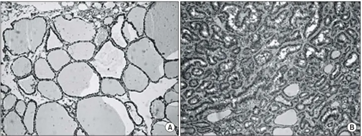
면역조직화학 염색 결과를 판정하기 위하여, 한 슬라이 드의 3 영역을 고배율(×100)에서 관찰한 후, 양성으로 염 색된 세포의 비율에 따라 염색 정도를 구별하였다. 양성으 로 염색된 세포가 전혀 없는 경우를 grade 0, 염색된 세포가 15% 이하인 경우를 grade I, 염색된 세포가 15∼50%인 경우 를 grade II, 50% 이상의 세포들이 염색된 경우를 grade III로 정의하였다(Fig. 1, 2). 최종적인 염색 정도는 3명의 독자적 인 관찰자가 판정 과정을 반복한 후 결정하였다.
결 과
Hsp70은 모든 검체 조직에서 발현되어 있었으나 조직에 따라 발현의 정도는 다양하였다. 정상 갑상선 조직은 75.0%
(12/17)가 grade I에 속해 있었으며, grade II 발현을 보인 경 우는 25.0% (4/17)였으나 grade III의 발현 정도는 한 예도 없었다(Table 1, Fig. 3). 양성 종양조직인 결절 증식증의 경 우 grade I 발현이 36.4% (4/11), grade II의 발현이 54.5%
(6/11), grade III 발현이 9.1% (1/11)를 보였으며, 또 다른 양 성 종양 조직인 여포성 선종의 경우에도 발현 정도의 양상 이 결절 증식증과 비슷하여 grade I 발현이 25.0% (3/12), grade II 발현이 58.3% (7/12), grade III 발현이 16.7% (2/12)를 보이면서, 정상 조직에 비하여는 grade I 발현보다 grade II
Fig. 1. Immunohistochemical staining for hsp70 in normal thyroid tissue (A) and papillary carcinoma tissue (B) (×100 for A and ×40 for B).
A B
발현이 많기는 하였으나 grade III의 발현이 되는 경우는 매 우 적었다(Table 1, Fig. 3). 반면, 유두상 암종의 경우는 grade I이 7.1% (1/14), grade II가 25.0% (4/14), grade III가 64.3%
(9/14)를 보여 정상 조직과 양성 종양 조직과는 대조적으로 grade III의 빈도가 매우 높았다.
정상조직에 대한 hsp90 발현 정도는 87.5% (14/16)가 grade 0에 속해 있었으며 나머지 12.5% (2/16)의 경우도
Fig. 2. Immunohistochemical staining for hsp90 in normal thyroid tissue (A) and papillary carcinoma tissue (B) (×100 for A and ×40 for B).
A B
Table 1. Results for hsp70 immunohistochemical staining in thyroid neoplasm
ꠚꠚꠚꠚꠚꠚꠚꠚꠚꠚꠚꠚꠚꠚꠚꠚꠚꠚꠚꠚꠚꠚꠚꠚꠚꠚꠚꠚꠚꠚꠚꠚꠚꠚꠚꠚꠚꠚꠚꠚꠚꠚꠚꠚꠚꠚꠚꠚꠚꠚꠚꠚꠚꠚꠚ Immunohistochemical grade
Pathologic type ꠏꠏꠏꠏꠏꠏꠏꠏꠏꠏꠏꠏꠏꠏꠏꠏꠏꠏꠏꠏꠏꠏꠏꠏꠏꠏ Total
0 I II III
ꠏꠏꠏꠏꠏꠏꠏꠏꠏꠏꠏꠏꠏꠏꠏꠏꠏꠏꠏꠏꠏꠏꠏꠏꠏꠏꠏꠏꠏꠏꠏꠏꠏꠏꠏꠏꠏꠏꠏꠏꠏꠏꠏꠏꠏꠏꠏꠏꠏꠏꠏꠏꠏꠏꠏ
Normal tissue 0 12 4 0 16
Nodular hyperplasia 0 4 6 1 11
Follicular adenoma 0 3 7 2 12
Papillary carcinoma 0 1 4 9 14
ꠏꠏꠏꠏꠏꠏꠏꠏꠏꠏꠏꠏꠏꠏꠏꠏꠏꠏꠏꠏꠏꠏꠏꠏꠏꠏꠏꠏꠏꠏꠏꠏꠏꠏꠏꠏꠏꠏꠏꠏꠏꠏꠏꠏꠏꠏꠏꠏꠏꠏꠏꠏꠏꠏꠏ
Table 2. Results for hsp90 immunohistochemical staining in thyroid neoplasm
ꠚꠚꠚꠚꠚꠚꠚꠚꠚꠚꠚꠚꠚꠚꠚꠚꠚꠚꠚꠚꠚꠚꠚꠚꠚꠚꠚꠚꠚꠚꠚꠚꠚꠚꠚꠚꠚꠚꠚꠚꠚꠚꠚꠚꠚꠚꠚꠚꠚꠚꠚꠚꠚꠚꠚ Immunohistochemical grade
Pathologic type ꠏꠏꠏꠏꠏꠏꠏꠏꠏꠏꠏꠏꠏꠏꠏꠏꠏꠏꠏꠏꠏꠏꠏꠏꠏ Total
0 I II III
ꠏꠏꠏꠏꠏꠏꠏꠏꠏꠏꠏꠏꠏꠏꠏꠏꠏꠏꠏꠏꠏꠏꠏꠏꠏꠏꠏꠏꠏꠏꠏꠏꠏꠏꠏꠏꠏꠏꠏꠏꠏꠏꠏꠏꠏꠏꠏꠏꠏꠏꠏꠏꠏꠏꠏ
Normal tissue 14 2 0 0 16
Nodular hyperplasia 11 0 0 0 11
Follicular adenoma 10 1 1 0 12
Papillary carcinoma 0 3 6 5 14
ꠏꠏꠏꠏꠏꠏꠏꠏꠏꠏꠏꠏꠏꠏꠏꠏꠏꠏꠏꠏꠏꠏꠏꠏꠏꠏꠏꠏꠏꠏꠏꠏꠏꠏꠏꠏꠏꠏꠏꠏꠏꠏꠏꠏꠏꠏꠏꠏꠏꠏꠏꠏꠏꠏꠏ
Fig. 4. Hsp90 expression in thyroid tissue by immnohistochemical grading.
Fig. 3. Hsp70 expression in thyroid tissue by immnohistochemical grading.
grade I의 발현만을 보였다(Table 2, Fig. 4). 양성 종양인 결 절 증식증은 100% (11/11) 모두 grade 0에 속해 있었으며, 여포성 선종 역시 대부분인 83.3% (10/12)가 grade 0에 속하 였고 grade I 과 grade II에 각각 8.3% (1/12)만이 분포되어 있었다(Table 2, Fig. 4). 반면, 유두상 암종은 정상 갑상선 조직과 양성 종양 조직과는 달리 hsp90 발현이 되지 않은 경우는 한 예도 없었으며, grade I에 21.4% (3/14), grade II에 42.9% (6/14), grade III에 35.7% (5/14)가 분포되어 다른 조직 의 발현 양상과 대조적인 양상을 보여주었다(Table 2, Fig. 4).
고 찰
Heat shock proteins은 분자량의 크기에 따라 small heat shock protein family, hsp70 family, hsp90 family, hsp90 family 로 크게 구분되며 1962년 처음 보고된 이래 열, 방사능, 저 산소증 등과 같은 스트레스 환경에 대응하여 세포를 보호 하는 역할을 하는 단백질로 알려져 왔다.(1) 임상적으로는 heat shock protiens과 면역 체계와의 관계를 이용한 암세포 면역 치료를 위한 물질로 많은 주목을 받아왔다.(11) 1995년 Jaattela(12)가 rodent cells을 이용한 실험 모델에서 heat shock proteins이 tumorigenic potential을 증가시키는 것을 보고한 이후, 최근에는 molecular chaperone으로서의 역할이 세포 내 신호전달경로, 세포조절 단백질과 연관이 있다는 사실 이 밝혀지고,(2,13) 몇몇 종양세포에서 이들 단백질 발현이 증가되어 있는 것이 보고되면서,(4-6,8,9) heat shock proteins 이 종양발생 과정에서도 일련의 역할을 하는 것으로 추측 되고 있다. Hsp70과 hsp90은 Ras/Raf-1을 통한 extracellular regulated kinases (ERK)의 활성화와 연관이 있는 것으로 추 측된다. 몇몇 연구 결과에 의하면 hsp90은 co-chaperone인 Cdc37을 통하여 Raf-1과 결합하며 Raf-1을 활성화시키는 데 중요한 역할을 한다.(14) Hsp70 역시 co-chaperone인 Bag 1 을 통하여 Raf-1을 활성화시킨다.(15) 또한, hsp70과 hsp90은 핵내 호르몬 수용체의 활성화에 필수적인 물질로 보고되고 있어 hsp70과 hsp90은 전사 과정의 조절 물질인 핵내 호르 몬 수용체(nuclear hormone receptors)의 활성 과정에서도 중 요한 역할을 한다고 생각한다.(16)
종양의 발생 기전을 계속적인 세포 성장 촉진에 초점을 두고 이해하여 왔던 경향은, 세포 성장을 촉진하는 기능 없 이 세포자멸사를 억제함으로써 종양 발생을 유도하는 bcl-2 의 역할이 밝혀지면서,(17) 종양발생 기전에 대한 다른 관 점에서의 접근을 시도하게 되는 계기를 맞이하게 되었으며 세포자멸사를 억제하는 유전자와 단백질에 대한 많은 연구 가 시작되었다. 세포자멸사 신호전달경로 과정의 결손은 악성종양에서 흔히 관찰되는 현상이며, 비정상적인 세포자 멸사 과정은 종양의 악성도와 연관이 있을 뿐 아니라 치료 약물에 대한 종양 세포의 저항성을 유도하기도 한다.(18) 지난 수년간의 세포자멸사에 대한 연구를 통하여 세포자멸
사 과정을 억제함으로써 종양발생을 초래하는 여러 단백질 들이 발견되었으며 Bcl-2 family와 inhibitor of apoptosis pro- tein (IAP) family 외에 heat shock protein 역시 이러한 기능을 갖고 있는 것으로 생각되고 있다. Hsp70은 heat shock proteins family 중 anti-apoptotic 기능을 가진 단백질로 분류된다.
Hsp70은 세포자멸사의 mitochondrial pathway 중 Apaf- 1과 결 합하여 apoptosome 형성을 억제하며, apoptosis inducing factor (AIF)를 억제하여 세포자멸사 과정을 차단한다.(19,20) 또한 death receptor pathway 과정에서 Fas를 통한 JNK pathway를 억제하며, proapoptotic 단백질인 p53, c-myc과도 연관이 있 는 것으로 보고되고 있다.(21,22) Hsp90 역시 hsp70과 마찬 가지로 Apaf-1과 결합하여 apoptosome의 형성을 억제한다 는 사실이 보고된 바 있으며, RIP (receptor- interacting protein), Akt/PKB 등을 활성화하여 NF-κB (nuclear factor-κ B)를 통한 세포 생존을 촉진시킨다는 사실이 보고되기도 하였다.(23-25)
임상적으로, heat shock proteins은 예후인자, 예측인자로 서의 가치뿐 아니라 항암치료제의 대상물질로서의 가능성 에 대하여 연구되고 있다. 유방암이나 자궁내막암 등에서 는 발현이 많이 될수록 악성도가 심하고, 생존율이 낮으며, 약물에 대한 반응률이 낮은 것으로 보고되었다.(26,27) 반 면, 골육종, 신장 종양의 경우에는 악성 정도와는 상관 관계 가 있으나 예후, 약물 반응 정도와는 역관계가 있는 것으로 보고된다.(5,28) Heat shock protein에 대한 억제제는 새로운 항암제로서의 가능성을 시험받고 있다. Hsp90에 대한 억제 제인 17-allylamino-17-demethyxy analogue of geldanymycin (17AAG)은 항암제로의 가능성을 인정받고 임상시험 단계 에 있다.(10,29)
본 연구는 아직 보고되지 않은 갑상선 조직과 heat shock proteins과의 관계를 알아보기 위하여 시행되었으며, 결과에 서 보여주듯이 갑상선의 정상조직, 양성종양조직, 악성종양 조직간의 heat shock proteins 발현 양상이 특징적인 것으로 확인되었다. Heat shock proteins에 대한 기존의 연구 결과와 본 연구 결과는 갑상선의 종양에 있어서의 heat shock pro- teins의 영향을 암시하는 것으로 생각된다. 따라서 이에 대 한 계속적이고 적극적인 연구는 종양발생 과정에 대한 이 해뿐 아니라 새로운 치료제의 가능성을 제시할 수 있을 것 으로 기대된다.
결 론
본 연구는 heat shock protien70과 heat shock protein90이 갑상선의 정상 조직, 양성 종양조직, 악성 종양조직간에 발 현양상의 차이를 보이는 지를 알아보기 위하여 시행되었 다. 한림대학교 성심병원에서 수술을 통하여 얻은 갑상선 의 각 조직에 대하여 hsp70과 hsp90의 발현 정도를 면역조 직화학 염색을 통하여 확인하였으며 각 조직에서의 발현
정도를 비교하였다. Hsp70은 모든 조직에서 발현이 되기는 하였으나, 정상 조직은 grade I의 발현 정도를 보이는 경우 가 가장 많았으며(75%), 양성 종양조직은 grade II 발현을 보이는 조직이 가장 많았으나(56.5%), 특이하게 정상조직과 양성종양조직은 grade III의 발현을 보이는 경우는 미미하 였다(정상조직; 0%, 양성종양조직; 13.0%). 반면에 악성 종 양조직의 경우 grade III 발현을 보이는 경우가 64.3%로 가 장 많아 정상 또는 양성 종양조직과는 대조를 이루었다.
Hsp90의 경우 이러한 차이는 더욱 현저하여, 정상조직과 양 성 종양조직의 대부분은 grade 0에 속하였으나(정상조직;
87.5%, 양성종양조직; 91.3%), 악성 종양조직의 경우 78.6%
가 grade II 또는 III의 발현을 보였다. 이러한 결과는 hsp70 과 hsp90의 종양발생 기전과의 개연성을 보여주는 것이며, 추가적인 연구를 통하여 종양 발생기전에 대한 이들의 역 할과 임상적 응용에 대한 가능성을 제시할 수 있을 것으로 생각된다.
REFERENCES
1) Ritossa F. A new puffing pattern incucedby temperature and DNP in Drosophila. Experimentia 1962;18:571-73.
2) Nollen EA, Morimoto RI. Chaperoning signaling pathways:
molecular chaperones as stress-sensing ‘heat shock' proteins.
J Cell Sci 2002;115:2809-16.
3) Shi Y, Thomas JO. The transport of proteins into the nucleus requires the 70-kilodalton heat shock protein or its cytosolic cognate. Mol Cell Biol 1992;12:2186-92.
4) Nanbu K, Konishi I, Mandai M, Kuroda H, Hamid AA, Komatsu T, et al. Prognostic significance of heat shock pro- teins HSP70 and HSP90 in endometrial carcinomas. Cancer Detect Prev 1998;22:549-55.
5) Santarosa M, Favaro D, Quaia M, Galligioni E. Expression of heat shock protein 72 in renal cell carcinoma: possible role and prognostic implications in cancer patients. Eur J Cancer 1997;33:873-7.
6) Jameel A, Skilton RA, Campbell TA, Chander SK, Coombes RC, Luqmani YA. Clinical and biological significance of HSP89 alpha in human breast cancer. Int J Cancer 1992;50:
409-15.
7) Li CY, Lee JS, Ko YG, Kim JI, Seo JS. Heat shock protein 70 inhibits apoptosis downstream of cytochrome c release and upstream of caspase-3 activation. J Biol Chem 2000;275:
25665-71.
8) Yufu Y, Nishimura J, Nawata H. High constitutive expression of heat shock protein 90 alpha in human acute leukemia cells.
Leuk Res 1992;16:597-605.
9) Hsu PL, Hsu SM. Abundance of heat shock proteins (hsp89, hsp60, and hsp27) in malignant cells of Hodgkin's disease.
Cancer Res 1998;58:5507-13.
10) Workman P. Pharmacogenomics in cancer drug discovery and
development: inhibitors of the Hsp90 molecular chaperone.
Cancer Detect Prev 2002;26:405-10.
11) Robert J. Evolution of heat shock protein and immunity. Dev Comp Immunol 2003;27:449-64.
12) Jaattela M. Over-expression of hsp70 confers tumorigenicity to mouse fibrosarcoma cells. Int J Cancer 1995;60:689-93.
13) Jaattela M. Escaping cell death: survival proteins in cancer.
Exp Cell Res 1999;248:30-43.
14) Grammatikakis N, Lin JH, Grammatikakis A, Tsichlis PN, Cochran BH. p50 (cdc37) acting in concert with Hsp90 is required for Raf-1 function. Mol Cell Biol 1999;19:1661-72.
15) Song J, Takeda M, Morimoto RI. Bag1-Hsp70 mediates a physiological stress signalling pathway that regulates Raf-1/
ERK and cell growth. Nat Cell Biol 2001;3:276-82.
16) Arbeitman MN, Hogness DS. Molecular chaperones activate the Drosophila ecdysone receptor, an RXR heterodimer. Cell 2000;101:67-77.
17) Vaux DL, Cory S, Adams JM. Bcl-2 gene promotes haemo- poietic cell survival and cooperates with c-myc to immortalize pre-B cells. Nature 1988;335:440-2.
18) Garrido C, Mehlen P, Fromentin A, Hammann A, Assem M, Arrigo AP, et al. Inconstant association between 27-kDa heat-shock protein (Hsp27) content and doxorubicin resistance in human colon cancer cells. The doxorubicin-protecting effect of Hsp27. Eur J Biochem 1996;237:653-9.
19) Saleh A, Srinivasula SM, Balkir L, Robbins PD, Alnemri ES.
Negative regulation of the Apaf-1 apoptosome by Hsp70. Nat Cell Biol 2000;2:476-83.
20) Beere HM, Wolf BB, Cain K, Mosser DD, Mahboubi A, Kuwana T, et al. Heat-shock protein 70 inhibits apoptosis by preventing recruitment of procaspase-9 to the Apaf-1 apopto- some. Nat Cell Biol 2000;2:469-75.
21) Jolly C, Morimoto RI. Role of the heat shock response and molecular chaperones in oncogenesis and cell death. J Natl Cancer Inst 2000;92:1564-72.
22) Park HS, Lee JS, Huh SH, Seo JS, Choi EJ. Hsp72 functions as a natural inhibitory protein of c-Jun N-terminal kinase.
Embo J 2001;20:446-56.
23) Pandey P, Saleh A, Nakazawa A, Kumar S, Srinivasula SM, Kumar V, et al. Negative regulation of cytochrome c-mediated oligomerization of Apaf-1 and activation of procaspase-9 by heat shock protein 90. Embo J 2000;19:4310-22.
24) Lewis J, Devin A, Miller A, Lin Y, Rodriguez Y, Neckers L, et al. Disruption of hsp90 function results in degradation of the death domain kinase, receptor-interacting protein (RIP), and blockage of tumor necrosis factor-induced nuclear factor- kappaB activation. J Biol Chem 2000;275:10519-26.
25) Cardone MH, Roy N, Stennicke HR, Salvesen GS, Franke TF, Stanbridge E, et al. Regulation of cell death protease caspase-9 by phosphorylation. Science 1998;282:1318-21.
26) Vargas-Roig LM, Fanelli MA, Lopez LA, Gago FE, Tello O, Aznar JC, et al. Heat shock proteins and cell proliferation in
human breast cancer biopsy samples. Cancer Detect Prev 1997;21:441-51.
27) Vargas-Roig LM, Gago FE, Tello O, Aznar JC, Ciocca DR.
Heat shock protein expression and drug resistance in breast cancer patients treated with induction chemotherapy. Int J Cancer 1998;79:468-75.
28) Trieb K, Lechleitner T, Lang S, Windhager R, Kotz R, Dirnhofer S. Heat shock protein 72 expression in osteosar- comas correlates with good response to neoadjuvant chemo- therapy. Hum Pathol 1998;29:1050-5.
29) Neckers L. Hsp90 inhibitors as novel cancer chemotherapeutic agents. Trends Mol Med 2002;8:S55-61.