ORIGINAL ARTICLE
위장관기질종양과 구별되는 위 신경초종의 초음파 내시경 특징
박형철, 손동준, 오형훈, 옥찬영, 김미영, 정조윤, 명대성, 김종선, 조성범, 이완식, 주영은
전남대학교 의과대학 내과학교실 소화기내과
Endoscopic Ultrasonographic Characteristics of Gastric Schwannoma Distinguished from Gastrointestinal Stromal Tumor
Hyung-Chul Park, Dong-Jun Son, Hyung-Hoon Oh, Chan-Young Oak, Mi-Young Kim, Cho-Yun Chung, Dae-Seong Myung, Jong-Sun Kim, Sung-Bum Cho, Wan-Sik Lee and Young-Eun Joo
Division of Gastroenterology, Department of Internal Medicine, Chonnam National University Medical School, Gwangju, Korea Background/Aims: Gastric schwannoma (GS), a rare neurogenic mesenchymal tumor, is usually benign, slow-growing, and asymptomatic. However, GS is often misdiagnosed as gastrointestinal stromal tumors (GIST) on endoscopic and radiological examinations. The purpose of this study was to evaluate EUS characteristics of GS distinguished from GIST.
Methods: A total of 119 gastric subepithelial lesions, including 31 GSs and 88 GISTs, who were histologically identified and underwent EUS, were enrolled in this study. We evaluated the EUS characteristics, including location, size, gross morphology, mucosal lesion, layer of origin, border, echogenic pattern, marginal halo, and presence of an internal echoic lesion by retrospective review of the medical records.
Results: GS patients comprised nine males and 22 females, indicating female predominance. In the gross morphology according to Yamada’s classification, type I was predominant in GS and type III was predominant in GIST. In location, GSs were predominantly located in the gastric body and GISTs were predominantly located in the cardia or fundus. The frequency of 4th layer origin and isoechogenicity as compared to the echogenicity of proper muscle layer was significantly more common in GS than GIST.
Although not statistically significant, marginal halo was more frequent in GS than GIST. The presence of an internal echoic lesion was significantly more common in GIST than GS.
Conclusions: The EUS characteristics, including tumor location, gross morphology, layer of origin, echogenicity in comparison with the normal muscle layer, and presence of an internal echoic lesion may be useful in distinguishing between GS and GIST. (Korean J Gastroenterol 2015;65:21-26)
Key Words: Endoscopy; Ultrasonography; Stomach; Schwannoma; Gastrointestinal stromal tumors
Received December 9, 2014. Revised January 2, 2015. Accepted January 6, 2015.
CC This is an open access article distributed under the terms of the Creative Commons Attribution Non-Commercial License (http://creativecommons.org/licenses/
by-nc/3.0) which permits unrestricted non-commercial use, distribution, and reproduction in any medium, provided the original work is properly cited.
Copyright © 2015. Korean Society of Gastroenterology.
교신저자: 주영은, 501-757, 광주시 동구 백석로 160, 전남대학교 의과대학 내과학교실 소화기내과
Correspondence to: Young-Eun Joo, Division of Gastroenterology, Department of Internal Medicine, Chonnam National University Medical School, 160 Baekseok-ro, Dong-gu, Gwangju 501-757, Korea. Tel: +82-62-220-6296, Fax: +82-62-225-8578, E-mail: yejoo@chonnam.ac.kr
Financial support: None. Conflict of interest: None.
INTRODUCTION
Subepithelial lesions of the stomach are found in- cidentally, occurring in approximately 0.36% of screening up- per endoscopy. Most gastric subepithelial lesions are mesen- chymal tumors. The entities responsible for mesenchymal tu-
mors of the stomach include gastrointestinal stromal tumor (GIST), leiomyoma, leiomyosarcoma, lipoma, schwannoma, and so on. Of these, GIST is the most common mesenchymal tumor of the stomach with a malignant potential.1,2
Gastric schwannoma (GS) is a rare neurogenic mesen- chymal tumor. This tumor is usually benign, slow-growing,
Table 1. Baseline Characteristics of the Patients with GSs and GISTs of the Stomach
Variable GS (n=31) GIST (n=88) p-value
Gender 0.092
Male 9 (29.0) 42 (47.7)
Female 22 (71.0) 46 (52.3)
Age (yr) 58.13±9.219 58.09±11.433 0.987
Gastric symptom 0.455
Absent 26 (83.9) 67 (76.1)
Present 5 (16.1) 21 (23.9)
Values are presented as n (%) or mean±SD.
GS, gastric schwannoma; GIST, gastrointestinal stromal tumor.
and asymptomatic, with extremely low malignant potential and an excellent prognosis after surgical resection.3,4 However, GS is often misdiagnosed as GIST on endoscopic and radio- logical examinations. Therefore, accurate differential diag- nosis of GS and GIST has important prognostic and ther- apeutic implications.
EUS is the most reliable procedure for assessing the tu- mor’s layer of origin, the exact size of the lesion, morphologic features, differential diagnosis, classification, and follow up of gastric subepithelial lesions. Therefore, a better under- standing of its unique features for differential diagnosis may be helpful in providing effective intervention strategies and guide selection of appropriate therapy.
To date, case series describing EUS features in only four GS patients, respectively, have been reported.5,6 The purpose of this study was to evaluate EUS characteristics of GS dis- tinguished from GIST.
SUBJECTS AND METHODS
We searched the pathologic database at Department of Gastroenterology, Chonnam National University Hwasun Hospital (Hwasun, Korea) to find patients with histologically proven gastric subepithelial lesions between January 2004 and December 2013. A total of 573 gastric subepithelial le- sions, including 283 GISTs (49.4%), 90 leiomyomas (15.7%), 60 ectopic pancreas (10.5%), 31 schwannomas (5.4%), and 15 carcinoids (2.6%) were identified histologically by surgical resection. Among them, patients with GS and GIST who un- derwent EUS examination were enrolled in this study;
EUS-guided fine-needle aspiration or trucut biopsy was not performed. The final study population consisted of 31 GS and 88 GIST patients. Subsequently, we searched the medical da- tabase and the following information was retrieved for analy- sis: (1) age, sex, and symptoms of the patients, (2) EUS char- acteristics including location, size, gross morphology classi- fied according to Yamada’s classification,7 mucosal lesion, layer of origin, border, echogenic pattern including echoge- nicity, homogenecity and comparison to the echogenicity of proper muscle layer, marginal halo, and presence of an in- ternal echoic lesion including cyst, hyperechogenic spot, and calcification. This study was reviewed and approved by the Institutional Review Board of Chonnam National University Hwasun Hospital, and written informed consent was ob-
tained from all participating subjects for retrospective review of the patients’ medical records and images. EUS examina- tion was performed using a mechanical radial-scan- ning-echoendoscope (GF-UM2000; Olympus, Tokyo, Japan).
The scanning frequency ranged from 5 to 20 MHz. All exami- nations were performed by 1 of 4 experienced endo- sonographers who had performed more than 150 diagnostic EUS examinations.
The SPSS software version 15.0 (SPSS Inc., Chicago, IL, USA) was used for data analysis employing the χ2 and Fisher’s exact test. A value of p<0.05 was considered stat- istically significant.
RESULTS
1. Baseline characteristics of GS and GIST patients The final enrolled population consisted of 31 GS and 88 GIST patients. The baseline characteristics of enrolled sub- jects, including age, sex, and gastrointestinal (GI) symptoms are described in Table 1. The mean age of GS patients was 58.1±9.2 (mean±SD) with a range from 40 to 73 years. GS patients comprised nine males (29.0%) and 22 females (71.0%), indicating female predominance. Most GS patients were asymptomatic (83.9%) and five patients (16.1%) pre- sented with epigastric discomfort. However, no significant differences in the baseline characteristics were observed be- tween the GS and GIST groups.
2. Endoscopic characteristics of GS and GIST
The endoscopic characteristics of GS and GIST patients are summarized in Table 2. All GSs were round to ovoid shape and there was no lobulated shape. The five GSs (16.1%) had
Table 2. Endoscopic Features of the Patients with GSs and GISTs of the Stomach
Variable GS (n=31) GIST (n=88) p-value
Shape 0.062
Round to ovoid 31 (100) 78 (88.6)
Lobulated 0 (0) 10 (11.4)
Mucosal lesion 1.000
Present 5 (16.1) 14 (15.9)
Absent 26 (83.9) 74 (84.1)
Morphology
(Yamada’s classification)a
0.030
Type I 16 (51.6) 26 (29.5)
Type II 12 (38.7) 35 (39.8)
Type III 3 (9.7) 27 (30.7)
Type IV 0 (0) 0 (0)
Location 0.001
Body, GC 19 (61.3) 21 (23.9)
Body, LC 9 (29.0) 24 (27.3)
Cardia/fundus 1 (3.2) 37 (42.0)
Antrum 2 (6.5) 6 (6.8)
Values are presented as n (%).
aYamada type I is elevated, with an indistinct border; Type II is elevated with a distinct border at the base but no notch; Type III is elevated, but no peduncle; Type IV is pedunculated and elevated.
GS, gastric schwannoma; GIST, gastrointestinal stromal tumor; GC, greater curvature; LC, lesser curvature.
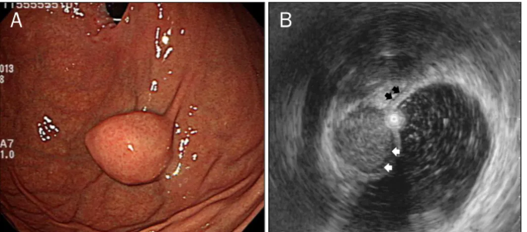
Fig. 1. Endoscopic (A) and endosono- graphic (B) findings in a 59-year-old woman with a gastric schwannoma. (A) Endoscopy shows a submucosal ele- vated lesion with type I morphology according to Yamada’s classification in the greater curvature of the body.
(B) On EUS, the mass is homogeneous and its echogenicity is similar to that of the normal proper muscle layer (black arrows). It measures 32.0×21.0 mm in size and a marginal halo (white arrows) is observed.
mucosal lesions such as central ulceration (n=2), central de- pression (n=1), erosion (n=1), and umbilication (n=1). In the gross morphology of GSs according to Yamada’s classi- fication, 16 (51.6%) were type I, 12 (38.7%) type II, and 3 (9.7%) type III; 28 (90.3%) GSs were located in the gastric body (19 [61.3%] in the greater curvature [GC] and 9 [29.0%]
in the lesser curvature of the body), followed by antrum (n=2) and cardia or fundus (n=1). In GISTs patients, the gross mor- phology according to Yamada’s classification was predom- inant type III (n=35, 39.8%) and the most common location
was the cardia or fundus. Comparing GSs with GIST patients, the gross morphology and location was significantly different (p=0.030 and p<0001, respectively) (Fig. 1).
3. Endosonographic characteristics of GS and GIST The endosonographic characteristics of GS and GIST pa- tients are summarized in Table 3. The mean size of GSs was 26.0±8.4 (mean±SD) with a range from 12 to 42 mm. All GSs originated from the fourth layer, with a connection between the tumor and the muscularis propria and had a distinct border. In echogenic patterns of the GSs, 17 (54.8%) ex- hibited homogeneous hypoechogenicity and 14 (45.2%) were heterogeneous hypoechogenicity; 22 GSs (71.0%) had a marginal hypoechoic halo; 22 GSs (71.0%) exhibited iso- echogenicity and 9 (29.0%) exhibited hyperechogenicity, compared to the echogenicity of surrounding proper muscle layer (Fig. 1B). Only three GSs had internal echoic lesions in- cluding cystic change (n=2) and hyperechogenic spot (n=1).
GISTs originated from the 4th layer in 76 patients (86.4%) and 3rd layer in 12 patients (13.6%); 42 GISTs (47.7%) exhibited isoechogenicity and 46 (52.3%) exhibited hyperechogenicity, compared to the echogenicity of the normal proper muscle layer (Fig. 2B). The frequency of 4th layer origin and iso- echogenicity compared to the echogenicity of the normal proper muscle layer was significantly more common in GS than GIST (p=0.035 and p=0.036, respectively); 44 GISTs (50.0%) had a marginal hypoechoic halo. Although not stat- istically significant, the frequency of marginal halo was great- er in GS than GIST (p=0.058). The presence of an internal echoic lesion including cystic change (n=14, 15.9%), hyper- echogenic spot (n=22, 25.0%), and calcification (n=9, 10.2%) was significantly more common in GIST than GS (p=0.036).
Fig. 2. Endoscopic (A) and endosono- graphic (B) findings in a 66-year-old woman with a gastrointestinal stromal tumor. (A) Endoscopy shows a submu- cosal elevated lesion with type III morphology according to Yamada’s classification in the cardia. (B) On EUS, the mass is homogeneous and its echogenicity is higher than that of the normal proper muscle layer (black arrows). It measures 20.0×16.0 mm in size and a marginal halo (white arrows) is observed.
Table 3. Endoscopic Ultrasonography Features of the Patients with GSs and GISTs of the Stomach
Variable GS (n=31) GIST (n=88) p-value
Size (mm) 26.03±8.373 25.20±15.665 0.780
Layer 0.035
3rd 0 (0) 12 (13.6)
4th 31 (100) 76 (86.4)
Border 0.110
Regular 31 (100) 80 (90.9)
Irregular 0 (0) 8 (9.1)
Marginal halo 0.058
Absent 9 (29.0) 44 (50.0)
Present 22 (71.0) 44 (50.0)
Echogenicity 0.408
Homogeneous hypoechoic 17 (54.8) 40 (45.5)
Heterogeneous hypoechoic 14 (45.2) 48 (54.5)
Echogenicity in comparison with the surrounding muscle echo
0.036
Isoechoic 22 (71.0) 42 (47.7)
Hyperechoic 9 (29.0) 46 (52.3)
Internal echoic lesion
Cystic change 1 (3.2) 14 (15.9) 0.112
Hyperechogenic spots 2 (6.5) 22 (25.0) 0.036
Calcification 0 (0) 9 (10.2) 0.110
Values are presented as mean±SD or n (%).
GS, gastric schwannoma; GIST, gastrointestinal stromal tumor.
DISCUSSION
Schwannomas are spindle cell mesenchymal tumors origi- nating from any nerve having a Schwann cell sheath. In the GI tract, GISTs comprise the largest group of mesenchymal tumors, whereas schwannomas are rare, with reported prev- alence ranging from 3.3-12.8% of all GI mesenchymal tumors.4,8,9 In our study, prevalence of GS was 5.4% (31 GS patients among a total of 573 patients) and the tumors oc- curred predominantly in older adults with a marked female predominance. The majority of GSs follow a benign clinical
course and malignant transformation is extremely rare, as a few cases have been reported in the literature.10,11
In clinical practice, preoperative differential diagnosis be- tween mesenchymal tumors of the stomach is usually difficult. GSs are rare benign mesenchymal tumors of the stomach. However, because of different prognostic and ther- apeutic implications, it is necessary to differentiate GSs from other mesenchymal tumors of the stomach, particularly GISTs with malignant potential. Endoscopically, GSs appear as elevated lesions, with or without central ulcers. In several studies, the location of the tumors was predominant in GC of
the body of the stomach.4,6 In our study, GSs showed statisti- cally significant predominance in the GC of the body, com- pared with GISTs. In addition, previous studies showed that GSs had an exophytic growth pattern rather than an endolu- minal growth pattern.12-14 In our study, the gross finding ac- cording to Yamada’s classification was a predominance of type I, classified by an elevated lesion with an indistinct bor- der and it was statistically significant, compared with GISTs.
There are no previous reports describing the gross finding ac- cording to Yamada’s classification in GSs or GISTs. Although our study population was relatively small, which might influ- ence some results, this finding may be the endoscopic fea- ture, reflecting GSs with an exophytic growth pattern.
The features of GSs on EUS have been described as round submucosal lesions with marginal halo, and homogeneous internal echogenicity without internal echogenic foci, arising from the 4th layer.6,15 Some reports have suggested that the echogenicity of GSs compared with the normal proper mus- cle layers may be helpful for differentiating them from GISTs.5,6 One study suggested that the echogenicity of GSs was much lower than that of the normal proper muscle layers.6 In other case reports, the internal echogenicity of GSs was heterogeneous and low, but slightly higher than that of muscularis propria, with internal patch high echo.5 According to our results, isoechogenicity in comparison with the normal proper muscle layer was predominantly found in 71.0% of GSs, whereas hyperechogenicity was predominantly found in 52.3% of GISTs. Pathologically, GISTs have high cellularity with a basophilic appearance on H&E, whereas GSs have moderate overall cellularity.4,16-18 In addition, the degener- ative changes, including hemorrhage, necrosis, and cystic change, which are often seen in soft-tissue schwannomas, were not the common features of GI schwannomas, although these tumors grossly resemble soft-tissue schwannomas.9 However, hemorrhage, necrosis, and cystic change are the common features in GISTs and calcification is seen in 6% of GISTs.19 In our study, the presence of an internal echoic le- sion including cystic change, hyperechogenic spot, and calci- fication was significantly more common in GIST than GS.
These differences in echogenicity and the presence of an in- ternal echoic lesion between GSs and GISTs might reflect the pathologic differences of cellularity and the structural com- ponents of the tumors.
In previous studies a marginal halo, corresponding to his-
topathologically lymphoid cuff was observed on EUS in GSs and GISTs.4,15 Recently, although small case series, a margin- al halo was found in the majority of GSs.5,6,15 In our study, mar- ginal halo was found in 71.0% of GSs and 50.0% in GISTs.
Although not statistically significant, the frequency of mar- ginal halo was greater in GSs than GISTs.
Our study had several limitations. First, this was a retro- spective study comparing the EUS features of GISTs and GSs.
In addition, there might have been a potential bias when ret- rospectively reviewing the EUS photos. Second, although EUS examinations were performed, patients were selected for surgery according to the clinical opinions of the many doc- tors including internist and surgeon. Third, in addition to the GS, preoperative differentiation between GISTs and gastric leiomyomas is also important. However, the gastric leiomyo- ma was not included in our study. In previous studies, a mar- ginal halo appeared more frequently in GISTs than in leiomyo- ma and the echogenicity of the leiomyoma was similar to that of the normal proper muscle layer.15,20 These results demon- strate that the EUS features of leiomyomas and GSs have many similarities. Therefore, in the future, conduct of large- scale studies should be considered in order to determine the differential diagnostic points of many gastric mesenchymal tumors.
In summary, GSs were predominantly type I according to Yamada’s classification and were predominantly located in the gastric body, compared with GISTs. The EUS features such as homogeneous hypoechogenicity, a well-demarcated margin, fourth-layer origination, and lack of cystic change, hy- perechogenic spot and calcification, isoechogenicity com- pared to the echogenicity of normal proper muscle layer may be helpful in differentiation of GSs from GISTs. In our study, a marginal halo was the common feature of GSs, but not an essential one for differentiation GSs from GISTs.
REFERENCES
1. Papanikolaou IS, Triantafyllou K, Kourikou A, Rösch T. Endosco- pic ultrasonography for gastric submucosal lesions. World J Gastrointest Endosc 2011;3:86-94.
2. Song JH, Kim JI, Kim HJ, et al. Endoscopic characteristics of up- per gastrointestinal mesenchymal tumors originating from mus- cularis mucosa or muscularis propria. Korean J Gastroenterol 2013;62:92-96.
3. Hou YY, Tan YS, Xu JF, et al. Schwannoma of the gastrointestinal tract: a clinicopathological, immunohistochemical and ultra-
structural study of 33 cases. Histopathology 2006;48:536-545.
4. Voltaggio L, Murray R, Lasota J, Miettinen M. Gastric schwanno- ma: a clinicopathologic study of 51 cases and critical review of the literature. Hum Pathol 2012;43:650-659.
5. Zhong DD, Wang CH, Xu JH, Chen MY, Cai JT. Endoscopic ultra- sound features of gastric schwannomas with radiological corre- lation: a case series report. World J Gastroenterol 2012;18:
7397-7401.
6. Jung MK, Jeon SW, Cho CM, et al. Gastric schwannomas: endo- sonographic characteristics. Abdom Imaging 2008;33:388-390.
7. Yamada T, Ichikawa H. X-ray diagnosis of elevated lesions of the stomach. Radiology 1974;110:79-83.
8. Daimaru Y, Kido H, Hashimoto H, Enjoji M. Benign schwannoma of the gastrointestinal tract: a clinicopathologic and immuno- histochemical study. Hum Pathol 1988;19:257-264.
9. Kwon MS, Lee SS, Ahn GH. Schwannomas of the gastrointestinal tract: clinicopathological features of 12 cases including a case of esophageal tumor compared with those of gastrointestinal stromal tumors and leiomyomas of the gastrointestinal tract.
Pathol Res Pract 2002;198:605-613.
10. Loffeld RJ, Balk TG, Oomen JL, van der Putten AB. Upper gastro- intestinal bleeding due to a malignant Schwannoma of the stomach. Eur J Gastroenterol Hepatol 1998;10:159-162.
11. Gennatas CS, Exarhakos G, Kondi-Pafiti A, Kannas D, Athanassas G, Politi HD. Malignant schwannoma of the stomach in a patient with neurofibromatosis. Eur J Surg Oncol 1988;14:261-264.
12. Levy AD, Quiles AM, Miettinen M, Sobin LH. Gastrointestinal schwannomas: CT features with clinicopathologic correlation.
AJR Am J Roentgenol 2005;184:797-802.
13. Hong HS, Ha HK, Won HJ, et al. Gastric schwannomas: radio- logical features with endoscopic and pathological correlation.
Clin Radiol 2008;63:536-542.
14. Choi JW, Choi D, Kim KM, et al. Small submucosal tumors of the stomach: differentiation of gastric schwannoma from gastro- intestinal stromal tumor with CT. Korean J Radiol 2012;13:425- 433.
15. Okai T, Minamoto T, Ohtsubo K, et al. Endosonographic evalua- tion of c-kit-positive gastrointestinal stromal tumor. Abdom Imaging 2003;28:301-307.
16. Pidhorecky I, Cheney RT, Kraybill WG, Gibbs JF. Gastrointestinal stromal tumors: current diagnosis, biologic behavior, and management. Ann Surg Oncol 2000;7:705-712.
17. Miettinen M, Sobin LH, Sarlomo-Rikala M. Immunohistochem- ical spectrum of GISTs at different sites and their differential di- agnosis with a reference to CD117 (KIT). Mod Pathol 2000;13:
1134-1142.
18. Sarlomo-Rikala M, Kovatich AJ, Barusevicius A, Miettinen M.
CD117: a sensitive marker for gastrointestinal stromal tumors that is more specific than CD34. Mod Pathol 1998;11:728-734.
19. Miettinen M, Sobin LH, Lasota J. Gastrointestinal stromal tu- mors of the stomach: a clinicopathologic, immunohistochem- ical, and molecular genetic study of 1765 cases with long-term follow-up. Am J Surg Pathol 2005;29:52-68.
20. Kim GH, Park do Y, Kim S, et al. Is it possible to differentiate gas- tric GISTs from gastric leiomyomas by EUS? World J Gastroenter- ol 2009;15:3376-3381.