Received November 4, 2014, Revised November 24, 2014, Accepted November 27, 2014 Corresponding author: Jin-bong Choi
Department of Oriental Rehabilitation Medicine, Dongshin University Oriental Medicine, 141 Wolsan-ro, Nam-gu, Gwangju 503-232, Korea Tel: +82-62-350-7114, Fax: +82-62-350-7551, E-mail: [email protected]
CCThis is an open access article distributed under the terms of the Creative Commons Attribution Non-Commercial License (http://creativecommons.org/licenses/ by-nc/3.0) which permits unrestricted non-commercial use, distribution, and reproduction in any medium, provided the original work is properly cited.
침 치료가 신경 재생 및 회복에 미치는 영향에 대한 연구 동향:
PubMed를 중심으로
양미성ㆍ김선종ㆍ최진봉
동신대학교 한의과대학 한방재활의학과
Current Research Trend on Acupuncture Treatment for Nerve Regeneration and Recovery:
Based on the Data of PubMed
Mi-sung Yang, Sun-jong Kim, Jin-bong Choi
Department of Oriental Rehabilitation Medicine, Dongshin University Oriental Medicine
Objectives : The purpose of this study is to explore the current research trend on acupuncture treatment for nerve regeneration and recovery effect. Methods : We investigated the researches so far, on acupuncture treatment for the nerve regeneration and recovery via searching Pubmed from 2005 up to October 2014. Data were extracted from the included studies regarding the authors, countries, type of nerve injury, type of acupuncture, treatment period, acupuncture points, assessment tool and results.
Results and Conclusions : Twenty-four research papers were included in the review. Outcomes were measured by immu- nohistochemical results, motor behavior scores, and electrocphysiological results. All but one study favored acupuncture and electroacupuncture treatment for nerve regeneration and recovery regardless of type of nerve injury and acupuncture modality.
Acupuncture treatment may have a potential for nerve regeneration and recovery and further research is required.
Key words : Acupuncture, Nerve recovery, Nerve regeneration, Neurotrophins, Pubmed
서 론
신경계는 크게 뇌, 척수를 포함하는 중추 신경계(Central Ner- vous System; CNS)와 말초 신경계(Peripheral Nervous System;
PNS)로 나눌 수 있다. 이러한 신경계는 노화, 질병, 상해 등에 의해 손상되기도 하는데 자연적, 혹은 약물을 이용한 재생이 힘들고 이 에 따라 치유가 어렵거나 후유증이 남는 경우가 많다1).
국내에서 허혈성 뇌손상은 가장 흔한 장애의 원인 중 하나이며2) 최근 들어 노년층 인구 급증으로 치매, 중풍, 파킨슨병과 같은 퇴행
성 신경계 질환이 빠른 속도로 증가하고 있다3).
또한, 2011년 장애인 실태조사의 결과를 재분석한 추정치로는 지체 장애인의 4.4%인 약 60,000명이 척수 손상이고, 그 중에서도 80%가 외상에 의한 손상으로 이러한 중추신경계의 손상은 대부분 비가역적이며 사실상 완전 회복이 어려워 경제적, 사회적 손실이 매우 크다4).
말초 신경의 손상은 뇌, 척수의 중추신경계를 제외한 나머지 신 경계의 손상을 통틀어 이르는 용어로 마찬가지로 외상이나 질병, 대사 장애, 약물, 독소, 유전적 원인 등 다양한 원인에 의해 손상될
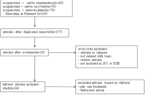
Fig. 1. Flow chart of selection process.
수 있다. 말초 신경 손상의 경우, 느리더라도 축삭의 느린 재생을 동반하여 회복을 일으키고5), 중추 신경계의 손상 또한 신경 재생이 불가능하다고 여겼던 과거와 달리, 신경가소성에 의해 신경재활이 가능하다고 여겨지고 있다6).
그러나 중추 신경이든, 말초 신경이든, 일단 손상된 후에 근본적 이고 완전한 신경의 재생을 일으키는 치료는 현재까지 딱히 없는데 일례로, 서양의학에서 척수손상의 치료는 수술 및 스테로이드 약물 을 이용한 합병증 방지와 기능회복이 주가 되며 치료 결과는 만족 할 만한 수준은 못되는 것으로 평가된다7,8). 최근 줄기세포를 이용 하거나 신경성장인자를 투여하고 축삭의 성장을 돕는 세포 이식 방법이 활용되고 있으나 아직은 널리 상용화되지는 못하고 있다9). 이처럼 아직까지 신경재생에 대한 근본적 치료가 없는 가운데 침 치료가 신경손상 후 재생 및 회복에 미치는 영향에 대한 국내연
구10-14)는 꾸준히 발표되어 왔으나 그 수가 많지 않아 결과를 논하
기에는 미흡한 실정이다. 이에 저자는 실제로 침 치료가 손상된 신 경을 재생하고 회복하는데 연관성이 있는지와 어떠한 측면에서 주 로 영향을 주는지에 대해 해외 연구 동향을 살펴보았다.
연구방법
1. 논문 검색
영문 문헌 검색을 위하여 미국국립의학도서관의 논문 및 자료 검색엔진인 PubMed(www.pubmed.gov)를 기본 대상으로 하였 다. 검색어는 ʻacupunctureʼ와 ʻnerve regenerationʼ을 기본 검색
어로 하고 ʻnerve recoveryʼ, ʻneurotrophinsʼ 등을 추가로 검색하 여 논문을 취합하였으며 최근 동향을 알아보기 위해 2005년 1월 1일 이후로 발표한 논문을 검색하였다.
검색을 통해 나온 논문들 중 중복되거나 침 치료와 신경의 재생 혹은 회복을 동시에 만족하지 않는 논문, 침 치료나 신경 회복에 관한 언급이 직접적인 주제가 아니라 간접적으로 언급된 논문, 침 치료가 단 1회로 설계된 논문, 원문이 영어 이외의 언어로 되어 있어 본문을 읽을 수 없는 논문, 실험 연구나 임상 연구가 아닌 리뷰논문, Journal Citation Report(JCR) edition 2013(http://
www.isiknowledge.com/JCR)에서 제공하는 2013년 SCI 혹은 SCIE에 포함되지 않은 논문들은 제외하고 원문을 찾아 총 24편의 논문을 연구하였다(Fig. 1).
2. 연구 방법
총 24편의 논문을 게재지, 1저자의 발표국가, 신경 손상의 유형, 연구의 종류 및 연구 대상, 침 치료의 종류 및 자극의 세기, 연구 기간과 치료횟수, 치료 혈자리의 선택, 연구 결과 요약 및 평가 도 구 부문으로 분류하고 살펴보았다.
결 과
1. 게재지 및 발표국가
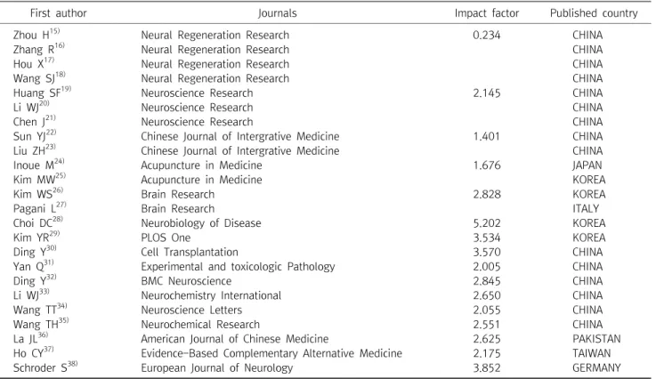
총 24편의 논문 중 Neural Regeneration Research(Neural Regen Res)에 4편15-18) Neuroscience Research(Neurosci Res)에
First author Journals Impact factor Published country
Zhou H15) Neural Regeneration Research 0.234 CHINA
Zhang R16) Neural Regeneration Research CHINA
Hou X17) Neural Regeneration Research CHINA
Wang SJ18) Neural Regeneration Research CHINA
Huang SF19) Neuroscience Research 2.145 CHINA
Li WJ20) Neuroscience Research CHINA
Chen J21) Neuroscience Research CHINA
Sun YJ22) Chinese Journal of Intergrative Medicine 1.401 CHINA
Liu ZH23) Chinese Journal of Intergrative Medicine CHINA
Inoue M24) Acupuncture in Medicine 1.676 JAPAN
Kim MW25) Acupuncture in Medicine KOREA
Kim WS26) Brain Research 2.828 KOREA
Pagani L27) Brain Research ITALY
Choi DC28) Neurobiology of Disease 5.202 KOREA
Kim YR29) PLOS One 3.534 KOREA
Ding Y30) Cell Transplantation 3.570 CHINA
Yan Q31) Experimental and toxicologic Pathology 2.005 CHINA
Ding Y32) BMC Neuroscience 2.845 CHINA
Li WJ33) Neurochemistry International 2.650 CHINA
Wang TT34) Neuroscience Letters 2.055 CHINA
Wang TH35) Neurochemical Research 2.551 CHINA
La JL36) American Journal of Chinese Medicine 2.625 PAKISTAN
Ho CY37) Evidence-Based Complementary Alternative Medicine 2.175 TAIWAN
Schroder S38) European Journal of Neurology 3.852 GERMANY
Table 1. Journals and Published Country of 24 Articles
3편19-21), Chinese Journal of Intergrative Medicine(Chin J Integr Med)에 2편22,23), Acupuncture in Medicine(Acu in Med)
에 2편24,25) Brain Research(Brain Res)26,27)순으로 게재되었고 나
머지 논문들은 각각 1편씩 총 16종의 다양한 분야의 저널에 게재되 었다.
1저자를 기준으로 발표 국가를 살펴보았을 때, 중국15-23,30-35)
이 15편으로 가장 많았고, 그 외에 한국25,26,28,29)
이 4편, 일본24), 이탈 리아27) 파키스탄36), 대만37), 독일38)이 각각 1편이었다(Table 1).
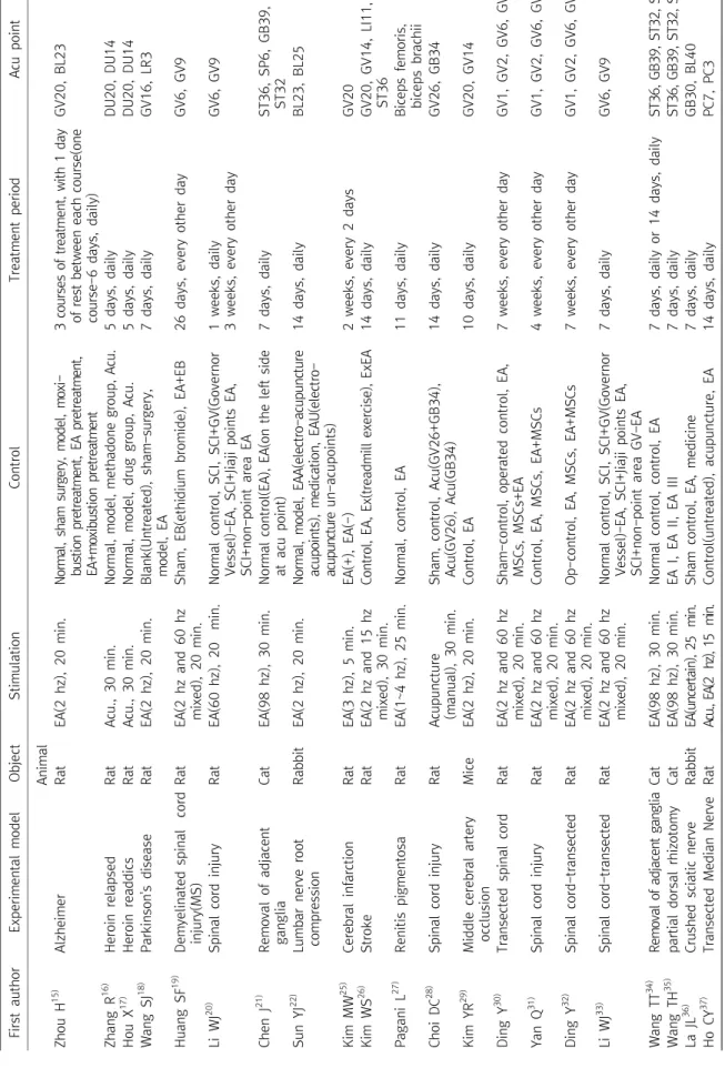
2. 신경손상의 유형 및 연구 대상
손상된 신경의 부위는 척수손상(Spinal cord injury; SCI)19-22,28,30-35)
가 11건으로 가장 많았고 뇌손상의 경우 각각 허혈성 뇌손상25,26,29), 알츠하이머15), 파킨슨병18), 헤로인 중독16,17), 뇌성마비23) 등 다양한 원인으로 총 8건이었다. 그 외 특발성 말초신경 손상24,38), 망막색소 변성증27), 좌골신경 손상36), 정중신경 손상37) 등의 경우가 있었다.
연구 대상은 실험용 rat 혹은 mice가 16편, cat 3건, rabbit이 2건, 환자를 대상으로 한 연구가 3건이었다(Table 2).
3. 침 치료의 방법
실험 논문의 18편이 전침15,18-22,24-27,29-36)
만을 사용했고, 침 치
료16,17,23,28,38)
를 단독 사용한 것이 5편, 침과 전침37)을 병용하여 비 교한 논문도 1편 있었다.
전침의 자극 정도는 2 Hz15,18,22,29)
, 3 Hz25), 60 Hz20), 98 Hz21,34,35), 100 Hz24), 2 Hz와 60 Hz19,30-33), 2 Hz와 15 Hz26)의 혼합된 정도로 다양하였으며 2 Hz의 저주파가 가장 많았고 100 Hz를 넘지 않는 정도의 주파수로 시행되었다.
침과 전침 치료의 유침 시간은 5분25), 15분37), 20분15,18-20,22,24,29-33,38)
, 25분27,36), 30분16,17,21,23,26,28,34,35)
으로 다양하였으며 침 치료의 경우 에는 수기자극23,28)이 들어가기도 혹은 들어가지 않고 유침만 을16,17,38) 하기도 하였다.
치료를 위한 경혈은 신경의 주행 경로나 경락에 의거하여 다양 하게 선택되었는데, 뇌신경 손상의 경우에는 백회(百會; GV20, DU20)15-17,23,25,26,29)
혈이 7회로 가장 많이 중복 선택되었고 척수 손 상의 경우에는 독맥경의 혈위가 가장 많이 선택되었으며 그 중에서 도 지양(至陽; GV9)과 척중(脊中; GV6)19,20,30-33)
, 요수(腰兪; GV2) 와 장강(長强; GV1)30-32)이 가장 많이 선택되었다. 그 외에 신수(腎 兪; BL23)15,22,23), 대추(大椎; GV14, DU14)16,17,26,29)
, 신정(神庭;
GV24)23), 전정(前頂; GV21)23), 뇌호(腦戶; GV17)23), 양릉천(陽陵 泉; GB34)23,28), 족삼리(足三里; ST36)21,23,26,34,35)
, 삼음교(三陰交;
SP6)21,23,34,35)
, 태계(太溪; KI3)23), 풍부(風府; GV16)18), 태충(太衝;
LR3)18), 대장수(大腸兪; BL25)22), 현종(懸鐘; GB39)21,34,35), 경외기 혈(四神聰; EX-HN1)23)이 고루 선택되었다.
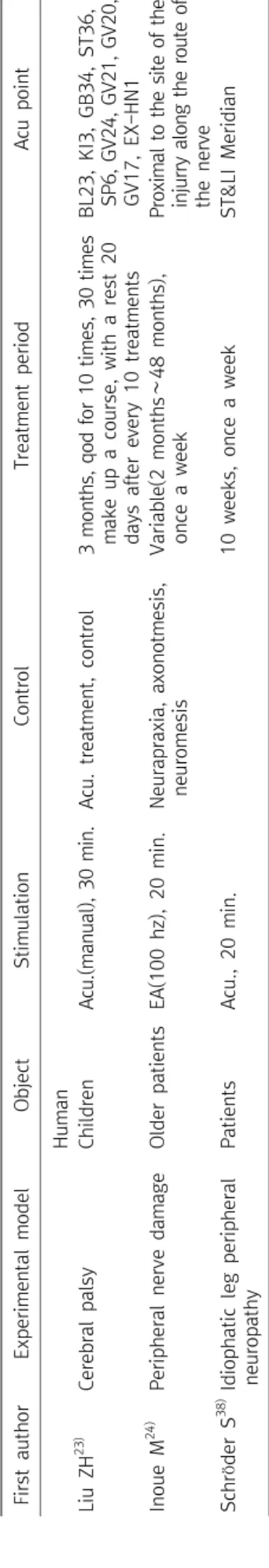
말초 신경 중에서 정중신경과 좌골신경이 손상된 경우는 각각 신경 주행 경로에 따라 대릉(大陵; PC7)과 곡택(曲澤; PC3)37), 환도 (環跳; GB30)36)와 위중(委中; BL40)36)혈이 선택되었고 족양명위경 이나 수양명대장경등 경락별로 변증에 맞게 선택하는 경우38)도 있 었다. 특정한 혈위나 경락보다는 손상된 신경의 주행을 따라 침 치 료를 선택한 경우24)도 있었으며 전통적인 경혈을 채택하지 않고 biceps femoris, biceps brachii27)와 같은 근육을 선택하는 경우도 있었다. 백회(百會; GV20) 단독 연구25)를 제외한 모든 연구에서 치료 경혈은 하나가 아니고 2개 이상이었다(Table 2).
임상 연구의 경우 뇌성마비 소아환자를 치료하는 경우에는 정해 진 경혈이 있었으나 다른 경우 대체적으로 침을 놓는 시술자의 판 단과 경락변증, 신경 주행에 따른 경혈등이 주관적으로 채택되었다.
4. 치료 기간 및 치료 횟수
침이나 전침치료의 치료 기간은 5∼7일16-18,21,33-36)
에서 1주 이 상 3주 이하15,22,25-29,34,37)
, 4주19,20,31), 7주30,32)까지 다양하게 설계 되었고 임상 연구에 있어서 10주38), 혹은 3개월23), 48개월24)까지 대체적으로 더 긴 치료기간을 보였다23,24,38). 치료 간격은 동물 실험 의 경우 매일15-18,21,22,26-29,33-37)
혹은 격일19,25,30-32)
이 가장 많았으 며 매일 치료와 격일 치료를 번갈아 하는 경우20)도 있었다. 임상 연구의 경우 주 1회로 치료하는 경우24,38)와 격일 치료 후 일정 기간 을 휴식한 후에 치료 결과를 평가한 경우23)로 나뉘었다(Table 2).
5. 다른 치료와 병행 및 비교 여부
거의 대부분의 논문이 침이나 전침 등을 단독 선택하여 연구하 였지만, 그 외에 타 치료와 병행하는 결과도 같이 연구한 경우도 있었다. 뜸 치료와 같이 병행한 경우15), 중간엽 줄기세포(mesen- chymal stem cells; MSCs) 치료와 같이 병행한 경우30-32), 기본적 인 재활치료와 병행한 경우23,26)가 있었으며 모두 병행한 경우 단독 침치료나 전침치료보다 유의한 결과를 보였다.
특히, MSCs 경우에는 전침의 자극이 MSCs 자체를 더욱 분화시 키는 결과를 보여주었다30-32).
특이하게, SCI 경우 전침과 NSAIDs(Nonsteroidal anti infla- mmatory drugs)의 일종인 diclofenac sodium 15 mg의 경우를 비교 연구한 경우36)도 있었는데 이 경우 전침치료는 원위조직의 유수신경섬유의 증가를 보여 유의한 결과를 나타냈으나 diclo- fenac sodium 15 mg의 경우에는 유수신경섬유의 유의한 변화가 없었다.
그러나 트레드밀 운동과 전침을 병행한 경우에서는 전침이 뇌유 래신경인자(Brain-derived neurotrophic factor; BDNF) 발현과 운동신경 기능회복에 유의한 결과를 보이지 않았다26).
6. 평가 도구
신경 재생에 대한 평가는 동물 실험에서는 크게 면역조직화학적 인 평가와 운동행동기능의 평가 두 가지 영역에서 이뤄진 것이 대 부분 이었고, 임상 연구에서는 소아 발달지수나 환자의 기능적 회 복, 전기생리학적으로 운동신경 전도의 속도 향상 등을 평가한 경 우가 있었다.
실험 연구에서는 면역조직화학적인 평가가 가장 많았으며, 신경 전달 인자(neurotrophin)16,18,19,21,25-27,31,34,35)
그 중에서도 특히 신 경 성장 인자(Nerve growth factor; NGF), neurotrophin-2 (NT-2), neurotrophin-3(NT-3), BDNF, 아교세포 신경인자(glial cell line-derived neurotrophic factor; GDNF)와 신경 섬유의 숫 자 및 새로운 수초의 생성19,27,28,30,36,37)
, 신경 재생에 관련된 단백질 발현 및 유전자 물질15,17,20,21,27-33)
등을 평가하였다.
운동행동 기능의 평가는 BBB 척도(Baso, Beattle, bresnahan locomotor rating scale)28,30-32), 유발전위 검사19), 운동신경 전도 속도37), Myotonometer(fast muscle state detector)를 이용한 근 육의 FDVs(force-displacement value)22)를 측정하여 평가하였다.
그 외, 임상 연구에서 환자의 주관적인 증상에 대한 문진 항목24) 과, 운동신경 전도 속도38) 뇌성마비 소아 환자의 Beijing Gesell Developmental Scale, GMFM(The Gross Motor Function Mea- sure), skull CT/MRI 전후 비교 하는 평가23)도 있었다.
7. 침 치료가 신경재생에 미치는 영향
전침은 손상된 조직에서 대표적인 신경 성장 인자인 NGF21,35), NT-319,31,35), BDNF16,21,25,27,35)
, GDNF16,34) 발현과 BDNF 수용체 인 trkB(tropomyosin-related kinase B)25,27)를 증가시켰으며 이러 한 작용은 새로운 수초 생성을 증가시켜 손상된 조직의 원위부위에 서 유수신경섬유가 관찰되었다19,27). 또한, ANXA5와 CRMP2 단백 질을 증가20)시키고 BDNF mRNA18,21,29)와 GDNF mRNA18)를 증가 시키며 GFAP(glial fibrillary acidic protein)와 CSPGs(chon- droitin sulfate proteoglycans) 단백질 발현을 억제30)하고 cAMP level32), 5-HT(5-hydroxytryptamine) positive32) 신경섬유와 CGRP (Calcitonin gene-related peptide) positive32,33) 신경섬유 숫자를 증가시킴으로써 축삭의 재생을 돕고 축삭의 퇴화를 방지하였다.
특히, 뇌 손상과 관련하여 Wnt 신호 전달 경로에서 axin을 감소 시키고 β-catenin을 증가시켜 뇌의 신경학적 부종을 감소시키고15)
First authorExperimental modelObjectStimulationControlTreatment periodAcu point Animal Zhou H15) AlzheimerRatEA(2 hz), 20 min.Normal, sham surgery, model, moxi- bustion pretreatment, EA pretreatment, EA+moxibustion pretreatment 3 courses of treatment, with 1 day of rest between each course(one course-6 days, daily)
GV20, BL23 Zhang R16) Heroin relapsedRatAcu., 30 min.Normal, model, methadone group, Acu.5 days, dailyDU20, DU14 Hou X17) Heroin readdicsRatAcu., 30 min. Normal, model, drug group, Acu.5 days, dailyDU20, DU14 Wang SJ18) Parkinson's diseaseRatEA(2 hz), 20 min.Blank(Untreated), sham-surgery, model, EA7 days, dailyGV16, LR3 Huang SF19) Demyelinated spinal cord injury(MS) RatEA(2 hz and 60 hz mixed), 20 min.Sham, EB(ethidium bromide), EA+EB26 days, every other day GV6, GV9 Li WJ20) Spinal cord injuryRatEA(60 hz), 20 min.Normal control, SCI, SCI+GV(Governor Vessel)-EA, SCI+Jiaji points EA, SCI+non-point area EA
1 weeks, daily 3 weeks, every other dayGV6, GV9 Chen J21) Removal of adjacent gangliaCatEA(98 hz), 30 min.Normal control(EA), EA(on the left side at acu point)7 days, dailyST36, SP6, GB39, ST32 Sun YJ22) Lumbar nerve root compressionRabbitEA(2 hz), 20 min.Normal, model, EAA(electro-acupuncture acupoints), medication, EAU(electro- acupuncture un-acupoints)
14 days, dailyBL23, BL25 Kim MW25) Cerebral infarctionRatEA(3 hz), 5 min.EA(+), EA(-)2 weeks, every 2 daysGV20 Kim WS26) StrokeRatEA(2 hz and 15 hz mixed), 30 min.Control, EA, Ex(treadmill exercise), ExEA14 days, dailyGV20, GV14, LI11, ST36 Pagani L27) Renitis pigmentosaRatEA(1~4 hz), 25 min.Normal, control, EA11 days, dailyBiceps femoris, biceps brachii Choi DC28) Spinal cord injuryRatAcupuncture (manual), 30 min.Sham, control, Acu(GV26+GB34), Acu(GV26), Acu(GB34)14 days, dailyGV26, GB34 Kim YR29) Middle cerebral artery occlusionMiceEA(2 hz), 20 min.Control, EA10 days, dailyGV20, GV14 Ding Y30) Transected spinal cordRatEA(2 hz and 60 hz mixed), 20 min.Sham-control, operated control, EA, MSCs, MSCs+EA7 weeks, every other dayGV1, GV2, GV6, GV9 Yan Q31) Spinal cord injuryRatEA(2 hz and 60 hz mixed), 20 min.Control, EA, MSCs, EA+MSCs4 weeks, every other dayGV1, GV2, GV6, GV9 Ding Y32) Spinal cord-transectedRatEA(2 hz and 60 hz mixed), 20 min.Op-control, EA, MSCs, EA+MSCs7 weeks, every other dayGV1, GV2, GV6, GV9 Li WJ33) Spinal cord-transectedRatEA(2 hz and 60 hz mixed), 20 min.Normal control, SCI, SCI+GV(Governor Vessel)-EA, SCI+Jiaji points EA, SCI+non-point area GV-EA
7 days, dailyGV6, GV9 Wang TT34) Removal of adjacent gangliaCatEA(98 hz), 30 min.Normal control, control, EA7 days, daily or 14 days, dailyST36, GB39, ST32, SP6 Wang TH35) partial dorsal rhizotomyCatEA(98 hz), 30 min.EA I, EA II, EA III7 days, dailyST36, GB39, ST32, SP6 La JL36) Crushed sciatic nerveRabbitEA(uncertain), 25 min.Sham control, EA, medicine 7 days, dailyGB30, BL40 Ho CY37) Transected Median NerveRatAcu., EA(2 hz), 15 min.Control(untreated), acupuncture, EA14 days, dailyPC7, PC3
Table 2. Experimental Design of 24 Articles
BDNF와 GDNF의 발현을 증가시켜 뇌 복부 외피 영역(ventral tegmental area) 신경세포의 미세구조를 개선하였다16). 또한, 뇌세 포의 자연사에 관련된 Bcl-2를 증가시키고 Bax발현을 억제하여 해 마와 전두엽의 손상을 줄이는 작용을 하였다17).
또한, 근육의 FDVs(force-displacement value)22), 유발전위 검사19), 운동신경 전도 속도37), BBB28,30-32) 등을 통한 운동기능 평가에 있어 서도 침의 자극이 신경 손상 후 회복에 유의하게 빠른 회복을 보였 다.
임상 연구에 있어서는 환자의 주관적인 증상에 대한 문진 항목24) 과, 뇌성마비 소아 환자의 Beijing Gesell Developmental Scale, GMFM, skull CT/MRI 전후 비교 하는 평가23), 운동신경 전도 속도38) 에서 침 치료를 하지 않은 대조군보다 유의한 기능 회복을 보였다 (Table 3).
고찰 및 결론
이상에서 침 치료가 신경의 재생과 회복에 미치는 영향에 대해 서 최근 10년간의 해외 연구의 동향을 파악해 보았다. 24편의 논문 의 게재지와 1저자의 국가별 분류를 보았을 때 16종의 학회지에서 중국과 한국을 비롯한 7개 나라에서 연구가 발표되었다.
신경 손상의 유형은 척수 손상이 11건, 다양한 원인에 의한 뇌손 상이 8건, 정중 신경 손상, 특발성 하지 말초 신경 손상, 망막색소변 성이 각각 1건 이었으며 연구 대상은 실험용 동물이 21건, 환자 대상 연구가 3건이었다.
침 치료와 전침 치료 중 전침 치료를 선택한 경우가 19편, 침 치료만을 선택한 경우가 5건으로 전침의 경우가 더 많았으며 주파 수는 2 Hz에서 100 Hz까지 다양하였고 혼용되기도 하였다. 침 치 료의 경우, 수기 자극을 하는 경우는 2건이었다. 유침 시간은 대부 분 15분에서 30분 내외였고 한 건을 제외한 모든 연구에서 2개 이상의 경혈이 선택되었다.
동물 실험의 경우, 4개 이하로 경혈이 선택되었고 환자를 대상으 로 한 연구에서는 10개 이상 혹은 경혈의 속성이나 변증에 따른 경락, 신경 주행의 경로에 따라 다양하게 선택되었다.
치료 기간은 5일, 7일, 2주∼7주, 10주, 48주까지 다양하였고 치료 간격은 매일, 격일, 주 1회 등 이었다. 동물 실험의 경우 최대 7주를 넘지 않는 치료기간과 매일 혹은 격일로 시행되는 치료방법 을 선택하였으며 임상 연구의 경우 최소 2개월에서 48개월까지의 상대적으로 장기간의 치료기간과 주1회의 치료방법을 선택하였다.
1건의 실험 연구를 제외한 모든 연구에서 유침 시간과 치료기간,
First authorExperimental modelObjectStimulationControlTreatment periodAcu point Human Liu ZH23) Cerebral palsyChildrenAcu.(manual), 30 min.Acu. treatment, control3 months, qod for 10 times, 30 times make up a course, with a rest 20 days after every 10 treatments
BL23, KI3, GB34, ST36, SP6, GV24, GV21, GV20, GV17, EX-HN1 Inoue M24) Peripheral nerve damageOlder patientsEA(100 hz), 20 min.Neurapraxia, axonotmesis, neuromesisVariable(2 months∼48 months), once a weekProximal to the site of the injurry along the route of the nerve Schröder S38) Idiophatic leg peripheral neuropathyPatientsAcu., 20 min.10 weeks, once a weekST&LI Meridian Acu : Acupuncture, EA : Electroacupuncture, min : minutes.
Table 2. Continued
First author Summary of results Animal
Zhou H15) - Compared with the model group, axin protein expession was lower(p<0.01), but β-catenin expession was higher (p<0.01) in the EA, moxibustion, EA+moxibustion group
- Neuronal cytoplasmic edema was visibly prevented
Zhang R16) - BDNF, GDNF expression increased in the EA group than normal, model, methadone group(p<0.05)
Hou X17) - In the acupuncture and drug groups, Bcl-2 expression increased compared with the model group(p<0.05)and in the acupuncture group, Bcl-2 expression increased compared with the drug group(p<0.01 or p<0.05) - In the acupuncture and drug groups, the number of Bax-positive cells and Bax expression significantly decreased
compared with the model group(p<0.05). In addition, the acupuncture group showed more Bax-positive cells and higher Bax expression than the drug group(p<0.01 or p<0.05)
Wang SJ18) - EA increased BDNF and GDNF mRNA expression in the substantia nigra compared with model group(p<0.01) Huang SF19) - EA+EB group promote NT-3(p<0.05), NG-2 expression(p<0.01), increase the cell number and differentiation of
endogenous OPCs(p<0.05)
- In the EA+EB day 30 group, the number of normal myeline or newborn myeline was higher than that in the EB group(p<0.05)
- Promote behavioural scores measured by SCEP and befavioural test(p<0.05)
Li WJ20) - GV-EA group increased ANXA5 and CRMP2 expression than the other groups(p<0.05)
- One month following EA treatment, survival of neurons in CN shows improvement, especially in GV-EA treatment group(p<0.01)
Chen J21) - NGF, NT-3 protein and mRNA, in the small and large neurons of spared L6 dorsal root ganlglion in EA side increased greatly more than that of control side(p<0.05 or p<0.01)
- BDNF and its mRNA, in the medium and smallneurons of spared L6 dorsal root ganlglion in EA side increased greatly more than that of control side(p<0.05 or p<0.01)
Sun YJ22) - The increase in FDVs of RSR in the EAA group was significantly higher than that in EAU(p=0.000) and medication groups(p=0.002)
- The EAA group had a greater MNCV recovery than the medication group(p=0.022) Kim MW25) - EA increased BDNF, trkB expression(p<0.05)
Kim WS26) - EA, Ex, Ex+EA, have no significantly effects in BDNF, trkB expression
Pagani L27) - EA increased BDNF, trkB, trkB-mRNA, blood vessels expression than that of control(p<0.05) - EA increased thickness of ONL, INL, GC layer than that of contol(p<0.01 or p<0.001)
Choi DC28) - Acu(GV26+GB34) group display more viable motor neurons compared to control(p<0.001), compared to Acu(GV26) or Acu(GB34)(p<0.05)
- Acu inhibits caspase-3(p<0.05), caspase-3 positive oligodendrocytes(CC1)(p<0.05), p38MAPK(p<0.001) and proNGF(p<0.001), proinflammatory cytokines(TNF-α, IL-1β, IL-6, cox-2, iNOS)(p<0.001), MMP-9(p<0.01) compared with sham and control
- Acu improves functional recovery(BBB test, grid walk test, inclined plane test, and footprint analysis) compared with control(p<0.05)
- Acu reduce axon loss and lesion volume compared with control(p<0.05)
Kim YR29) - EA treatment increased total number of BrdU and Dcx or NeuN double-positive cells, the mRNA level of BDNF and VEGF in the ipsilateral hemisphere, number of pPI3K/BrdU double-positive cells, VEGF positive cells in the hippocampus and ipsilateral subventicular zone versus middle cerebral artery occlusion mice(p<0.05, p<0.01 or p<0.001)
Ding Y30) - MSCs+EA group significantly improved BBB scales(p<0.01) and enhanced motor evoked potentials(MEPs)(p<0.05), number of NF-Positive Fibers(p<0.01), preserved more surviving neurons(p<0.05) than that of EA or MSCs - The levels of GFAP protein in the EA group and MSCs+EA group were significantly downregulated(p<0.05) Yan Q31) - MSCs and EA+MSCs group increased endogenous NT-3 than EA(p<0.05)
- EA increased differentiation of MSCs(EA+MSCs increased MMP2 positive cells, MOSP positive cells than MSCs)(p<0.05) - EA+MSCs group increased endogenous NF positive nerve fibers, 5-HT positive nerve fibers than EA, MSCs group(p<0.05) - BBB score increased in EA+MSCs than EA or MSCs(p<0.05)
Ding Y32) - EA+MSCs group increased NT-3(p<0.01), cAMP level(p<0.001), the differentiation of MSCs(p<0.05), the 5-HT positive and CGRP positive nerve fibers(p<0.001) than op-control, EA, MSCs groups.
- EA+MSCs group promoted BBB score(p<0.01) and recovered spinal cord evoked potentials(SCEP) Li WJ33) - GV-EA increased CGRP expression compared with normal and SCI(p<0.05)
Wang TT34) - EA increased GDNF, FGF-2 expression than that of control(p<0.05)
Wang TH35) - Acupunctured side increased NGF, BDNF, NT-3 expression than that of non-acupunctured side(p<0.05 or p<0.01) La JL36) - EA increased myelinated fibers in the distal parts of crushed nerve than the medicine(p<0.01), the control(p<0.001) Ho CY37) - In the acupuncture and EA groups, axon number, endoneurial area, total nerve area, blood vessel number, and
blood vessel area were larger than those in the control group(p<0.05) - EA treatment improved grasping capacity compared with the controls(p<0.05) Table 3. Summary of Results of 24 Articles
First author Summary of results Human
Liu ZH23) - Children's total effective rate(development quotient; DQ) in Acu treatment group was 87%, significantly higher than the 55% in the control group(p<0.01)
Inoue M24) - Two cases with neurapraxia and two with axonotmesis was obseved completely funcional recovery - One axonotmesis case achieved improvement
- One axonotmesis case showed reinnervation potential without functional recovery - One neuromesis case was observed no improvement
Schröder S38) - 16 patients(76%) in the Acu. group improved symptomatically and objectively as measured by NCS while only 4 patients(15%) in the control group did so(p=0.001)
VEGF(vascular endotherial growth factor).
Table 3. Continued
간격, 경혈에 상관없이 모두 유의한 효과를 보여주었다.
21편의 실험 연구 중, 침 치료의 신경 재생에 대한 효과를 측정 하는 도구는 면역 조직화학적, 운동행동 기능적으로 이루어진 것이 대부분이었고 임상 연구의 경우 주관적 증상, 신경 전도 속도, 유발 전위, FDVs 등의 측면에서 평가 되었다.
면역 조직화학적으로는 침 치료가 neurotrophin, 그 중에서도 NGF를 매개한다는 것이 많은 실험 연구 설계의 토대가 되고 있었 다. Manni et al39)에 따르면, 상피조직의 침의 자극은 trkA (tyro- sine kinase A) 활동을 조정하여 CNS와 PNS에 영향을 주고 neu- rotrophin, NGF를 활성화한다. 실제로 실험 연구의 면역 조직화학 적 평가에서 침이나 전침의 자극은 신경 성장인자인 NGF, NT-3, BDNF, GDNF와 trkB, BDNF mRNA, GDNF mRNA 발현을 증가 시켰고 cAMP level, 5-HT positive, CGRP positive 신경 섬유 숫자를 증가시켜 손상된 조직의 새로운 수초 생성을 증가시켰다.
또한, 침 치료는 ANXA5와 CRMP2 단백질을 증가시키고 GFAP와 CSPGs 단백질 발현을 억제하여 축삭의 재생은 돕고 퇴화는 방지하 는 작용을 하였다. 신경인자와 상관없이 조직학적으로 보았을 때도, Huang19), Pagani27), Choi28) 등의 연구에서 신생 수초의 숫자가 증가함을 알 수 있었다.
신경성장인자나 수초 생성처럼 직접적으로 신경 재생을 돕는 이 외에도 뇌세포에 있어서는 뇌세포의 자연사에 관련된 Bcl-2를 증 가시키고 Bax 발현을 억제하여 해마와 전두엽의 손상을 줄이는 작 용을 하였고 axin을 줄이고 β-catenin을 증가시켜 뇌의 신경학적 인 부종을 감소시켜 신경세포 재생에 도움을 주었다.
근육의 FDVs, 유발전위 검사, 운동신경 전도 속도, BBB등을 통 한 운동기능 평가에 있어서도 침의 자극이 신경 손상 후 회복에 유의하게 빠른 회복을 보였으며 환자의 주관적인 증상에 대한 문진 항목과, 뇌성마비 소아 환자의 Beijing Gesell Developmental Scale, GMFM, skull CT/MRI 전후 비교 하는 평가에서도 침 치료 를 하지 않은 대조군보다 유의한 기능 회복을 보였다.
그러나 stroke 처치된 Rat의 14일간의 2 Hz와 15 Hz의 혼합된 전침 자극 연구에서 BDNF와 trkB 발현은 대조군에 비해 유의할 만한 증가를 보여주지 못했는데26) 이는 유사한 다른 실험 연구의 결과와는 완전히 상반되는 결과이다. 다른 유사한 연구에서 cere- bral infarction 처치된 Rat을 2주간 3 Hz의 전침 자극 했을 때, BDNF와 trkB 발현은 증가25)되었다. 실험 결과에 대해서 Kim et al26)은 연구의 초기 설계에서 Rat의 운동신경 손상이 너무 미약해 서 중등도 이상의 운동신경 손상의 연구결과가 필요할 것이라는 점, 그리고 2주의 평가기간에 있어서 뇌졸중 발생시기와 치료종료 시기가 너무 가깝다는 두 가지 한계점을 들어 전침이 뇌졸중의 초 기회복 단계에서 운동신경 회복을 보이지 못했다는 결과를 도출하 면서 새로운 연구의 설계가 필요하다고 하였다. 그러나 평가기간의 짧음에 있어서 Zhang et al16), Chen et al21)의 rat 연구에서는 신경 손상 후 각각 5일과 7일의 평가기간만을 가졌는데도 각각 헤로인 재발로 인한 뇌손상과 척수 손상 후 BDNF 발현에 있어서 유의한 효과를 보였다. 만약 Kim et al26)의 지견대로 중등도 이상의 신경 손상에서만 침 치료가 효과적이라면 다양한 신경손상의 정도에 따 른 비교 연구도 필요하리라 여겨진다.
환자를 대상으로 한 연구에서 Inoue et al24)은 neurapraxia, axonotmesis, neuromesis로 나뉜 7명의 신경 손상 회복을 위한 침치료 연구에서 neuromesis와 axonotmesis 1건을 제외하고는 모두 완전 회복 혹은 호전을 보였다. 임상연구에서는 오히려 미약 한 신경 손상에서 더 좋은 효과를 보인 셈이다. 이 경우에 있어서 신경 손상 후 즉시 치료가 들어간 것이 아니고 짧게는 신경 손상 1개월 후부터, 길게는 신경 손상 14개월이 지난 환자 군에게 침 치료를 했다. Inoue et al24)의 연구에서는 신경 손상 후 1년이 지난 환자의 경우도 호전을 보였으나, 몇 례에 불과한 연구라서 일반화 시키기는 어려운 면이 있으며 만성 신경 손상의 침 치료 연구가 더욱 필요하리라 사료된다.
Schröder et al38)은 하지의 특발성 말초신경병증 환자 47명을