군 훈련 이후 발생한 전경골근 탈출에 있어 근막하 합성 그물 이식술을 이용한 치료
- 증례 보고 -
김경호ㆍ신영수*
국군대전병원 정형외과, 한림대학교 춘천성심병원 정형외과*
Surgical Repair of Tibialis Anterior Muscle Herniation Using a Synthetic Mesh That Was Beneath the Fascia after a Military
Training Program - A Case Report -
Kyoung-Ho Kim, M.D., Young-Soo Shin, M.D., Ph.D.*
Department of Orthopedic Surgery, Armed Forces Daejeon Hospital, Daejeon,
Department of Orthopedic Surgery, Hallym University Chuncheon Sacred Heart Hospital*, Chuncheon, Korea
Received January 7, 2019 Revised February 19, 2019 Accepted March 13, 2019 Correspondence to:
Young-Soo Shin, M.D., Ph.D.
Department of Orthopedic Surgery, Hallym University Chuncheon Sacred Heart Hospital, 77 Sakju-ro, Chuncheon 24253, Korea Tel: +82-33-240-5000 Fax: +82-33-255-6244 E-mail: [email protected] Financial support: None.
Conflict of interests: None.
Tibialis anterior muscle herniation is the most common type of skeletal muscle herniation of the lower legs. The treatment of muscle herniation relies on the patient’s symptoms. For patients with chronic large fascial defects, fascial grafting with synthetic mesh can be considered. In this case of a patient who was exposed to excessive strain on his lower legs during a military training program, the use of a secure repair technique with synthetic mesh was required. This paper presents a case of tibialis anterior muscle herniation that was treated successfully with a monofilament knitted polypropylene mesh cov- ered by the tibialis anterior fascia. The advantages of this technique include early rehabilitation and an early return to work. No significant difference in the clinical results compared to other methods were observed and there were no complications. The military training program appeared to have aggravated the patient’s symptoms of tibialis anterior muscle herniation. On the other hand, larger scale study will be needed to determine if this program actually affects the clinical outcomes.
Key Words: Mesh, Fascial grafting, Muscle herniation, Tibialis anterior muscle
Copyright © 2019 The Korean Fracture Society. All rights reserved.
This is an Open Access article distributed under the terms of the Creative Commons Attribution Non-Commercial License (http://creativecommons.org/licenses/by-nc/4.0) which permits unrestricted non-commercial use, distribution, and reproduction in any medium, provided the original work is properly cited.
Traumatic symptomatic herniation of the tibialis ante- rior muscle requires surgical treatment.1) Previously, several authors reported repairing fascial defects using mesh placed over the fascia to reduce the strain between the tibialis ante- rior muscle and the mesh.2,3) Here we present a case of tibi-
alis anterior muscle herniation that was successfully treated with synthetic mesh that was beneath the fascia. Believ- ing that the military training program likely worsened the symptoms of tibialis anterior muscle herniation, we designed the present study to investigate the correlation between the
military training program and muscle herniation.
Case Report
A 19-year-old male military recruit visited our out- patient clinic in Armed Forces Daejeon Hospital with vague pain and a palpable mass in the anterolateral aspect of the right lower leg. Four months prior, he suffered blunt trauma to the same area induced by a sharp metal fitness equip- ment. At that time, because the pain was feeble and there was no visible wound, he did not visit the hospital. Howev- er, as the military training program progressed, the palpable mass and vague pain developed and the size of the mass increased considerably. The mass also increased in size upon prolonged standing, squatting, or walking uphill. A physical examination revealed that the mass was located in the right lower leg along the lateral border in the middle of the tibia at 10 cm from the tibial tuberosity. The mass was prominent when the ankle joint was dorsiflexed (Fig. 1).
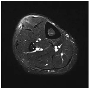
Based on history and clinical findings, we suspected that the mass was a tibialis anterior muscle herniation; magnetic resonance imaging (MRI) findings confirmed our suspi- cions. The MRI scans revealed a 3×5 cm oval-shaped focal herniation of the tibialis anterior muscle overlying a fascial defect. The margin of the fascial defect was thickened, and the muscle belly protruded through it (Fig. 2).
We attempted conservative treatments including lifestyle
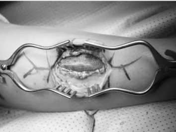
modification, compression stockings, and oral medications for a 1-month period. After conservative treatment failure, we provided surgical treatment. During surgery, we used a longitudinal skin incision overlying the mass to reveal a 3×
5 cm fascial defect. The tibialis anterior muscle was bulging out through the fascial defect (Fig. 3).
Direct closure was not sufficient to repair the fascial defect because the margin was hypertrophied and it was difficult to approximate the margin without tension. There- fore, we applied fascial grafting using synthetic mesh placed under the fascia. We chose to cover the fascial defect with a monofilament knitted polypropylene mesh (Bard® mesh;
Fig. 1. Clinical photograph of the mass over the right leg.
Fig. 2. Magnetic resonance imaging axial cut showing herniation of muscle through the fascia.
Fig. 3. Intraoperative photo showing a fascial defect with herniating muscle of the right leg.
C. R. Bard Inc., Warwick, RI, USA) (Fig. 4). The mesh was prepared to match the shape of the fascial defect (Fig.
5). Using an Army Navy retractor and Freer elevator, the protruded muscle belly was reduced. The mesh was placed under the fascia and the anterior margin of the fascia was initially repaired with a Prolene 4-0 monofilament. The posterior margin of the fascia was retracted with Allis tissue forceps to reduce the gap in the margin. The posterior mar- gin was then repaired (Fig. 6). After a meticulous and secure repair was completed, friction and irritation between the mesh, suture knots, and the muscle were checked for during passive ankle movement.
After the surgery, we applied a short leg splint with the ankle in plantar flexion. Since a tight repair was performed,
early rehabilitation was started. One week after surgery, we removed the splint and weight bearing was started. We recommended that the patient use compression stockings.
Two weeks after surgery, full weight bearing was started.
One month after surgery, the patient returned to work. At the 2-month follow-up, the patient returned to his normal sports activities. At the 6-month follow-up, there were no complications including irritation or cosmetic problems.
Discussion
Skeletal muscle herniations are classified as constitutional or traumatic.4,5) Constitutional herniations are caused by chronic stress on the fascia of the underlying muscle, while traumatic defects are classified as those caused by direct or indirect trauma. In the present case, the blunt trauma to the right lower leg caused by a sharp metal fitness instrument may have contributed to the weakening of the fascia overly- ing the tibialis anterior muscle that led to the symptomatic muscle herniation later on.
Most cases to date have been described in athletes and young adults experiencing excessive strain on the lower legs.6,7) Due to being tight and superficial, the anterior tibial compartment is most commonly affected by muscle hernia- tion. Therefore, here we investigated whether the military training program and excessive strain on the lower legs
Fig. 4. Intraoperative photograph of polypropylene non-absorbable synthetic surgical mesh.
Fig. 5. Intraoperative photo showing the fascial defect and the mesh beneath the fascia.
Fig. 6. Intraoperative photograph showing the fascial defect covered with polypropylene non-absorbable synthetic surgical mesh beneath the fascia.
were correlated. We found that the military training pro- gram could have worsened the symptoms of tibialis anterior muscle herniation. These results confirm those of a previous study8) in which the primary strength of military-related physical activities was shown to be derived more from the lower extremities than from the upper extremities. The weight-based training program is used to strengthen the lower extremities, while the calisthenics program trains the leg and hip muscles. Furthermore, working as a sentry who maintains a standing position for a long time can be a factor of the acquired excessive strain on the lower legs.
Since treatment relies mainly on patient symptoms, as- ymptomatic muscle herniation requires no specific treat- ment. Symptomatic muscle herniation has various treat- ments ranging from conservative management to surgical procedures including direct repair, fasciotomy, and fascial grafting using an autologous graft or synthetic mesh. For patients with chronic and large fascial defects, fascial graft- ing with an autologous graft or synthetic mesh can be con- sidered. Fascial grafting using an autologous graft such as a tensor fascia lata9) or periosteal graft10) can be useful. There are several advantages to this, such as a lack of skin irrita- tion and low cost. However, this procedure requires care- ful postoperative observation for compartment syndrome and donor site morbidity. Considering the patient’s concern about the cosmetic result of the surgery and secondary damage that would occur at the donor site, an autologous graft was not considered.
In this case, the patient was exposed to excessive strain on the lower legs during the military training program and his work as a sentry. This meant that he maintained a long-term standing position of more than 4 hours a day.
He wanted to achieve early recovery and an early return to work. Therefore, secure surgical repair with synthetic mesh was required. Some authors previously suggested that the mesh should be fixed above rather than under because it al- lows the underlying muscle to slide without any friction by the mesh or suture materials.2,3) However, the fascial defect margin was hypertrophied and thickened in this case, and we were concerned that the bulky margin could interrupt the tight repair and cause skin irritation. Thus, we de-
cided to fix the mesh beneath the fascia. After fixation, we checked the grafting site during passive ankle movement.
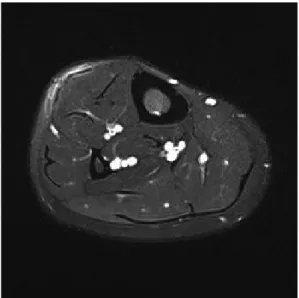
There was no irritation around the mesh or suture materials and the underlying muscle was sliding without any inter- ruption. There were no abnormal findings on the immediate postoperative and 3-month follow-up MRI scans (Fig. 7).
Since a tight repair was performed, early rehabilitation was started and an early return to work enabled. To the best of our knowledge, this is the first case report to focus on South Korean military recruits. Although only one case was de- scribed in this report and the follow-up period was short, fascial grafting using synthetic mesh provided satisfactory outcomes without serious complications and could be an alternative option to conventional surgical treatments.
In conclusion, tibialis anterior muscle herniations should be considered a rare differential diagnosis whenever patients present with persistent vague leg pain after a military train- ing program. Furthermore, in cases of chronic and large fascial defects, fascial grafting using synthetic mesh under the fascia can be a good option for managing tibialis ante- rior muscle herniation, as this simple procedure can provide good functional outcomes without complications.
Fig. 7. Three-month follow-up magnetic resonance imaging scan showing well repaired fascial defect using synthetic Mesh.
ORCID
김경호, https://orcid.org/0000-0002-0539-223X 신영수, https://orcid.org/0000-0003-1030-9979
References
1. Miniaci A, Rorabeck CH: Tibialis anterior muscle hernia: a ra- tionale for treatment. Can J Surg, 30: 79-80, 1987.
2. Siliprandi L, Martini G, Chiarelli A, Mazzoleni F: Surgical re- pair of an anterior tibialis muscle hernia with Mersilene mesh.
Plast Reconstr Surg, 91: 154-157, 1993.
3. Marques A, Brenda E, Amarante MT: Bilateral multiple muscle hernias of the leg repaired with Marlex mesh. Br J Plast Surg, 47:
444-446, 1994.
4. Nguyen JT, Nguyen JL, Wheatley MJ, Nguyen TA: Muscle
hernias of the leg: a case report and comprehensive review of the literature. Can J Plast Surg, 21: 243-247, 2013.
5. Ihde H: On muscular hernia of the leg. Acta Chir Scand, 65: 97- 120, 1929.
6. Kim M, Hong SP, Hwang SM, Park H, Ahn SK: Tibialis ante- rior muscle herniation developed after trauma. Int J Dermatol, 47: 845-847, 2008.
7. Rho NK, Kim WS, Kim YJ, Yoo KH, Kim BJ, Kim MN: The use of dynamic ultrasonography for the confirmation of lower leg muscle herniation. Ann Dermatol, 20: 190-192, 2008.
8. Harman EA, Gutekunst DJ, Frykman PN, et al: Effects of two different eight-week training programs on military physical per- formance. J Strength Cond Res, 22: 524-534, 2008.
9. Simon HE, Sacchet HA: Muscle hernias of the leg: review of lit- erature and report of twelve cases. Am J Surg, 67: 87-97, 1945.
10. Choi YR, Hong CG: Repair of tibialis anterior muscle herniation using periosteum. Orthopedics, 37: 748-750, 2014.