I. 서 론
의치구내염은 의치 장착 환자의 지지점막에 발생 하는 만성 염증성 변화로서 특히 상악 구개부위에 빈번히 나타나며, 증상으로 염증성 발적, 부종, 과립 형 증식 등이 있으며 자각증상은 거의 없으나 출혈, 작열감, 동통 등이 나타날 수 있다.1,2)의치 구내염에 대한 병인은 아직 완전하게 밝혀지지 않았으나 다양 한 인자가 관계되는 것으로 알려져 있다. 주원인으 로는 적합이 좋지 않은 의치 장착, 이갈이 습관 및 교합부조화로 인한 기계적 외상이 있으며, 그 외에 도Candida 균주에 의한 점막 감염, 의치의 치태 침 착, 야간의 의치장착 습관, 이장재의 사용, 영양결핍, 전신적 소모성질환, 방사선요법, 부적절한 구강위 생, 알레르기성 반응, 타액감소 및 미생물의 감염 등 이 보고되었다.1,3-5)
세균 부착은 군락형성과 감염 진행의 중요한 단계 로 인지되고 있으며, Theilade와 Budtz-Jorgensen3), Sanger 등6)및 Hans와 Hendrick7)은 플라스틱 인공 보철물, 혈관 도관 및 의치상 표면에서 세균 부착을 보고하였다. 의치상에 세균이 부착해서 증식하면 치 태의 성숙을 촉진시켜 충치, 치은염 및 치주염의 유 병율을 증가시키며, 인접한 구강점막에 염증 반응을 일으킨다. Samar-anayake 등8)에 의하면 자당이나 포도당이 함유된 배지에서 아크릴릭 레진 표면에 세 균 부착이 촉진된다고 하였으며, McCourtie와 Douglas9), McCourtie 등10)은 자당과Candida albi-
cans를 함께 배양 후 생성된 부가적인 표면층이 아 크릴릭 레진의 세균 부착에 관여한다고 하였다.
Minagi 등11)은 레진 표면에 있어서Candida 등의 부 착과 세균표면의 소수성 결합과는 밀접한 관련이 있 다고 보고하였으며, Taylor 등12)은 코발트-크롬 합금 이나 의치상 레진의 표면 거칠기와 세균 부착과는 밀접한 관련이 있다고 하였다.
구강내에 존재하는 정상 균주 중 Streptococcus mutans는 치태형성의 주원인 균으로 초기 치태 형 성에 관여하며, Lactobacillus casei는 노년층에서 치 경부 우식증의 주원인 균으로 보고되었다.13-17)
키토산은 천연에 존재하는 다당류로서 갑각류의 외피에 있는 키틴이라는 물질을 탈아세틸화 (deace- tylation)함으로써 얻을 수 있다.18)키토산은 안전하 고 무독성이며 여러 가지 생리적 활성이 있는 것으 로 알려져 이에 대한 연구개발이 활발히 진행되고 있으며, 약리학적으로 혈압을 유지해주고 항세균성, 항진균성 및 항암성이 있고 콜레스테롤 조절 작용이 있다고 보고되었다.18)
본 연구는 현재 널리 사용되는 PMMA(polymethyl methacrylate) 의치상 레진 및 4-META(4- Methacryloxyethyl trimellitate anhydride) 의치상 레진의 세균 부착과 의치상 레진에 키토산 첨가 시 세균 부착 정도를 관찰함으로써, 위생적인 의치상 레진을 선택하고 의치상 레진에 키토산 첨가의 가능 성을 알아보기 위하여 시행되었다.
대한치과보철학회지:Vol. 40, No. 5, 2002
키토산을 첨가한 의치상 재료의 세균 부착에 관한 연구
정승환∙방몽숙∙박하옥 전남대학교 치과대학 보철학교실
Ⅱ. 연구재료 및 방법
1. 키토산 첨가농도에 따른Streptococcus mu- tans와 Lactobacillus casei의 생균수 검사
키토 올리고당(5%)을 M17 broth와 MRS broth에 각각 첨가하여 0.8%, 0.4%, 0.2%, 0.1%, 0.05%, 0.025%, 0.0125%, 0.00625%, 0.003125%, 0.0015625%의 농도가 되도록 하였다.
Strptococcus mutans (Ingbritt strain)는 M17 broth, Lactobacillus casei KCTC 3260(ATCC 334) 는 MRS broth 2㎖에 배양액 20λ를 접종하고 37℃에 서 24시간 배양하여 10-5배로 배양액을 희석하여 BHI(brain heart infusion) agar(Difco, USA)와 MRS agar (Difco, USA)에 각각 접종하여 37℃에서 48시 간 배양한 후 생균수를 계수하였다.
2. 의치상 재료 표면에서 Streptococcus mu- tans와 Lactobacillus casei의 흡착도 검사
본 연구에서는 PMMA 의치상 레진으로 열중합 레진인 Vertex-RS (Dentimex Co., Holland)와 자 가중합 레진인 Vertex-SC (Dentimex Co., Holland), 4-META 의치상 레진으로 열중합 레진인 META- DENT (Sun Medical Co., Japan)와 자가중합 레진 인 META-FAST (Sun Medical Co., Japan)를 사 용하였다(Table Ⅰ).
(1) 시편의 제작
지름 5mm, 길이 6mm 크기의 가압할 수 있는 주 형을 만들고 여기에 각 레진을 제조회사의 지시대로 Vertex-RS는 2.18 g : 1 ㎖, Vertex-SC는 1.60 g :
1 ㎖, META-dent는 2 g : 1 ㎖, META-fast는 2 g : 1 ㎖의 비율로 혼합하고, 키토산을 첨가하는 경우 각 의치상 재료에 키토산을 monomer의 1/10의 양을 첨가하여 병상 상태에서 유압식 가압기(Samki Co., korea)로 가압 전입하였다. 열중합 레진인 Vertex- RS는 20분, META-dent는 40분 동안 온성조 (Hanau curing unit, Teledyne, USA)에 넣어 가열 중합하였으며 자가중합 레진인 Vertex-SC와 META-dent는 실온에서 경화시킨 후 분리하였다.
분리한 시편을 지그를 이용하여 #1200 silicone carbide paper로 장축에 수직되게 연마하여 각 군당 6개씩 총 24개의 레진시편을 제작하였다. 제작된 시 편을 증류수에 48시간 보관하여 여분의 단량체에 의 한 독성에 의해 세균의 성장이 영향을 받지 않도록 하였으며, 에틸렌 옥사이드 가스 소독을 시행하였 다.
(2) 세균 배양 및 측정
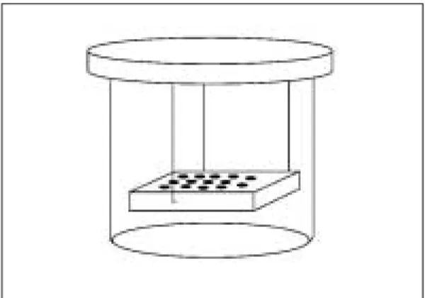
베이스 플레이트 왝스를 녹여 각 시편을 포매하고 윗면만을 노출하여 하나의 왝스 블록으로 준비하였 다. 200㎖ 비이커에 M17 broth 125㎖를 첨가한 후 18시간 배양한Streptococcus mutans 배양액을 접 종하였다(Fig. 1). 비이커 덮개에 wire로 고정시킨 왝 스 블록을 비이커 내에 넣고 37℃ shaking incubator 에서 24시간 배양하였다. 왝스 블록을 꺼낸 후 ster- ile cutter로 각 시편 주위의 왝스를 제거하였다. 각 시편을 M17 broth 1㎖를 넣은 시험관에 넣고 vortex 혼합기에서 10초간 shaking하여 이를 10-2, 10-3배로 희석 후 BHI agar에 접종하여 48시간 배양한 다음 생균수를 계수하였다. Lactobcillus casei는 MRS broth 125㎖에 배양액 20λ를 접종하여 wax block을 덮개에 wire를 이용하여 고정한 후 37℃에서 24시간
Table Ⅰ. Denture base materials
PMMA/MMA (heat curing) Vertex-RS Dentimex Co., Holland PMMA/MMA (self curing) Vertex-SC Dentimex Co., Holland 4-META (heat curing) META-DENT Sun Medical Co., Japan 4-META (self curing) META-FAST Sun Medical Co., Japan Type (curing type) Trade name Manufacturer
동안 shaking incubator에서 배양하였다. 위에서와 같은 방법으로 시편을 분리하고 MRS broth 1㎖가 담겨진 시험관에 각 시편을 넣고 vortex 혼합기에서 30초간 shaking 후 10-2, 10-3배로 희석하여 MRS agar에 접종하여 48시간 배양한 다음 생균수를 계수 하였다.
3. 레진 표면의 주사전자현미경적 관찰
4종의 의치상 레진과 키토산을 첨가한 의치상 레진 시편의 표면을 Ion sputter (E-1030, Hitachi Co., Japan)를 이용하여 백금 코팅한 후, 주사전자 현미경(S-4700, Hitachi Co., Japan)으로 관찰하 였다.
Ⅲ. 연구결과
1. 키토산 첨가 농도에 따른 Streptococcus mutans와 Lactobacillus casei의 생균수
키토산을 M17 broth와 MRS broth에 첨가했을 경 우Streptococcus mutans와 Lactobacillus casei의 증식 정도를 검사하였다. Streptococcus mutans의 경 우 0%에서는 1.12 ×10⁹/㎖ 였고 0.015%에서는 1.28×10³/㎖로 급격히 감소하여 0.0255%에서는 1.14×10²/㎖로 거의 자라지 않았다(Table Ⅱ).
Lactobacillus casei는 0%에서는 1.86×10⁹/㎖였 는데, 0.4%에서는 2.0×10³/㎖로 급격히 감소하여
0.8%에서는 2×10²/㎖이하로 증식이 억제되었다 (Table Ⅲ).
2. 의치상 재료에서 Streptococcus mutans와 Lactobacillus casei의 흡착도
1) Streptococcus mutans의 흡착도
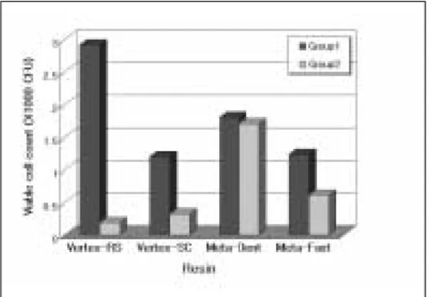
4종의 레진 시편상에서Streptococcus mutans의 생 균수를 검사한 결과 키토산을 첨가하지 경우에는 Vertex-RS가 6.9×10⁴CFU, Vertex-SC가 8.41×
105CFU, Meta-dent가 1.42×10⁶CFU, 그리고 Meta fast가 3.32×10⁵CFU 였다. 키토산을 첨가한 경우에는 Vertex-RS가 4.3×10⁴CFU, Vertex-SC가 1.14×10⁵CFU, Meta-dent가 1.02×10⁵CFU, 그리 고 Meta fast가 1.09×10⁵CFU 였다(Table Ⅳ)(Fig.
2.). 이상의 결과에서 키토산을 첨가한 레진에 비해 키토산을 첨가한 레진에서 생균수가 감소하였다.
2) Lactobacillus casei의 흡착도
4종의 레진 시편상에서Lactobacillus casei의 생균 수를 검사한 결과 키토산을 첨가하지 않은 경우에는 Vertex-RS가 2.91×10⁴CFU, Vertex-SC가 1.2×
104CFU, META-DENT가 1.8×10⁴CFU, 그리고 META-FAST가 1.23×10⁴CFU였다. 키토산을 첨가 한 경우에는 Vertex-RS가 1.9×10³CFU, Vertex-SC 가 3.2×10³CFU, META-DENT가 1.7×10⁴CFU, 그리고 META-FAST가 6.1×10³CFU였다(Table 5)(Fig. 3). 이상의 결과에서 키토산을 첨가한 레진에 비해 키토산을 첨가한 레진에서 생균수가 감소하였 다.
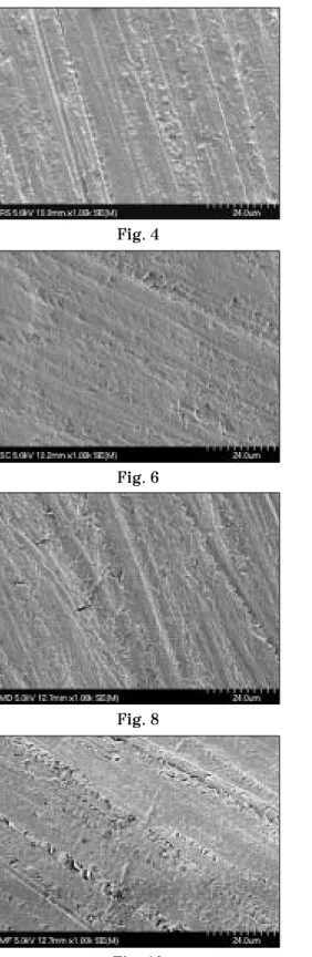
3. 주사전자현미경적 관찰 소견
레진 시편의 표면 형태를 주사전자 현미경하에서 1000배의 배율로 비교 관찰한 결과 키토산을 첨가하 지 않은 군에서는 기포가 적고 긴밀하게 밀착되어 매끄러운 표면을 나타내었으며, No. 1200 실리콘 카 바이드 페이퍼의 연마 흔적이 남아 있었다. 키토산 을 첨가한 군에서는 연마면에 따라서 돌기 및 다공 성 기포가 발견되어 거친 양상을 나타내었다(Figs.
4-11.).
Fig. 1. Schematic diagram of wax block containing the resin specimens in the beaker.
Table Ⅲ. Effect of chitosan on the replication of Lactobacillus casei
0 1.86×10⁹
0.025 1.68×10⁹
0.05 5.68×10⁸
0.1 8.80×10⁷
0.2 1.48×10⁷
0.4 2.0×10³
0.8 <102
Concentration of Viable cell count of chitosan (%) L. casei (/㎖) Table Ⅱ. Effect of chitosan on the replication of
Streptococcus mutans
0 1.12×10⁹
0.0015625 7.45×10⁸
0.003125 5.49×10⁸
0.00625 3.08×10⁷
0.0125 1.28×10³
0.025 1.14×10²
Concentration of Viable cell count of chitosan (%) S. mutans (/㎖)
Table Ⅳ. Viable cell count of Streptococcus mutans on the surface of 4 kinds of resin (CFU)
Group 1 6.9×10⁴ 8.41×10⁵ 1.42×10⁶ 3.32×10⁵ Group 2 4.3×10⁴ 1.14×10⁵ 1.02×10⁵ 1.09×10⁵ Group 1: resin without chitosan
Group 2: resin with chitosan
Vertex- Vertex- Meta- Meta-
RS SC Dent Fast
Fig. 2. Viable cell count of Streptococcus mutans on the surface of 4 kinds of resin.
Table Ⅴ. Viable cell count of Lactobacillus casei on the surface of 4 kinds of resin (CFU)
Group 1 2.91×10⁴ 1.2×10⁴ 1.8×10⁴ 1.23×10⁴ Group 2 1.9×10³ 3.2×10³ 1.7×10⁴ 6.1×10³ Group 1: resin without chitosan
Group 2: resin with chitosan
Vertex- Vertex- Meta- Meta-
RS SC Dent Fast
Fig. 3. Viable cell count of Lactobacillus casei on the surface of 4 kinds of resin.
Ⅳ. 총괄 및 고찰
아크릴릭 중합체의 사용에 대하여 Wright가 보고 한 이후, 의치상은 PMMA 레진과 4-META 레진으 로 제작되어 사용되고 있다. 아크릴릭 레진은 심미 적으로 양호하고 흡수도와 용해도가 낮으며 인체에 독성이 없고 비교적 정확한 재현성을 가질 뿐 아니 라 조작과 수리가 간편한 장점이 있는 반면, 마모성 이 높고 표면강도 및 충격저항이 낮은 단점이 있다.
또한 아크릴릭 레진은 중량체(polymer)와 단량체 (monomer)가 합쳐져서 중합되는 형태로 중합과정 중에 기포가 많이 발생하며, 이러한 기포는 세균의 서식처가 될 수 있으므로 의치 장착자의 구강 상태 는 불량하게 되기 쉽다. 의치장착자중 의치구내염의 비율은 Nyquist19)는 27%, Love 등20)은 43%, Budtz- jorgensen21)은 67%라고 보고하였다.
의치구내염의 원인은 다양하다. 국소적인 인자로 는 세균3), 진균4), 잘 맞지 않은 의치, 불량한 구강위 생1), 탄수화물이 풍부한 음식의 섭취5), 의치상 재료 자체의 영향4)등이 있다. 의치상의 세균이나 진균 치 태는 특히 소모성 질환이 있는 환자에서 심각한 감 염을 일으킬 수 있다.4,22)
의치상 재료에 미생물의 부착 양상에 대한 연구에 서 Samaranayake 등8)는 아크릴 판형에 혈청을 도포 시 캔디다(Candida)의 부착이 증가하였으나, 클로르 헥시딘(chlorhexidine)과 타액의 존재 하에서는 캔디 다의 부착이 감소한다고 하였다. Nikawa와 Samaranayake23)는 배지에 포도당과 유당을 첨가하 면 아크릴릭 레진 표면에 세균의 부착이 촉진된다고 하였는데, McCourtie와 Douglas9)는 첨가하는 당의 종류에 따라 캔디다의 부착정도에 차이가 있다고 하 였다. McCourtie 등10)은 첨가하는 당의 종류에 따라 Candida albicans의 클로르헥시딘에 대한 저항정도 가 다르다고 보고하였다. Minagi 등11)과 Miyake 등24)은 레진 표면에Candida 등의 부착과 세균표면 의 소수성 결합과는 밀접한 관계가 있다고 보고하였 다. Taylor 등12)는 세포 크기와 표면 거칠기가 미생물 의 부착에 많은 영향을 미친다고 하였고, Verran과 Maryan25)은 아크릴릭 레진과 실리콘 표면의 거칠기 가 미생물의 부착과 감염을 증가시킨다고 하였으며, Radford 등26)과 Edgerton 등27)은 표면 거칠기와 유리 표면 에너지가Candida의 부착을 증진시키지만 타
액이 이것을 감소시킨다고 하였다.
구강내 정상 균주로 여러 가지 세균이 존재한다.
Morris 등28)은 의치상 레진의 초기 치태를 조사한 결 과Streptococci의 비율이 높다고 하였고, 이들 세균 의 초기 침착은 표면의 기포부분에서 일어난다고 하 였다. Hayes 등13)은 충치 이환율이 높은 사람에서 산 형성세균 비율이 높다고 보고하였으며, Sansone 등14)은 노령자에서 치경부 우식증과Streptococcus가 강한 상관관계를 가진다고 보고하였다. Ellen 등15)은 노인 입원 환자에서 치근 우식증과 미생물 조성을 검사한 결과Streptococcus mutans와 Lactobacillus 가 강한 상관 관계가 있다고 보고하였다. Boue 등16) 은 다발성 치아우식증 환자의 미생물 조성에서 비착 색성 우식증의 치태에는Streptococcus mutans와 Actinomyces가 많고, 착색성 우식증에서는 Lactobacillus와 Actinomyces가 많이 분포한다고 보고하였다. 그러므로 본 연구에서는Streptococcus mutans와 Lactobacillus casei를 아크릴릭 레진의 부 착 정도를 관찰할 세균으로 선택하였다. 의치상에 위의 세균의 부착을 저지할 수 있다면 결과적으로 치태의 침착을 방지하여 이와 관련된 구내병소 억제 효과를 얻을 수 있을 것이다.29)
키토산은 이런 측면에서 봤을 때 아주 흥미로운 물 질이다. 키토산은 천연에 존재하는 다당류로써 갑각 류의 껍질에 있는 키틴을 탈아세틸화(deacetyla- tion) 시킴으로써 얻을 수 있다. 키토산은 처음에는 정수 처리용 금속 흡착제로서 이용되어 왔는데18), 여 러 가지 생리활성이 있는 것으로 알려지면서, 기능 성 신소재로서 연구개발이 활발히 진행되고 있다.
키토산은 인체에 안전하고 무독성이며 항세균성, 항 진균성 및 항암성이 있으며 혈압 유지, 콜레스테롤 조절 작용 등의 생물학적 특성을 가지고 있다18). Muzzarelli 등30,31)은 키토산이 구강 연조직의 재생에 관여하고 특정 수술 상황에서는 골조직 재생 능력을 가진다고 보고하였다. Muzzarelli 등32)은 키토산이 Streptococcus mutans에 살균효과를 나타낸다고 하였고, Tarsi 등33,34)은 키토산이 Streptococcus mutans의 hydroxyapatite에 대한 흡착을 방지한다 고 보고하였다. 키토산에 의한 살균효과의 기전은 효소의 비활성화, 필수 금속이온의 치환, 그리고 세 균막 성분과의 polyelectrolyte complex 형성에 의한 것으로 생각된다.33)
본 연구결과에서 키토산을 배양액에 첨가하여 세 균을 배양하였을 때 세균증식이 확실히 억제되는 것 을 나타내어 Muzzarelli 등32)과 Tarsi 등33,34)의 연구결 과와 일치하였다. 의치상 레진에 키토산을 배합하여 만든 시편에서의 미생물의 증식정도는 키토산을 배 양액에 직접 첨가시킨 경우에 비해서 효과가 감소하 였다. 이는 아마도 키토산이 단량체와 반응하는 과 정이나, 키토산이 의치상 레진에 파묻혀서 그 효과 가 감소하였다고 생각된다. 주사전자현미경적 관찰 결과에서는 키토산을 첨가한 군에서 표면이 더 거칠 게 나타났다. 이러한 점도 항균 효과감소의 원인이 되었을 것으로 생각된다.
본 연구에서 의치상 레진의 세균 부착 정도는 Streptococcus mutans에 대해서는 Meta-Dent, Vertex-SC, Meta-Fast, Vertex-RS의 순으로, Lactobacillus casei에 대해서는 Vertex-RS, Meta- Dent, Meta-Fast, Vertex-SC 순으로 나타났다.
이로 미루어 볼 때 아크릴릭 레진의 종류에 따른 미 생물 부착은 레진 성분이나 중합 방식과는 상관관계 가 없는 것으로 생각된다.
본 연구에서는 의치상 레진에 키토산을 포함시켜 만든 시편에서 세균 부착을 본 것이라 할 수 있는데, 의치 세척제나 저장액의 용도로 키토산의 사용을 고 려해볼때, 향후 의치상 레진에 키토산을 흡착시킨 경우에서의 세균 부착에 대하여 관찰해보는 것도 필 요할 것으로 생각된다.
Ⅴ. 결 론
본 연구는 의치상 레진의 세균 부착 정도와 레진에 키토산을 첨가했을 경우 이것이 세균의 부착에 미치 는 영향을 알아보기 위하여 시행되었다. PMMA의치 상 레진으로는 열중합 레진인 Vertex-RS와 자가중합 레진인 Vertex-SC, 4-META 의치상 레진으로는 열중합 레진인 Meta-Dent와 자가중합 레진인 Meta- Fast를 사용하여, 키토산을 첨가하지 않은 경우와 첨 가한 경우에Streptococcus mutans와 Lactobacillus casei의 부착정도를 비교하고, 각 시편의 표면을 주 사전자현미경으로 관찰하였다.
M17 broth와 MRS broth에 키토산 첨가 시 세균의 생균수가 감소하였다.
각 시편에서 세균 흡착 정도는 Streptococcus
mutans에 대해서는 Meta-Dent, Vertex-SC, Meta- Fast, Vertex-RS의 순으로, Lactobacillus casei에 대해서는 Vertex-RS, Meta-Dent, Meta-Fast, Vertex-SC 순으로 나타났다. 키토산을 첨가한 레진 은 첨가하지 않은 레진에 비해서 세균 부착이 적었 으며 주사전자현미경으로 관찰한 결과 키토산을 첨 가한 시편의 표면이 더 거칠었다.
이상의 결과에서 의치상 재료에 키토산 첨가 시 표 면이 거칠어짐에도 불구하고 세균 부착이 감소함을 알았으며, 이로써 키토산을 첨가한 항균 의치상의 사용 가능성을 시사하였다.
참고문헌
1. Arendorf TM, Walker DM : Denture stom- atitis: a review. J Oral Rehab 14 : 217-27, 1987.
2. Jennings KJ, MacDonald DG : Histological, microbiological and hematological inves- tigations in denture-induced stomatitis. J Dent 18: 102-6, 1990.
3. Theilade E, Budtz-Jorgensen E : Predominant cultivable microflora of plaque on removable dentures in patients with den- ture-induced stomatitis : Arch Oral Biol 28:
675-80, 1983.
4. Budtz-Jorgensen E : The significance of C.
albicans in denture sore mouth. Scand J Dent Res 82 : 151-1.1974;
5. Smarayake LP. Nutritional factors and oral candidiasis. J Oral Pathol 1986; 15: 61- 5.
6. Sanger JR, Sheth NK, Franson TR.
Adherence of microorganisms to breast prosthesis : an in vitro study. Ann Plast Surg 1989; 22: 337-41.
7. Hans FM, Hendrik KF, Candida vegeta- tions on silicone voice prosthesis. Arch Otolaryngol Head Neck Surg. 1986; 112:
321-25.
8. Samaranayake LP, McCourtie J, MacFarlane TW. Factors affecting th in-
vitro adherence of Candida albicans to acrylic surfaces. Arch Oral Biol 1980;
25: 611-5.
9. McCourtie J, Douglas LJ. Relationship between cell surface composition of Candida albicans and adherence to acrylic after growth on different carbon sources. Infect Immun 1981; 32(3): 1234-41.
10. McCourtie J, MacFarlane TW, Sam- aranayake LP. Effect of chlorhexidine gluconate on the adherence of Candida species to denture acrylic. J Med Microbiol 1985; 20(1): 97-104.
11. Minagi S, Miyake Y, Inagaki K, Tsuru H.
Hydrophobic interaction in Candida albi- cans and Candida tropicalis adherence to various denture base resin materials.
Infect. Immun. 1985; Jan: 11-14.
12. Taylor R, Maryan C, Verran J. Retention of oral microorganisms on cobalt-chromi- um alloy and dental acrylic resin with different surface finishes. J Prosthet Dent 1998; 80: 592-7.
13. Hayes ML, Carter EC, Griffiths SJ. The acidogenic microbial composition of dental plaque from caries-free and caries-prone people. Archs oral Biol 1983; 28: 381-386.
14. Sansone C, Van Houte K, Joshipura K, Kent R, Margolis HC. The association of mutans streptococci and non-mutans streptococci capable pf acidogenesis at a low pH with dental caries on enamel and root surfaces. J Dent Res 1993; 72(2): 508- 516.
15. Ellen RP, Banting DW, Fillery ED.
Streptococcus mutans and Lactpbacillus de- tection in the assessment of dental root sur- face caries risk. J Dent Res 1985; 64(10):
1245-9.
16. Boue D, Armau E, Tiraby G. A bacterio- logical Study of rampant caries in children.
J Dent Res 1987; 66(1): 23-28.
17. Fure M, Romaniec M, Emilson CG, Krasse B. Proportions of Streptococcus mutans, Lactobacilli and Actinomyces spp in root sur- face plaque. Scand J Dent 1987; 95:
119-23.
18. Suheyla kas H. Review Chitosan: properties, preparations and application to mi- croparticulate systems. J microencapsulation 1997; 14(6): 689-711.
19. Nyquist G. A study of denture sore mouth.
An investigation of traumatic, allergic and toxic lesions of the oral mucosa aris- ing from the use of full denture. Acta Odont Scand 1952; 10: supp 19: 11-4.
20. Love WD, Gusta FA, Nixon RJ. The eti- ology of mucosal inflammation associated with dentures. J Prosthet Dent 1967;
117: 515-27.
21. Budtz-Jorgensen E. Denture stomatitis V.
Candida agglutinins in human sera. Acta Odontol Scand 1972; 30: 313-25.
22. Budtz-Jorgensen E. Clinical aspects of Candida infection indenture wearers JA- DA 1978; 96: 474-9.
23. Nikawa H, Samaranayake LP. Effects of dietary sugars and, saliva and serum on Candida bioflim formation on acrylic sur- faces. Mycopathologia. 1997; 39(2): 87- 91.
24. Miyake Y, Fujita Y, Minagi S, Suginaka H.
Surface hydrophobicity and adherence of Candida to acrylic surfaces. Microbios 1986; 46(186): 7-14.
25. Verran J, Maryan CJ. Retention of Candida albicans on acrylic resin and silicon of different surface topography. J Prosthet Dent 1997; 77: 535-9.
26. Radford DR, Sweet SP, Challacombe SJ, Walter JD. Adherence of Candida albicans to denture-base materials with different sur- face fisishes. J Dent 1998; 26: 577-583.
27. Edgerton M, Raj PA, Levine MJ. Surface-
modified poly (methyl methacrylate) en- hances adsortion and retains anticandidal activities of salivary histatin 5. J Biomed Mater Res 1995; 29: 1277-86.
28. Morris IJ, Wade WG, Aldred MJ, Walker DM. The early bacterial colonization of acrylic plates in man. J OraL Rehabil 1987; 14(1): 13-21.
29. Quirynen M, Bollen CML. The influence of surface roughness and surface-free ener- gy on supra- and subgingival plaque for- mation in man. A review of the literature.
J Clin Periodontol 1995; 22: 1-14.
30. Muzzarelli RA, Biagini G, Pugnaloni A, Filippini O, Baldassare V, Castaldini C, et al. Reconstruction of periodontal tissue with chitosan. 1989; 10: 598-603.
31. Muzzarelli RA, Mattioli-Belmonte M, Tietz C, Biagini R, Ferioli G, Brunelli
MA et al. Stimulatory effect on bone for- mation exerted by a modified chitosan.
Biomaterials 1994; 15: 1075-81.
32. Muzzarelli RA, Tarsi R, Filippini O, Giovanetti E, Biagini G, Varaldo PE.
Antimicrobial properties of N-carboxy- butyl chitosan. Antimicrob Agents Chemother 1990; 34: 2019-23.
33. Tarsi R, Muzzarelli RA, Guzman CA, Pruzzo C. Inhibition of Streptococcus mu- tans adsorption to hydroxyapatite by low- molecular- weight chitosans. J Dent Res 1997; 76(2): 665-72.
34. Tarsi R, Corbin B. Pruzzo C, Muzzarelli RA.
Effect of low-molecular- weight chitosans on the adhesive properties of oral Streptococci. Oral Microbiol Immunol 1998; 13: 217-24.
Reprint request to:
Mong-Sook Vang, D.D.S., Ph.D
Department of Prothodontics, College of Dentistry, Chonnam National University hak-1-dong, Dong-gu, Gwang-ju, 501,757,Korea
Tel. 82-62-220-4430
E-mail : [email protected]
Explanation of Figures
Fig. 4. The surface image of Vertex-RS by SEM (×1000).
Fig. 5. The surface image of Vertex-RS with chitosan by SEM (×1000).
Fig. 6. The surface image of Vertex-SC by SEM (×1000).
Fig. 7. The surface image of Vertex-SC with chitsan by SEM (×1000).
Fig. 8. The surface image of Meta-Dent by SEM (×1000).
Fig. 9. The surface image of Meta-Dent with chitosan by SEM (×1000).
Fig. 10. The surface image of Meta-Fast by SEM (×1000).
Fig. 11. The surface image of Meta-Fast with chitosan by SEM (×1000).
사진부도
Fig. 4 Fig. 5
Fig. 6 Fig. 7
Fig. 8 Fig. 9
Fig. 10 Fig. 11
The purposes of this study were to evaluate the adherence of bacteria on various denture base resin materials and effects of chitosan, added to denture base materials on bacterial adherence.
PMMA denture base resin such as heat-cured Vertex-RS, self-cured Vertex-SC and 4-META den- ture base resin such as heat-cured Meta-Dent , self-cured Meta-Fast were used in this study.
Samples were divided into two groups : the denture base resin with chitosan, without chitosan.
Streptococcus mutans and Lactobacillus casei were used in this study. The surface of samples was observed by SEM.
When chitosan was added to M17 and MRS broth, viable cell count of bacteria was reduced. Viable cell count of Streptococcus mutans on the samples decreased as follows: Meta-Dent, Vertex-SC, Meta-Fast, Vertex-RS. Viable cell count of Lactobacillus casei on the samples decreased as fol- lows: Vertex-RS, Meta-Dent, Meta-Fast, Vertex-SC.
The resin with chitosan showed lower adherence of bacteria than without chitosan. The images of SEM showed that the surface of the resin with chitosan was rougher than that of without chi- tosan.
These results showed that the denture base resin materials with chitosan have rougher surface than without chitosan, but less bacteria adhered on them.
ADHERENCE OF ORAL BACTERIA ON CHITOSAN-ADDED DENTURE BASE MATERIALS IN VITRO
Sung-Hwan Chung, D.D.S., Mong-Sook Vang, D.D.S.,Ph.D., Ha-Ok Park, D.D.S.,Ph.D.
Department of Prosthodontics, College of Dentistry, Chonnam National University ABSTRACT
Key words : Chitosan, PMMA, 4-META, Streptococcus mutans, Lactobacillus casei