INTRODUCTION
Radical hysterectomy with pelvic lymphadenectomy is con- sidered the standard surgical treatment for early stage cervical cancer. The term radical hysterectomy has been widely used to define operations with striking variation in radicality among surgeons. It is known that the risk of skip metastasis to the para-aortic area is negligible in early stage cervical cancer (1).
The proper indications for radical hysterectomy and the role of extreme radicality in effectiveness of cure in patients with cervical cancer still remain debatable, and attempts have been made to identify a subset of pa-tients with early stage cervi- cal cancer eligible for less extensive radical surgery (2). The sentinel lymph node is defined as the first node of a regional lymphatic basin that receives the lymphatic drainage from a tumor (3), thus representing an elective site of lymph node metastasis (4). According to the sentinel lymph node hypoth- esis, histologically negative sentinel lymph nodes can guar- antee the histological negativity of the remaining regional lymph nodes (5). It has been demonstrated that there is an orderly and predictable pattern of lymphatic flow in cervical cancers, and there is a sequential progression of tumor cells passing by means of lymphatic vessels to the primary draining lymph node; the first lymph node filters the afferent lymph, whereby the tumor cells become entrapped. The evolution of the tumor cells is influenced by the immunologic response
generated within the lymph node. These assumptions suggest that the sentinel node can be a suitable marker of regional lymph node status. Thus sentinel node biopsy may be a rea- sonable alternative to unnecessary pelvic lymph nodes dissec- tion and a suitable method for limited control of early stage cervical cancer. To improve the sentinel node detection in surgical procedures, we investigated lymphatic mapping of sentinel lymph nodes with isosulfan blue dye (lymphazur 1%) and technetium-99m colloid albumin. The sentinel lymph node frozen biopsy for early stage cervical cancer is gaining a widespread interest due to its accurate prediction of the re- gional lymph node status (95-100%) that could safely avoid extensive dissection of the regional lymph nodes.
The concept of the sentinel lymph node was introduced by Cabanas in 1977 with regard to the management of penile carcinoma (6). He proposed that the lymph nodes that first receive lymphatic drainage from a tumor could be removed by limited surgery and examined to determine whether more extensive lymph node dissection should be performed (7). The technique of intraoperative lymphatic mapping for early stage melanoma was described by Morton and others and has been freely adapted to the cervical cancer with different routes of injection, such as intradermal, subdermal, intraparenchymal, or intratumoral, for marking the sentinel nodes (7-9).
This study was aimed to evaluate the feasibility of lymphat- ic mapping with a combined approach -isosulfan blue dye
Chae-Chun Rhim, Jong-Sup Park, Seog-Nyeon Bae, Sung-Eun Namkoong
Department of Obstetrics & Gynecology, Kangnam St. Mary's Hospital, Catholic University of Korea Medical College, Seoul, Korea
Address for correspondence Sung-Eun Namkoong, M.D.
Professor and Chairman of Obstetrics & Gynecology Department, Kangnam St. Mary's Hospital, Catholic University of Korea Medical College, 505 Banpo-dong, Seocho-gu, Seoul 137-040, Korea Tel : +82-2-590-1361, Fax : +82.2-595-1549 E-mail : [email protected]
507
Sentinel Node Biopsy as an Indicator for Pelvic Nodes Dissection in Early Stage Cervical Cancer
The purpose of this study was to investigate the feasibility of sentinel node frozen biopsy to minimize the extensive pelvic lymph nodes dissection in early stage cervical cancer patients on the basis that the risk of skip metastasis to the para- aortic area is negligible. Twenty-six patients with early stage cervical cancer were enrolled in this study. Technetium-99m colloid albumin (Tc99m) was injected intradermally around the tumor for allowing preoperative lymphoscintigraphy and intraoperative hand-held gama probe detection of seninel nodes. For visual detec- tion, isosulfan blue dye was injected into the peritumoral sites before peritoneal opening. Postoperative morbidity and negative predictive value were the endpoints of this study. The 26 patients, ranging in age from 32 to 71 yr, underwent intra- operative sentinel nodes mapping. All the patients underwent complete pelvic lymph nodes dissection including para-aortic nodes. There was one case with positive non-sentinel nodes despite the negative sentinel node by frozen biopsy (negative predictive value, 95.2%). This new technique of sentinel node map- ping is safe and simple to perform. Further clinical trials using the combination of Tc99mand isosulfan blue dye are warranted and this technique will make a true advance for less aggressive management of patients with early stage cervical cancer.
Key Words : Sentinel Lymph Node Biopsy; Cervix Neoplasms
Received : 6 March 2002 Accepted : 15 April 2002
and technetium 99m colloid albumin- and the correlation between the sentinel node and non-sentinel nodes status and their statistical significance in early stage cervical cancer.
MATERIALS AND METHODS Patients selection
Between April 2001 and January 2002, 26 patients with early stage cervical cancer successfully underwent sentinel lymph node frozen biopsy. Lymphoscintigraphy was per- formed in 24 of 26 cases and 2 of 26 cases received only iso- sulfan blue dye injection during this period. Informed con- sents about lymphatic mapping and sentinel lymph node biopsy were obtained before surgical procedures. We per- formed a prospective study on 26 consecutive patients un- dergoing sentinel node frozen biopsy by isosulfan blue dye and technetium-99m colloid albumin. In all patients, the sentinel lymph node frozen biopsies were followed by exten- sive pelvic lymph nodes dissection, regardless of the sentinel lymph nodes status.
The lymphatic system of uterine cervix
There are two sets of lymphatic systems in the uterus (10, 11). One is the superficial lymphatic system and the other is the deep one. The superficial lymphatic system is placed beneath the peritoneum, and the deep one in the parenchyme of the organ. The lymphatic vessels of the uterine cervix run
in three directions: transversely to the external iliac glands, postero-laterally to the hypogastric glands, and posteriorly to the common iliac glands. The majority of the vessels of the body and fundus of the uterus pass lateralward through the broad ligaments, and continue up with the ovarian ves- sels to the lateral and para-aortic glands. A few, however, run to the external iliac glands, and one or two run to the super- ficial inguinal glands.
Lymphoscintigraphy and isosulfan blue dye administra- tion
Technetium-99m colloid albumin was used for lymphatic mapping. The procedure was as follows; about 3 hr before surgery, the patients were transported to the nuclear medicine unit and subjected to large field scintillation camera-guided peritumoral injection of 10-20 MBq technetium-99m colloid albumin at 2 or 3 sites around the primary tumor (Fig. 1).
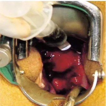
Dynamic scintigraphy was commenced immediately using a large field scintillation camera. Static scintigraphy was pro- longed for 20 to 40 min based on the visualization of sentinel lymph node (Fig. 2). The scintigraphic hot spots in vivo were detected using a hand-held gamma probe (Neoprobe 1500� Neoprobe Corp, Dublin, OH, U.S.A.). About 3 hr after tracer injection, the patients were sent to the operation room and radical hysterectomy with pelvic lymphadenectomy was per- formed following sentinel lymph node frozen biopsy. After induction of general anesthesia, isosulfan blue dye was inject- ed in the peritumoral area with a 20-gauge needle (Fig. 3).
The sentinel lymph node identification was always followed
Fig. 1.Intradermal technetium-99m colloid albumin injection in an early stage cervical cancer.
Fig. 2.Lymphoscintigraphy using a scintillation camera (arrow indicates scintigraphic hot spot and shows the location of lymph node).
by radical hysterectomy. The probe was used before making the abdominal skin incision to identify the area of greatest activity. Background activity was obtained in four areas of abdomen. Usually, sentinel lymph nodes had 6-to 8- fold radioactivity compared to non-sentinel lymph nodes as the background. Careful dissection was performed to identify the blue stained afferent lymphatic vessels, and they were followed up to the first blue-stained lymph node. The hand- held gamma probe was used to confirm the location of the sentinel lymph nodes, as well as to guide the dissection when- ever the afferent lymphatic channels were not detectable (Fig. 4).
Removal of sentinel nodes and pathologic examination
The sentinel node was excised together with a rim of sur- rounding tissue and sent as a separate specimen for intra- operative histologic examination. A pathologist examined all the surgically removed sentinel node specimens using a standard technique. The nodes were isolated fresh from the fat tissue. One section was taken per 5 mm of the greatest dimen- sion. Nodes greater than 5 mm in diameter were divided into two parts along the longitudinal axis. Nodes less than 5 mm in the major axis were totally embedded. Three histological sections were obtained from each specimen at different cut levels and were stained with hematoxylin and eosin.
RESULTS
The 26 patients who underwent radical hysterectomy with
pelvic lymph nodes dissection using preoperative technetium- 99m colloid albumin and intraoperative isosulfan blue dye injection were analyzed for the location of sentinel lymph nodes and for the sensitivity and negative predictive value for the purpose of clinical application of only sentinel lymph node frozen biopsy for less extensive pelvic lymph nodes dis- section in early stage cervical cancer. The order of sentinel
Fig. 3.Peritumoral injection of isosulfan blue dye around the cer- vical lesion.
Fig. 4.Detection of sentinel lymph node using an isosulfan blue dye and hand-held gamma probe (A: hand-held gamma probe.
Arrow indicates the sentinel lymph node).
A
External iliac 18
Obturator 12
Internal iliac 8
Parametrium 8
Common iliac 2
Inguinal 1
Locations of sentinel lymph node Number of detected sentinel lymph node Table 1.Distribution of locations of sentinel lymph node in 26 cervical cancer patients
Frozen biopsy (-)* 20 cases 1 case 21 cases
Frozen biopsy (+)� 1 case 4 cases 5 cases
Total 21 cases 5 cases 26 cases
Permanent pathology (-)�
Permanent
pathology (+)� Total Table 2. Relationship between frozen biopsy of sentinel lymph node and permanent pathology of pelvic lymph node
Frozen biopsy (-)* denotes the benign sentinel lymph node. Frozen biop- sy (+)�signifies the nodal status is malignant. �means there was no meta- static lesion in pelvic lymph nodes and �implies there were metastatic nodes in pelvic cavity.
node detection sites were external iliac, obturator, and inter- nal iliac areas (Table 1). The location of the primary cancer lesion had an influence on the detection sites of sentinel lymph nodes. The average number of sentinel lymph nodes per pa- tient was two. Among 21 cases where sentinel lymph nodes were negative, pelvic lymph nodes were all negative except for in one case. Among 5 cases where sentinel lymph nodes were positive, 4 were positive in pelvic lymph nodes and 1 was negative. Sensitivity and specificity of sentinel lymph node frozen biopsy were 95.2% (20/21) and 80% (4/5), re- spectively. The negative predictive value of the technique was 95.2% (20/21) (Table 2).
DISCUSSION
Sentinel lymph node biopsy in cervical cancer is a promis- ing surgical technique to confirm nodal status and minimize postoperative morbidity (5, 9, 12). There are many different ideas concerning the site of injection of the isosulfan blue dye and radionuclides, the dose of radioactivity used, the inter- val between dye injection or lymphoscintigram and sentinel node frozen biopsy (13-15). Isosulfan blue dye is used for dye- guided sentinel lymph node biopsy in western countries for visual detection. According to the initial experience of only dye-guided sentinel node biopsy, the identification rates of sentinel lymph nodes ranged between 66% and 82% (12).
Radionuclides for lymphatic mapping should be a few hun- dred nanometers in size, to permeate the lymphatic vessels and remain in the sentinel lymph nodes. However, techne- tium-99m HAS is too small (≤5 nm) to be retained in the sentinel lymph nodes and technetium-99m TC is too big (≥500 nm) to migrate through the lymphatic vessels com- pared with technetium-99m colloid albumin (16). These fac- tors contributed to low success rate of sentinel node biopsy with the hand-held gamma probe detector. As for the rela- tive contribution of each method to the detection of the sen- tinel node, it should be underlined that the detection rate was increased by means of combining isosulfan blue dye and technetium-99m colloid albumin. Hence, whenever it is af- fordable, isosulfan blue dye in combination with technetium- 99m colloid albumin should be used for the lymphatic map- ping. The combination of isosulfan blue dye and technetium- 99m colloid albumin can improve the rate of identification of sentinel lymph nodes up to 100%, and seems to be the best way to accurately detect sentinel lymph nodes. If sen- tinel lymph nodes are histologically negative by immediate frozen section examination, extensive pelvic lymph nodes dissection can be omitted. However, it may be difficult to detect micrometastasis of cancer cells by the frozen section of sentinel lymph nodes. To compensate for this false nega- tives, it will be helpful to examine multiple sections of sen- tinel lymph nodes especially with immunohistochemical staining. In conclusion, sentinel node frozen biopsy with iso-
sulfan blue dye and radionuclide was found to be the best way to identify sentinel lymph nodes in early stage cervical cancer.
The acceptable false-negative rate remains unclear, but sen- tinel node biopsy in a multicenter validation study will prove feasible for predicting pelvic lymph nodes metastases. Our findings confirm that the detection rate of the sentinel node (49 of 53, 94%), the overall accuracy (48 of 49, 97%) and false negative rate (1 of 21, 4.76%) are satisfactory for clini- cal application. This prospective study provides evidence for the necessity of coupling lymphoscintigraphy and isosulfan blue dye and hand held gamma probe detector in order to enhance the identification rate of sentinel lymph node. Sen- tinel lymph node biopsy is less invasive than complete pelvic lymph nodes dissection and lowers both postoperative mor- bidity and cost. It is expected that sentinel node frozen biopsy in early stage cervical cancer will be performed as a standard practice for less extensive pelvic lymph nodes dissection in the near future.
REFERENCES
1. Patsner B, Sedlacek TV, Lovecchio JL. Para-aortic node sampling in early stage Ib invasive cervical cancer. Gynecol Oncol 1992; 44:
53-4.
2. Landoni F, Maneo A, Cormio G, Mangioni C. Class II versus class III radical hysterectomy in stage IB-IIA cervical cancer. a prospective randomized study. Gynecol Oncol 2001; 80: 3-12.
3. Ramon M, Cabanas RM. Anatomy and biopsy of sentinel lymph nodes.
Urol Clin of North America 1992; 19: 267-76.
4. Borgstein P, Meijer S. Histological perspective of lymphatic tumor spread and the emergence of the sentinel node concept. Eur J Surg Oncol 1998; 24: 85-9.
5. Cox CE, Pendas S, Cox JM, Joseph E, Shons AR, Yeatman T, Ko NN, Lyman GH, Berman C, Haddad F, Reintgen DS. Guidelines for sentinel node biopsy and lymphatic mapping of patients with breast cancer. Ann Surg 1998; 227: 645-53.
6. Cabanas RM. An approach for the treatment of penile carcinoma.
Cancer 1977; 39: 456-66.
7. Morton DL, Wen DR, Wong JH, Economou JS, Lagle LA, Storm FK, Foshag LJ, Cochran AJ. Technical details of intraoperative lymphatic mapping for early stage melanoma. Arch Surg 1992; 127:
392-9.
8. Singletary E. Management of the axilla in early stage breast cancer.
In Peri MC: Educational Book 34th Annual Meeting ASCO. Alexan- dria VA 1998: 132-41.
9. Veronesi U, Paganelli G, Viale G, Galinberti V, Luini A, Zurrida S, Robertson C, Sacchini V, Veronesi P, Orvieto E, De Cicco C, Intra M, Josi G, Scarpa D. Sentinel lymph node biopsy and axillary dissec- tion in breast cancer. J Natl Cancer Inst 1999; 91: 368-73.
10. Gray Henry. Anatomy of the Human body. The lymphatic system 2000: 1918-50.
11. Sabin FR. The Development of the Lymphatic System. Springer, Hei- delberg 1996: 347-63.
12. Giuliano AE, Jones RC, Brennam M, Statman R. Sentinel lym- phadenectomy in breast cancer. J Clin Oncol 1997; 15: 2345-50.
13. Giuliano AE, Kirgan DM, Guenther JM, Morton DL. Lymphatic mapping and sentinel lymphadenectomy for breast cancer. Ann Surg 1994; 220: 391-401.
14. Noguchi M, Tsugawa K, Kawahara F, Bando E, Miwa K, Minato H, Nonomura A. Dye-guided sentinel lympha-denectomy in clinically
node-negative and node-positive breast cancer patients. Breast can- cer 1998; 5: 381-7.
15. Flett MM, Going JJ, Stanton PD, Cooke TG. Sentinel node localiza- tion in patients with breast cancer. Br J Surg 1998; 85: 991-3.
16. Krag D, Weaver D, Ashikaga T. The sentinel node in breast cancer;
A multicerter validation study. N Eng J Med 1998; 339: 941-6.